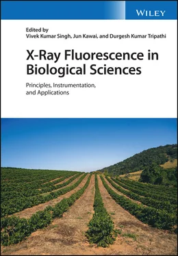
X-Ray Fluorescence in Biological Sciences
Здесь есть возможность читать онлайн «X-Ray Fluorescence in Biological Sciences» — ознакомительный отрывок электронной книги совершенно бесплатно, а после прочтения отрывка купить полную версию. В некоторых случаях можно слушать аудио, скачать через торрент в формате fb2 и присутствует краткое содержание. Жанр: unrecognised, на английском языке. Описание произведения, (предисловие) а так же отзывы посетителей доступны на портале библиотеки ЛибКат.
- Название:X-Ray Fluorescence in Biological Sciences
- Автор:
- Жанр:
- Год:неизвестен
- ISBN:нет данных
- Рейтинг книги:5 / 5. Голосов: 1
-
Избранное:Добавить в избранное
- Отзывы:
-
Ваша оценка:
- 100
- 1
- 2
- 3
- 4
- 5
X-Ray Fluorescence in Biological Sciences: краткое содержание, описание и аннотация
Предлагаем к чтению аннотацию, описание, краткое содержание или предисловие (зависит от того, что написал сам автор книги «X-Ray Fluorescence in Biological Sciences»). Если вы не нашли необходимую информацию о книге — напишите в комментариях, мы постараемся отыскать её.
Discover a comprehensive exploration of X-ray fluorescence in chemical biology and the clinical and plant sciences X-Ray Fluorescence in Biological Sciences: Principles, Instrumentation, and Applications
X-Ray Fluorescence in Biological Sciences: Principles, Instrumentation, and Applications
X-Ray Fluorescence in Biological Sciences — читать онлайн ознакомительный отрывок
Ниже представлен текст книги, разбитый по страницам. Система сохранения места последней прочитанной страницы, позволяет с удобством читать онлайн бесплатно книгу «X-Ray Fluorescence in Biological Sciences», без необходимости каждый раз заново искать на чём Вы остановились. Поставьте закладку, и сможете в любой момент перейти на страницу, на которой закончили чтение.
Интервал:
Закладка:
Acknowledgment
Authors thank Department of Biotechnology (No. BT/RLF/Re‐entry/32/2017),Government of India for funding this project.
References
1 1 Bruins, M.R., Kapil, S., and Oehme, F.W. (2000). Microbial resistance to metals in the environment. Ecotoxicol. Environ. Saf. 45 (3): 198–207.
2 2 Geoffrey, M.G. (2010). Metals, minerals and microbes: geomicrobiology and bioremediation. Microbiology 156 (3): 609–643.
3 3 Lengke, M. and Southam, G. (2006). Bioaccumulation of gold by sulfate‐reducing bacteria cultured in the presence of gold(I)‐thiosulfate complex[J]. Geochim. Cosmochim. Acta 70 (14): 3646–3661.
4 4 Silver, S. and Phung, I.T. (2005). A bacterial view of the periodic table: genes and proteins for toxic inorganic ions. J. Ind. Microbiol. Biotechnol. 32 (11–12): 587–605.
5 5 Dabonne, S., Koffi, B.P.K., Kouadio, E.J.P. et al. (2010). Traditional utensils: potential sources of poisoning by heavy metals. Br. J. Pharmacol. Toxicol. 1 (2): 90–92.
6 6 Griffiths, B.S. and Philippot, L. (2013). Insights into the resistance and resilience of the soil microbial community. FEMS Microbiol. Rev. 37 (2): 112–129.
7 7 Wuana, R.A. and Okieimen, F.E. (2011). Heavy metals in contaminated soils: a review of sources, chemistry, risks and best available strategies for remediation. ISRN Ecol. 2011 (2090–4614): 1–20.
8 8 Abdu, N., Abdullahi, A.A., and Abdulkadir, A. (2017). Heavy metals and soil microbes. Environ. Chem. Lett. (15): 65–84.
9 9 Behera, M., Dandapat, J., and Rath, C.C. (2015). Effect of heavy metals on growth response and antioxidant defense protection in Bacillus cereus. J. Basic Microbiol. 54 (11): 1201–1209.
10 10 Sanyal, S.K., Shuster, J., and Reith, F. Cycling of biogenic elements drives biogeochemical gold cycling. Earth‐Sci. Rev.
11 11 Dong, D.T., Yamaguchi, N., Makino, T., and Amachi, S. (2014). Effect of soil micro‐organisms on arsenite oxidation in paddy soils under oxic conditions. Soil Sci. Plant Nutr. 60 (3): 377–383.
12 12 Dixit, S. and Hering, J.G. (2003). Comparison of arsenic(V) and arsenic(III) sorption onto iron oxide minerals: implications for arsenic mobility. Environ. Sci. Technol. 37 (18): 4182.
13 13 Burton, E.D., Hockmann, K., Karimian, N., and Johnston, S.G. (2019). Antimony mobility in reducing environments: the effect of microbial iron(III)‐reduction and associated secondary mineralization. Geochim. Cosmochim. Acta 245: 278–289.
14 14 Ohnuki, T., Sakamoto, F., Kozai, N. et al. (2004). Mechanisms of arsenic immobilization in a biomat from mine discharge water. Chem. Geol. 212 (3–4): 279–290.
15 15 Toshihiko, O., Noriko, Y., Tomoyuki, M. et al. (2013). Arsenic dissolution from Japanese paddy soil by a dissimilatory arsenate‐reducing bacterium Geobacter sp. OR‐1. Environ. Sci. Technol. 47 (12): 6263–6271.
16 16 Gwynne, P. (2013). Microbiology: there's gold in them there bugs. Nature 495 (7440): 12–13.
17 17 Kaksonen, A.H., Mudunuru, B.M., and Hackl, R. (2014). The role of micro‐organisms in gold processing and recovery – a review. Hydrometallurgy 142: 70–83.
18 18 Nies, D.H. (1999). Microbial heavy‐metal resistance. Appl. Microbiol. Biotechnol. 51 (6): 730–750.
19 19 Reith, F., Etschmann, B., Grosse, C. et al. (2009). Mechanisms of gold biomineralization in the bacterium C. metallidurans. Proc. Natl. Acad. Sci. U. S. A. 106 (42): 17757.
20 20 Das, S.K., Liang, J., Schmidt, M. et al. (2012). Biomineralization mechanism of gold by zygomycete fungi Rhizopus oryzae. ACS Nano 6 (7): 6165.
21 21 Johnston, C.W., Wyatt, M.A., Li, X. et al. (2013). Gold biomineralization by a metallophore from a gold‐associated microbe. Nat. Chem. Biol. 9 (4): 241–243.
22 22 Lengke, M.F., Fleet, M.E., and Southam, G. (2006). Morphology of gold nanoparticles synthesized by filamentous cyanobacteria from gold(I)‐thiosulfate and gold(III) – chloride complexes. Langmuir ACS J. Surf. Colloids 22 (6): 2780.
23 23 Lengke, M.F., Ravel, B., Fleet, M.E. et al. (2006). Mechanisms of gold bioaccumulation by filamentous cyanobacteria from gold(III)‐chloride complex. Environ. Sci. Technol. 40 (20): 6304–6309.
24 24 Stylo, M., Alessi, D.S., Shao, P.Y. et al. (2013). Biogeochemical controls on the product of microbial U(VI) reduction. Environ. Sci. Technol. 47 (21): 12351–12358.
25 25 Fomina, M., Charnock, J.M., Hillier, S. et al. (2010). Fungal transformations of uranium oxides. Environ. Microbiol. 9 (7): 1696–1710.
26 26 Kenneth, K.M., Kelly, S.D., Lai, B. et al. (2004). Elemental and redox analysis of single bacterial cells by X‐ray microbeam analysis. Science 306 (5696): 686–687.
27 27 Etschmann, B., Brugger, J., Fairbrothe, L. et al. (2016). Applying the Midas touch: differing toxicity of mobile gold and platinum complexes drives biomineralization in the bacterium C. metallidurans. Chem. Geol. 438: 103–111.
28 28 Zeng, X., Su, S., Feng, Q. et al. (2015). Arsenic speciation transformation and arsenite influx and efflux across the cell membrane of fungi investigated using HPLC–HG–AFS and in‐situ XANES. Chemosphere 119: 1163–1168.
6 Use of Energy Dispersive X‐Ray Fluorescence for Clinical Diagnosis
Yeasmin Nahar Jolly
Atmospheric and Environmental Chemistry Laboratory, Chemistry Division, Atomic Energy Centre, Dhaka, Bangladesh Atomic Energy Commission
6.1 Introduction
X‐ray fluorescence (XRF) analysis is a method of qualitative and quantitative multi‐elemental detection which is non‐destructive. It is a nuclear analytical instrumental technique designed based on the determination of characteristic fluorescent radiation of a particular element. A suitable point source is applied to de‐excite the inner shell vacancies of sample elements by means of radiation. Of the numerous variants of XRF analysis, two major approaches are wavelength dispersive‐XRF (WDXRF) and energy dispersive‐XRF (EDXRF). They are distinguished from each other by the type of detector used to detect emission spectra, which is particular for each element. The characteristic wavelength of emitted X‐rays from elements obtained by the use of a diffracting crystal is the main focus of WDXRF whereas EDXRF relies on direct measurement of the energy of the X‐rays by collecting ionization produced in a suitable detecting medium. After the emergence of silicon drift detectors (SDD), EDXRF became the most widely used of the two. As compared with EDXRF, WDXRF is quite expensive and has few applications for testing materials in the steel or ceramics industries. In recent years, EDXRF has led over WDXRF and is a powerful tool for elemental analysis to determine major, minor, and trace elements in biological samples. The sample preparation for the EDXRF technique is much easier and therefore less prone to contamination. Generally, no chemicals are used, so system loss is relatively very small. A special feature of EDXRF is that only a very small amount of sample is required, which is in the range mg ‐ μg of a material. Hence it is suitable for clinical measurement of toxic elements in human body tissue, as the collection of human tissue in large volume/quantity is a big problem. Arsenicosis and autism spectrum disorder (ASD) are the conditions which are associated with the accumulation of toxic elements arsenic (As) and lead (Pb) in the human body respectively. Determining their concentrations in human tissues can give a clear picture of their presence (qualitatively and quantitatively) and evaluate their association with those diseases. In this chapter, EDXRF techniques for the determination of As concentration in human scalp hair for the diagnosis of arsenicosis, and determination of Pb concentration in whole blood samples for evaluating the relationship between lead exposure and blood lead concentration as a function of ASD have been discussed.
Читать дальшеИнтервал:
Закладка:
Похожие книги на «X-Ray Fluorescence in Biological Sciences»
Представляем Вашему вниманию похожие книги на «X-Ray Fluorescence in Biological Sciences» списком для выбора. Мы отобрали схожую по названию и смыслу литературу в надежде предоставить читателям больше вариантов отыскать новые, интересные, ещё непрочитанные произведения.
Обсуждение, отзывы о книге «X-Ray Fluorescence in Biological Sciences» и просто собственные мнения читателей. Оставьте ваши комментарии, напишите, что Вы думаете о произведении, его смысле или главных героях. Укажите что конкретно понравилось, а что нет, и почему Вы так считаете.
