Sean Gallagher - Musculoskeletal Disorders
Здесь есть возможность читать онлайн «Sean Gallagher - Musculoskeletal Disorders» — ознакомительный отрывок электронной книги совершенно бесплатно, а после прочтения отрывка купить полную версию. В некоторых случаях можно слушать аудио, скачать через торрент в формате fb2 и присутствует краткое содержание. Жанр: unrecognised, на английском языке. Описание произведения, (предисловие) а так же отзывы посетителей доступны на портале библиотеки ЛибКат.

- Название:Musculoskeletal Disorders
- Автор:
- Жанр:
- Год:неизвестен
- ISBN:нет данных
- Рейтинг книги:5 / 5. Голосов: 1
-
Избранное:Добавить в избранное
- Отзывы:
-
Ваша оценка:
Musculoskeletal Disorders: краткое содержание, описание и аннотация
Предлагаем к чтению аннотацию, описание, краткое содержание или предисловие (зависит от того, что написал сам автор книги «Musculoskeletal Disorders»). Если вы не нашли необходимую информацию о книге — напишите в комментариях, мы постараемся отыскать её.
Hands-on guidance and tools for the prevention of musculoskeletal injuries in the workplace Musculoskeletal Disorders: The Fatigue Failure Mechanism,
Musculoskeletal Disorders: The Fatigue Failure Mechanism
Musculoskeletal Disorders — читать онлайн ознакомительный отрывок
Ниже представлен текст книги, разбитый по страницам. Система сохранения места последней прочитанной страницы, позволяет с удобством читать онлайн бесплатно книгу «Musculoskeletal Disorders», без необходимости каждый раз заново искать на чём Вы остановились. Поставьте закладку, и сможете в любой момент перейти на страницу, на которой закончили чтение.
Интервал:
Закладка:
Cartilage
Cartilage is unique in all of the body tissues in that it is typically avascular, aneural, and alymphatic (Tortora & Derrickson, 2010). Because of these properties, injuries and damage to cartilage can be difficult to detect until they are quite severe, and difficult from which to heal. The general features of cartilage are summarized in Table 3.4.
Structure
Cells
Chondrocytes are the prevalent cells of cartilage, with the number per matrix ratio differing with cartilage type. Chondrocytes arise from chondroblasts, which are proliferating cells that originate from mesenchymal cells after exposure to the transcription factor SOX9 [sex determining region Y (SRY)‐box 9]. Chondroblasts produce type II collagen, aggrecan, proteoglycans, and glycosaminoglycans, and therefore, the cartilaginous matrix. They become embedded in individual lacunae within the matrix that they produce ( Figure 3.15); once embedded, they become chondrocytes. Chondrocytes then maintain the cartilage matrix throughout life, although the numbers of chondrocytes reduce with age. Joint trauma, inflammation, and stress fractures that extend into the cartilage can lead to chondrocyte damage and severe structural damage of the cartilage (Xiong & O'Brien, 2012).
Table 3.4 Summary of Cells, Extracellular Matrix (ECM), Subtypes, and Function of Cartilage and its Subtypes Under Normal Conditions
| Characteristic | Description |
|---|---|
| Tissue type | Dense pliable connective tissue |
| Cells | Main cell types: Chondrocytes, chondroblastsAdditional cell types: Mesenchymal stem cells (low in number) |
| ECM | Hyaline cartilage: Collagen II (15–20%), water (60–80%), GAGs (e.g., hyaluronic acid)Fibrocartilage: High collagen content, lower water content than hyalineElastic cartilage: High elastin fiber content |
| Subtypes | Hyaline (and its subtype, articular cartilage), fibrocartilage, elastic |
| Function | Hyaline: Protection of bony surfaces, especially at points of movementFibrocartilage: Strength and rigidity, joint support and fusionElastic cartilage: Resilience and pliability |
Extracellular matrix
In general, the extracellular material of each type of cartilage is firm but pliable. Cartilage consists of a dense network of collagen fibers (the type dependent on the subtype of cartilage) and sometimes elastic fibers, each embedded in chondroitin sulfate (a jelly‐like substance). The collagen fibers add great strength to cartilage, while the ability of cartilage to assume its original shape after deformation is due to the chondroitin sulfate. The three primary types of cartilage, namely hyaline (articular) cartilage, fibrocartilage, and elastic cartilage, can be distinguished from each other by the type of fibers within the matrix.
Organization
While the cell types are similar in each type of cartilage, the organization of cells and collagen/elastin fibrils differ extensively between types. The characteristics of each are described in detail next.
Hyaline Cartilage
Structure
Hyaline cartilage is the most abundant type of cartilage ( Figure 3.14). It is located at the ends of long bones (where it is called articular cartilage; Figure 3.14a,b), epiphyseal growth plates ( Figure. 3.14c,d), ribs (where it is called costal cartilage), and in parts of the larynx, trachea, bronchi, and bronchial tubes. During growth, chondrocytes and hyaline cartilage are also present within the epiphyseal plate, becoming hypertrophic and releasing factors necessary for osteoblast, osteoclast, and endothelial cell invasion needed for bone lengthening ( Figure 3.14c,d). Hyaline cartilage contains numerous chondrocytes responsible for manufacturing, secretion, organization, and maintenance of the organic components of the extracellular matrix (Nordin & Frankel, 2012). The ground substance is homogeneous and amorphous and is composed of fine collagen type II fibrils embedded in a concentrated solution of proteoglycans. Specifically, the collagen content of hyaline cartilage ranges from 15 to 20% of the wet weight. The matrix of hyaline cartilage contains three types of glycosaminoglycans (hyaluronic acid, chondroitin sulfate, and keratin sulfate). The chondroitin and keratin sulfates are joined together by a core protein to form a proteoglycan monomer. The proteoglycans account for 4–7% by wet weight (Nordin & Frankel, 2012; Ross, Romrell, & Kaye, 1995). About 80 proteoglycans are associated with each hyaluronic acid molecule in large aggregates reinforced by linking‐type proteins. These aggregates are bound to the thin collagen fibrils by electrostatic interactions and cross‐linking glycoproteins. The remainder of the matrix is composed of water (60–78%), inorganic salts, and small amounts of link proteins, glycoproteins, and lipids. Some of the water is loosely bound, allowing diffusion of small metabolites to the chondrocytes, which is key in this typically avascular tissue.
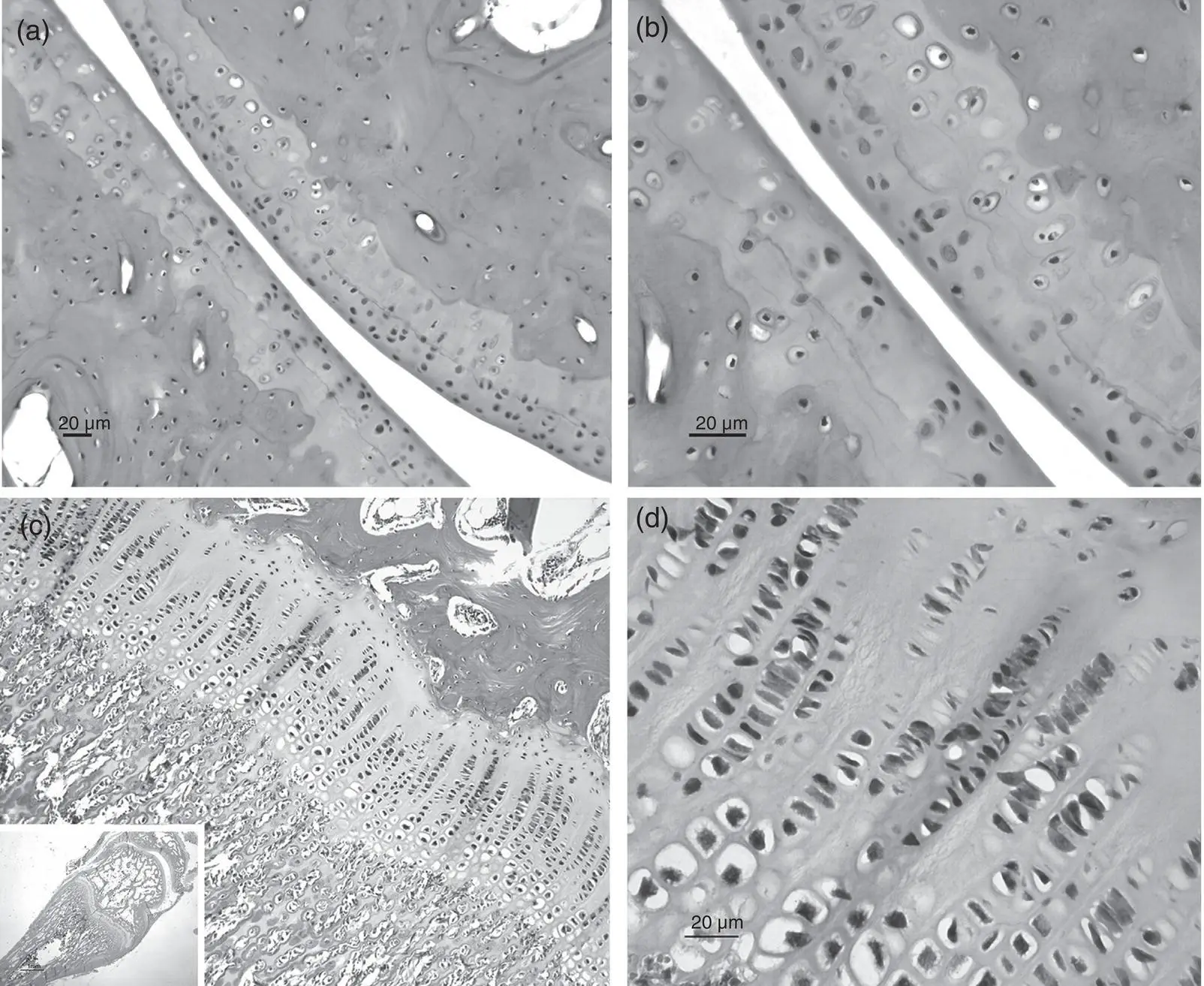
Figure 3.14 Hyaline cartilage. (a and b) Hyaline cartilage in the articular ends of the distal radius and a carpal bone of the radiocarpal joint. This is a plane‐type joint. The higher power image of panel B shows chondrocytes clustering at several sites. (c and d) Hyaline cartilage in an epiphyseal plate (growth plate) of a radial bone. Low to higher power images are shown.
Articular cartilage is a type of hyaline cartilage located in freely moving joints ( Figure 3.14a,b) in which the joint is encased by an articular capsule and the bones connect with each other in a fluid‐filled cavity known as the synovial cavity. Articular cartilage is organized into zones termed superficial, middle, deep, and a calcified zone (deepest). The most superficial zone is in contact with the synovial fluid that contains nutrients that diffuse into the cartilage. This zone protects the deeper layers from shear stresses and constitutes 10–20% of the thickness of articular cartilage. The chondrocytes in this upper zone are relatively high in number and flattened in shape, and the collagen fibers are tightly packed and are aligned in parallel to the articular surface. This combined structure resists the sheer, tensile, and compressive forces imposed by articulation and aids the protection and maintenance of the deeper structures. The middle zone represents 40–60% of the total cartilage volume. In this zone, the chondrocytes are spherical in shape and low in density and the collagen is obliquely organized (to help resist compressive forces). The deep zone of articular cartilage zone represents around 30% of the total cartilage volume. The collagen fibrils in this zone are arranged perpendicular to the articular surface and are large in diameter. The chondrocytes are arranged in columns ( Figure 3.14a,b) that are in parallel to the collagen fibers and perpendicular to the joint line. As a consequence, this zone provides the greatest resistance to compressive forces. The deepest zone is the calcified cartilage layer (and is separated from the deep zone by a histological stain detectable “tide mark”). Like the deep zone, the collagen fibrils are arranged perpendicular to the articular surface. Unlike the other zone, calcification is present and the cell population is scare and hypertrophic. Its main purpose is to anchor collagen fibrils to the underlying subchondral bone.
Function
The large amounts of hyaluronic acid and other components in hyaline cartilage help retain water in the extracellular matrix. As a consequence, this type of cartilage provides resilience and pliability and is well adapted to serve in a weight‐bearing joint, especially at points of movement. Hyaline cartilage also reduces friction and protects bony surfaces.
Читать дальшеИнтервал:
Закладка:
Похожие книги на «Musculoskeletal Disorders»
Представляем Вашему вниманию похожие книги на «Musculoskeletal Disorders» списком для выбора. Мы отобрали схожую по названию и смыслу литературу в надежде предоставить читателям больше вариантов отыскать новые, интересные, ещё непрочитанные произведения.


![Ally Carter - [Gallagher Girls 02 ] - Cross My Heart & Hope To Spy](/books/262178/ally-carter-gallagher-girls-02-thumb.webp)


![John Bruce - The Lettsomian Lectures on Diseases and Disorders of the Heart and Arteries in Middle and Advanced Life [1900-1901]](/books/749387/john-bruce-the-lettsomian-lectures-on-diseases-and-disorders-of-the-heart-and-arteries-in-middle-and-advanced-life-1900-1901-thumb.webp)
Обсуждение, отзывы о книге «Musculoskeletal Disorders» и просто собственные мнения читателей. Оставьте ваши комментарии, напишите, что Вы думаете о произведении, его смысле или главных героях. Укажите что конкретно понравилось, а что нет, и почему Вы так считаете.