Sean Gallagher - Musculoskeletal Disorders
Здесь есть возможность читать онлайн «Sean Gallagher - Musculoskeletal Disorders» — ознакомительный отрывок электронной книги совершенно бесплатно, а после прочтения отрывка купить полную версию. В некоторых случаях можно слушать аудио, скачать через торрент в формате fb2 и присутствует краткое содержание. Жанр: unrecognised, на английском языке. Описание произведения, (предисловие) а так же отзывы посетителей доступны на портале библиотеки ЛибКат.

- Название:Musculoskeletal Disorders
- Автор:
- Жанр:
- Год:неизвестен
- ISBN:нет данных
- Рейтинг книги:5 / 5. Голосов: 1
-
Избранное:Добавить в избранное
- Отзывы:
-
Ваша оценка:
Musculoskeletal Disorders: краткое содержание, описание и аннотация
Предлагаем к чтению аннотацию, описание, краткое содержание или предисловие (зависит от того, что написал сам автор книги «Musculoskeletal Disorders»). Если вы не нашли необходимую информацию о книге — напишите в комментариях, мы постараемся отыскать её.
Hands-on guidance and tools for the prevention of musculoskeletal injuries in the workplace Musculoskeletal Disorders: The Fatigue Failure Mechanism,
Musculoskeletal Disorders: The Fatigue Failure Mechanism
Musculoskeletal Disorders — читать онлайн ознакомительный отрывок
Ниже представлен текст книги, разбитый по страницам. Система сохранения места последней прочитанной страницы, позволяет с удобством читать онлайн бесплатно книгу «Musculoskeletal Disorders», без необходимости каждый раз заново искать на чём Вы остановились. Поставьте закладку, и сможете в любой момент перейти на страницу, на которой закончили чтение.
Интервал:
Закладка:
Bone Function
Bones serve numerous functions in the human body. Bones provide strength and structural stability to the body and provide a means by which loads can be transferred from one part of the body to another. Biomechanically, they are critical structures in that they provide levers and points of attachment for tendons (driven by muscle contraction) that are essential in permitting movement of the human body. Bones also play an important structural protective role for important organs of the body (most notably as the skull around the brain and the rib cage around the heart and lungs).
However, there is much more to bone than its structural and protective roles. Bones themselves are living dynamic organs and are quite complex in nature. These organs are constantly changing and remodeling to adapt to the stresses imposed them. This attribute permits both restorative and/or adaptive repair and helps to prevent fracture development when damage accumulation is not excessively rapid. Bone remodeling and maintenance is a normal, homeostatic process mediated by chondrocytes, bone‐forming osteoblasts, bone‐resorbing osteoclasts, and the mechanosensing osteocytes. Bone remodeling is triggered by growth, mechanical loading (muscular and/or cyclical), injuries (microcracks or fractures), and various local or systemic cytokines, chemokines, and hormones. During active remodeling, bone matrix is resorbed and replaced where needed in response to the loading demands on the bone. Many studies have demonstrated an anabolic effect of loading on bones in case studies or animal models (Schriefer, Warden, Saxon, Robling, & Turner, 2005; Srinivasan et al., 2003; Warden, Fuchs, Castillo, Nelson, & Turner, 2007; Zhang, Sun, Turner, & Yokota, 2007). Other studies show that depending on the intensity, frequency, and form of loading, persistent intense bone loading can also result in long‐term damage, excessive resorption, and detrimental changes to the cartilage and bone [reviewed in (Barbe & Popoff, 2020)]. This topic will be explored further in Chapter 11.
Bone is also the site of a number of hematopoietic cells and blood cell formation—all essential to the body’s nutrition and protection against infection and damaged tissue. Bones are a major site for the storage of minerals, especially calcium and phosphorus. Lastly, bone plays a critical role in storing nutrients and lipids that serve as an energy reserve for the body.
Ligament
Ligaments are structures that hold bones together at joints ( Figure 3.20). They are made up of dense regularly arranged connective tissues with a predominance of collagenous fibers arranged into bundles, and a small number of fibroblastic‐like or synoviocyte‐like cells. Ligaments can be either extrinsic ligaments located outside a synovial joint or strategic reinforcing thickenings of a joint capsule (intrinsic ligaments). These features are summarized in Table 3.6.
Ligament Structure
Ligaments have similarities yet differences from tendons in important respects. Ligaments have a similar hierarchical organization of collagen structures as tendons, although the collagen fibers tend to be more loosely packed in ligaments (the collagen fibrils are slightly less in volume fraction and organization than tendon). However, ligaments have a higher percentage of proteoglycan matrix than tendons. Another significant difference in structure between ligaments and tendons is the amount of elastin found in ligaments. Elastin comprises less than 4% of the dry weight of tendons; in contrast, typical ligaments exhibit 4–9% elastin as dry weight, while some highly extensible ligaments have over 70% elastin as dry weight. The elastin network in the ligament resides along and between the collagen fibers (Zitnay & Weiss, 2018).
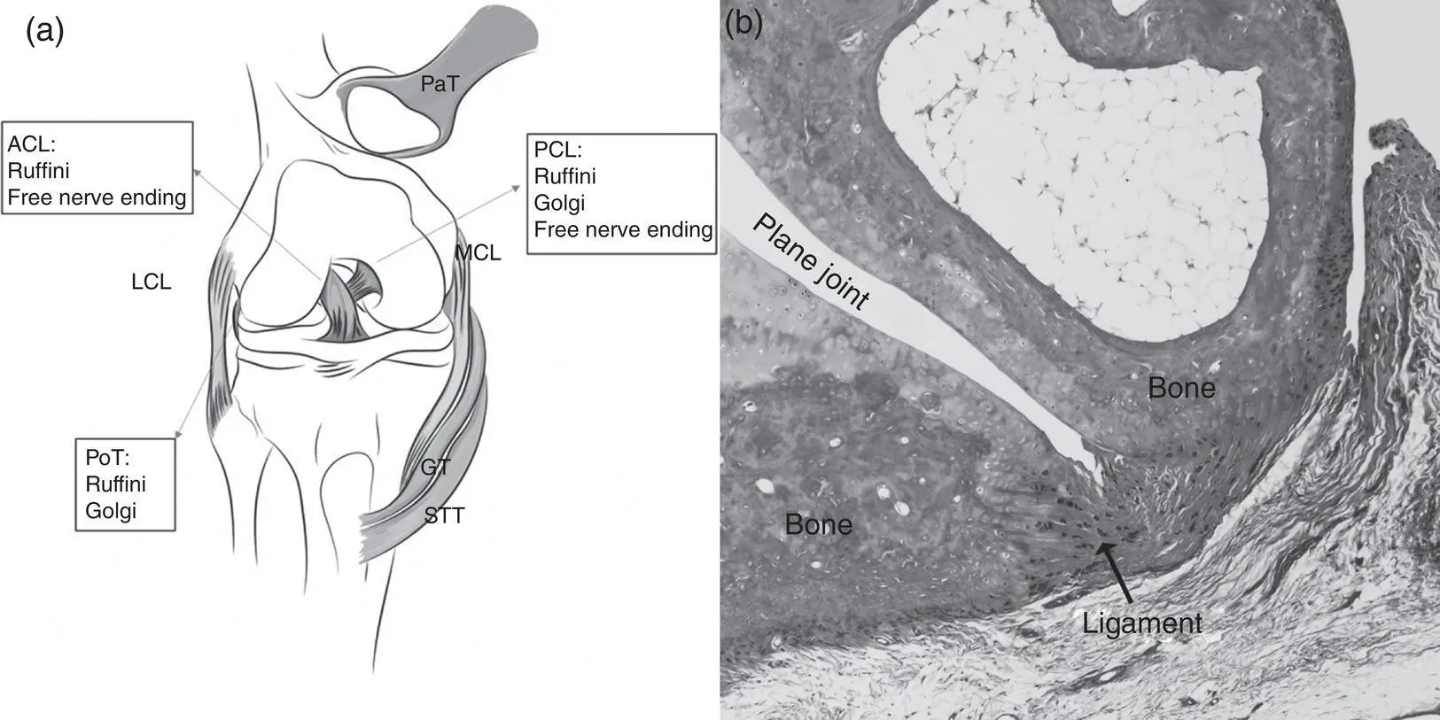
Figure 3.20 Ligaments. (a) A diagram showing the ligament of the knee: lateral collateral ligament (LCL), medial collateral ligament (MCL), anterior and posterior cruciate ligaments (ACL and PCL), and several other tendons. Sensory receptors present in the ligaments are listed and include free nerve endings (pain endings), Ruffini endings (pressure receptors), and Golgi tendon organs (tension receptors).
From Cabuk, H., & Ku şku Çabuk, F. (2016). Mechanoreceptors of the ligaments and tendons around the knee. Clinical Anatomy , 29 ,(6),789–795. doi: 10.1002/ca.22743.
(b) An image of a ligament joining carpal bones.
Table 3.6 Summary of Cells, Extracellular Matrix (ECM), Subtypes, and Function of Ligaments Under Normal Conditions
| Characteristic | Description |
|---|---|
| Tissue type | Dense regular connective tissues |
| Cells | Fibroblastic synoviocyte‐like type cells |
| ECM | Collagen, proteoglycans, elastin (varies from 4 to 70% of dry weight dependent on ligament) |
| Subtypes | Typical (4–9% elastin) to highly extensible (up to 70% extensible) |
| Function | Support and strength to joints |
Ligament Function
The function of ligaments is to provide support and structure to the joints (while providing some flexibility in joint motion). They also prevent excessive motion and provide feedback on positional information of the joints through sensory endings in the ligaments. Ligaments serve to join two or more bones together; both at rest and during the normal range of motion of the joint in question. Ligaments limit the range of motion through passive stabilization and guiding joints through their normal range of motion under tensile load. Thus, ligaments function to transfer forces from bone to bone in a particular joint, both at rest and during movement, and help to guide motion while providing structural stability to the joint. The range of motion of different joints can vary widely; thus, ligaments can assume a wide variety of shapes and sizes. Ligaments are pliant and flexible, but they also provide strength and support to the joint resulting from forces that may be applied.
Joint ligaments are also highly innervated near the site of attaching tendons and in the outer layer of the joint capsule. This innervation will be discussed further in Chapter 4.
Joints
Bones are too rigid to bend without damage. Fortunately, there are multiple bones throughout the body joined together by joints (the articulation or union between two or more bones or parts of bones). This segmentation of the skeleton allows for movement and segmental growth. As is the case with all aspects of the musculoskeletal system, the structure of a joint, specifically the manner in which the joints are held together, determines its function. Although there are many ways to classify joints, the simplest is a functional classification of little to no movement (synarthroses, syndesmoses, and synchondroses) versus freely movable (diarthroses) ( Table 3.7).
Structure of Synarthroses
Synarthroses are bound together by either fibrous (symphyses and syndesmoses) or cartilaginous tissue (synchondroses). Symphyses are joints joined by fibrocartilage, in which the two opposing surfaces are covered by hyaline cartilage (thus, symphyses are also considered cartilaginous joints). The strength of the fibrocartilage allows for only a little movement but much stability, while the hyaline cartilage on the articulating surfaces allows for shock absorption. The pubic symphyses and intervertebral joints are examples of symphyses ( Figure 3.15). The adjoining bones of fibrous syndesmoses are bound together by a thin sheet of fibrous tissue, either a ligament or a fibrous membrane. Since the fibrous tissue is flexible, these joints allow partial movement. The amount of movement allowed depends on the length of the fibers uniting the bones. Examples of syndesmoses are the suture joints of the skull and the union of the radius and ulna in the forearm by an interosseous membrane. Synchrondroses are joints joined together by cartilage and permit slight bending in early life. A key example is the joining of the epiphysis of a long bone with the metaphysis by a cartilaginous growth plate (the physis) ( Figure 3.14). This is a temporary synchondrosis since the growth plate eventually ossifies in the mature adult.
Читать дальшеИнтервал:
Закладка:
Похожие книги на «Musculoskeletal Disorders»
Представляем Вашему вниманию похожие книги на «Musculoskeletal Disorders» списком для выбора. Мы отобрали схожую по названию и смыслу литературу в надежде предоставить читателям больше вариантов отыскать новые, интересные, ещё непрочитанные произведения.


![Ally Carter - [Gallagher Girls 02 ] - Cross My Heart & Hope To Spy](/books/262178/ally-carter-gallagher-girls-02-thumb.webp)


![John Bruce - The Lettsomian Lectures on Diseases and Disorders of the Heart and Arteries in Middle and Advanced Life [1900-1901]](/books/749387/john-bruce-the-lettsomian-lectures-on-diseases-and-disorders-of-the-heart-and-arteries-in-middle-and-advanced-life-1900-1901-thumb.webp)
Обсуждение, отзывы о книге «Musculoskeletal Disorders» и просто собственные мнения читателей. Оставьте ваши комментарии, напишите, что Вы думаете о произведении, его смысле или главных героях. Укажите что конкретно понравилось, а что нет, и почему Вы так считаете.