Sean Gallagher - Musculoskeletal Disorders
Здесь есть возможность читать онлайн «Sean Gallagher - Musculoskeletal Disorders» — ознакомительный отрывок электронной книги совершенно бесплатно, а после прочтения отрывка купить полную версию. В некоторых случаях можно слушать аудио, скачать через торрент в формате fb2 и присутствует краткое содержание. Жанр: unrecognised, на английском языке. Описание произведения, (предисловие) а так же отзывы посетителей доступны на портале библиотеки ЛибКат.

- Название:Musculoskeletal Disorders
- Автор:
- Жанр:
- Год:неизвестен
- ISBN:нет данных
- Рейтинг книги:5 / 5. Голосов: 1
-
Избранное:Добавить в избранное
- Отзывы:
-
Ваша оценка:
Musculoskeletal Disorders: краткое содержание, описание и аннотация
Предлагаем к чтению аннотацию, описание, краткое содержание или предисловие (зависит от того, что написал сам автор книги «Musculoskeletal Disorders»). Если вы не нашли необходимую информацию о книге — напишите в комментариях, мы постараемся отыскать её.
Hands-on guidance and tools for the prevention of musculoskeletal injuries in the workplace Musculoskeletal Disorders: The Fatigue Failure Mechanism,
Musculoskeletal Disorders: The Fatigue Failure Mechanism
Musculoskeletal Disorders — читать онлайн ознакомительный отрывок
Ниже представлен текст книги, разбитый по страницам. Система сохранения места последней прочитанной страницы, позволяет с удобством читать онлайн бесплатно книгу «Musculoskeletal Disorders», без необходимости каждый раз заново искать на чём Вы остановились. Поставьте закладку, и сможете в любой момент перейти на страницу, на которой закончили чтение.
Интервал:
Закладка:
Fibrocartilage
Structure
Fibrocartilage is located at the intervertebral discs between the vertebrae ( Figure 3.15), pubic symphysis, and in the menisci of knees. Fibrocartilage is distinct from hyaline cartilage in composition and structure. Fibrocartilage has a higher dry weight of collagen and less water, making it tougher and less resilient than articular cartilage. Its collagen fibers are thick (and therefore visible) and densely deposited in the tissue. In fact, the collagen fibers are so tightly packed that there is little evidence of an extracellular matrix. The chondrocytes are scattered among the many bundles of collagenous fibers.
Function
The structure of fibrocartilage combines both strength and rigidity; therefore, it is key to the support and fusion of joints in which it is present. Fibrocartilage is an anisotropic material that exhibits different strength capacities depending on the direction of loading (Murphy, Black, & Hastings, 2016). For example, it has been shown that fibrocartilage is strong during tension occurring in parallel to the orientation of the collagen fibers, but weaker during shear loading (Mansour, 2009).
Elastic Cartilage
Structure
The ligamentum flava joining the laminae of vertebrae is a type of elastic cartilage. Elastic cartilage is also present in the epiglottis, external ear, and auditory tubes. It is distinguished from other types of cartilage by the presence of elastic fibers in the matrix, in addition to the normal components of hyaline cartilage. As shown in Figure 3.16, a thread‐like network of elastic fibers surrounds chondrocytes in this type of cartilage.

Figure 3.15 Fibrocartilage in the intervertebral discs between the vertebrae. (a) Low power image showing the disc between two rat lumbar level vertebrae. (b) Mid‐power image showing fibrocartilage encircling the nucleus pulposus (the gelatinous interior of intervertebral discs) located in the middle of the disc. (c) High power image showing the presence of chondrocytes within thick collagen fibers in the fibrocartilage.
Function
The elastic materials in this type of cartilage give it elastic properties in addition to the resilience and pliability of its other hyaline cartilage components. Related to musculoskeletal tissue function, the elastic fibers in the ligamentum flava of the vertebra aid in the rebound of vertebrae from a flexed position to an upright position. Thus, elastic cartilage gives support and maintains the shape of its inclusionary structure. Unlike hyaline cartilage, the matrix of elastic cartilage does not calcify.
Bone
Bone is a complex and dynamic tissue that continuously undergoes renewal and repair throughout life to fulfill its functions. It bears the weight of the body and provides the framework (i.e., helps the body maintain its shape). The anchoring of muscles to bone also permits mechanical movement and locomotion by providing levers for the muscles (Clarke, 2008). Bone is composed of several cell types and a predominantly collagenous extracellular matrix (the osteoid) that becomes mineralized by the deposition of calcium hydroxyapatite. Bone also hosts hematopoietic cells and provides the environment for hematopoiesis within marrow spaces (which is also the storage site of iron) (Yang, 2010). Its general characteristics are summarized in Table 3.5.
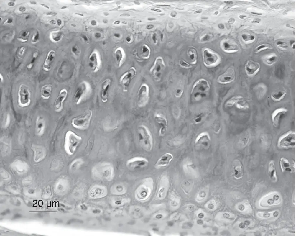
Figure 3.16 Elastic cartilage.
Table 3.5 Summary of Cells, Extracellular Matrix (ECM), Subtypes, and Function of Bone Under Normal Conditions
| Characteristic | Description |
|---|---|
| Tissue type | Dense mineralized connective tissue |
| Cells | Main cell types: Osteoblasts, bone lining cells, osteocytes, osteoclasts, bone marrow–derived mesenchymal stem cellsAdditional cell types: Hematopoietic cells in marrow spaces |
| ECM | Collagen type I (~70%), 25% water, inorganic minerals (e.g., calcium, phosphorus) |
| Subtypes | Cortical bone (compact bone), trabecular (cancellous/spongy bone) |
| Function | Strength, stability, lever at points of attachment, storage of minerals/lipids/nutrients, blood cell formation |
Bone Structure
Cells
The cells in bone include osteoblasts, bone lining cells, osteocytes, and osteoclasts ( Figure 3.17). They also contain nervous tissue, epithelial cells, and various types of hematopoietic cells. During bone growth and remodeling, there is a delicate balance between osteoblast and osteoclast function. This balance is orchestrated by osteocyte signaling, which dictates the outcome of loading on bone.
Osteoblasts—the producers of bone matrix
Osteoblasts are derived from pluripotent mesenchymal stem cells of the osteochondroprogenitor lineage that have the potential to proliferate and differentiate into several connective tissue cell types depending on the stimulus within the local microenvironment. Given the appropriate stimuli, osteoprogenitor cells will first give rise to preosteoblasts and then osteoblasts (Caplan, 1991; Friedenstein, Chailakhyan, & Gerasimov, 1987; Owen, 1988; Pittenger et al., 1999; Yamaguchi et al., 1991). Osteoblasts are cuboidal mononucleated cells normally found at the bone surface ( Figure 3.17a). They are responsible for the synthesis of the organic components of the bone matrix (the osteoid) and mediate its mineralization. Osteoblasts also produce a variety of other noncollagenous proteins, including osteocalcin (which is often used as a serum marker of bone formation). Osteoblasts are an important source of receptor‐activated nuclear factor kappa B ligand (RANKL) that stimulates the differentiation and activation of osteoclasts (Boyce & Xing, 2007). Once osteoblasts become trapped and encased within the mineralized matrix, they are called osteocytes ( Figure 3.17a,c).
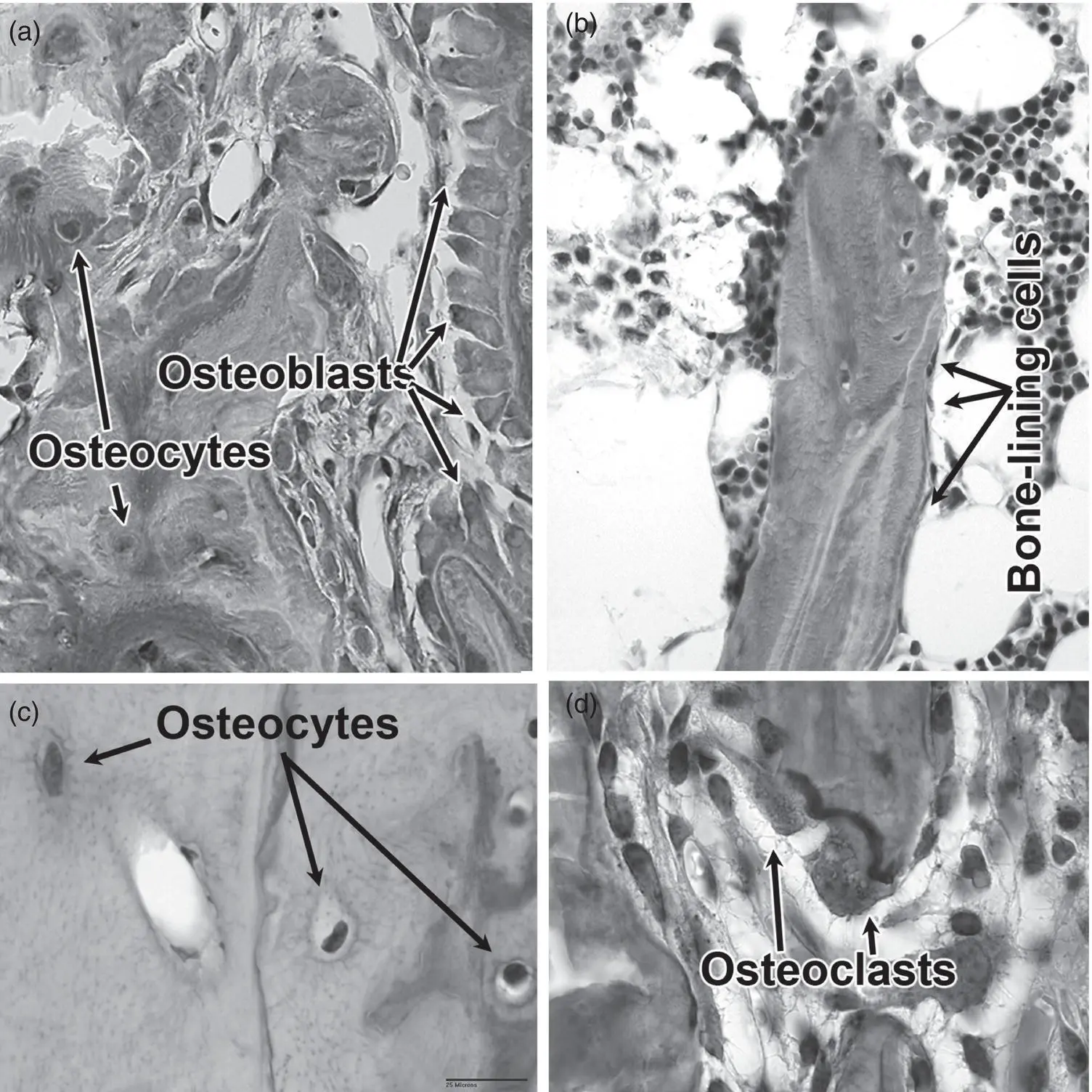
Figure 3.17 Bone cells. (a) Osteoblasts on the surface of trabeculae (larger cuboidal cells) and osteocytes that are embedded within the trabecular bone are depicted. (b) Bone lining cells are flat cells located on the surface of trabeculae. (c) A high power image of osteocytes, this time embedded within cortical bone. (d) Osteoclasts on the surface of trabecular bone.
Bone lining cells—effector cells on standby
Bone lining cells originate from the same lineage as osteoblasts. They are mononucleated, flattened cells found lining surfaces in areas where there is no active bone formation ( Figure 3.17b). They are believed to be a source of already committed osteogenic cells, known as dormant osteoblasts. They can be reactivated to the secreting form with the proper stimulus and under conditions that warrant active bone formation such as during mechanical stimulation, remodeling, and fracture repair (Miller, de Saint‐Georges, Bowman, & Jee, 1989).
Osteocytes—the mechanotransducers and maintainers of bone
Osteocytes are mature bone cells that live within the substance of bone and comprise 90–95% of all bone cells (Bonewald, 2007). Osteocytes are embedded in spaces (lacunae) in the interior of bone and are connected to adjacent cells by long cytoplasmic processes radiating from the cell body that lie within channels (canaliculi) throughout the mineralized matrix of bone ( Figure 3.17) (Hirose et al., 2007; Lian & Stein, 2008). The processes of adjacent osteocytes make contact via gap junctions as well as with the osteoblasts and bone lining cells, maintaining the vitality of osteocytes by passing nutrients and metabolites between blood vessels and distant osteocytes (Jiang, Siller‐Jackson, & Burra, 2007).
Читать дальшеИнтервал:
Закладка:
Похожие книги на «Musculoskeletal Disorders»
Представляем Вашему вниманию похожие книги на «Musculoskeletal Disorders» списком для выбора. Мы отобрали схожую по названию и смыслу литературу в надежде предоставить читателям больше вариантов отыскать новые, интересные, ещё непрочитанные произведения.


![Ally Carter - [Gallagher Girls 02 ] - Cross My Heart & Hope To Spy](/books/262178/ally-carter-gallagher-girls-02-thumb.webp)


![John Bruce - The Lettsomian Lectures on Diseases and Disorders of the Heart and Arteries in Middle and Advanced Life [1900-1901]](/books/749387/john-bruce-the-lettsomian-lectures-on-diseases-and-disorders-of-the-heart-and-arteries-in-middle-and-advanced-life-1900-1901-thumb.webp)
Обсуждение, отзывы о книге «Musculoskeletal Disorders» и просто собственные мнения читателей. Оставьте ваши комментарии, напишите, что Вы думаете о произведении, его смысле или главных героях. Укажите что конкретно понравилось, а что нет, и почему Вы так считаете.