Sean Gallagher - Musculoskeletal Disorders
Здесь есть возможность читать онлайн «Sean Gallagher - Musculoskeletal Disorders» — ознакомительный отрывок электронной книги совершенно бесплатно, а после прочтения отрывка купить полную версию. В некоторых случаях можно слушать аудио, скачать через торрент в формате fb2 и присутствует краткое содержание. Жанр: unrecognised, на английском языке. Описание произведения, (предисловие) а так же отзывы посетителей доступны на портале библиотеки ЛибКат.

- Название:Musculoskeletal Disorders
- Автор:
- Жанр:
- Год:неизвестен
- ISBN:нет данных
- Рейтинг книги:5 / 5. Голосов: 1
-
Избранное:Добавить в избранное
- Отзывы:
-
Ваша оценка:
Musculoskeletal Disorders: краткое содержание, описание и аннотация
Предлагаем к чтению аннотацию, описание, краткое содержание или предисловие (зависит от того, что написал сам автор книги «Musculoskeletal Disorders»). Если вы не нашли необходимую информацию о книге — напишите в комментариях, мы постараемся отыскать её.
Hands-on guidance and tools for the prevention of musculoskeletal injuries in the workplace Musculoskeletal Disorders: The Fatigue Failure Mechanism,
Musculoskeletal Disorders: The Fatigue Failure Mechanism
Musculoskeletal Disorders — читать онлайн ознакомительный отрывок
Ниже представлен текст книги, разбитый по страницам. Система сохранения места последней прочитанной страницы, позволяет с удобством читать онлайн бесплатно книгу «Musculoskeletal Disorders», без необходимости каждый раз заново искать на чём Вы остановились. Поставьте закладку, и сможете в любой момент перейти на страницу, на которой закончили чтение.
Интервал:
Закладка:
Extracellular matrix
The dry mass of a tendon accounts for about 30% of the total tendon mass, with water making up the remainder (Sharma & Maffulli, 2005). This dry mass portion is 65–80% collagen, 0.2% proteoglycans and inorganic substances, 1–2% elastin, and 4.5% other proteins (O'Brien, 1997). The most abundant type of collagen in tendons is collagen I (95%), with the remaining being collagen III and IV. In immature and healing tendons, collagen III is the initial collagen deposited by tenocytes (and occurs in a disorganized manner) and is subsequently replaced by collagen I. The direction of the collagen fibers is aligned linearly with the stresses exerted on the tendon. Several extracellular proteins cross‐link and act as structural scaffolds for the larger collagen I interdigitating fibrils. These include decorin, fibromodulin, laminin 2, and tenascin C. The inorganic components (calcium and magnesium) are involved in growth, development, and normal metabolism of tendon tissue (Kannus, 2000). There is also an interstitial matrix ( Figure 3.13) that contains ground substance, such as mucin (Ali et al., 2015).
Organization
Like muscle, tendons exhibit a hierarchical bundling structure ( Figure 3.13). At the smallest level, collagen molecules (tropocollagen) are bundled into collagen fibrils, which are bundled together into interdigitating collagen fibrils. These are then further bundled into primary fiber bundles (subfascicles) by an endotenon connective tissue layer. The subfascicles are bundled together into a larger fascicle that is also surrounded by endotenon. At the fascicle level, a characteristic “crimp” pattern can be seen histologically. Several fascicles are then bundled together to form the whole tendon, all of which are surrounded by an outer denser epitenon wrapping ( Figures 3.12and 3.13). This nested structure allows the bundles to slide independently from one another. As mentioned above, there are several components to the extracellular matrix of tendons that are hierarchically arranged and cross‐linked together. This highly ordered structure provides strength, durability, high tensile strength, and stability during force transmission.
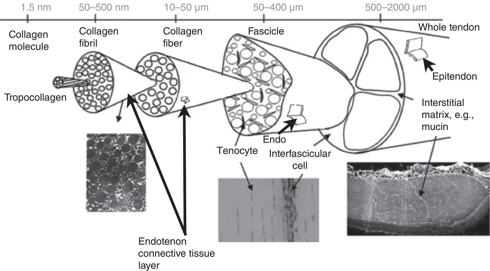
Figure 3.13 Schematic depicting the hierarchical structure of tendon, with inset images: Transverse sections show fibril and fascicle packing. The longitudinal histological section (hematoxylin and eosin) shows the tendon cell populations.
Modified from: Screen, H. R., Berk, D. E., Kadler, K. E., Ramirez, F., & Young, M. F. (2015). Tendon functional extracellular matrix. Journal of Orthopaedic Research , 33 (6), 793–799, First published: 30 January 2015, doi: 10.1002/jor.22818. Wiley.
At the macroscopic level, tendons are not homogeneous structures. They can be broadly divided into three regions along their axis. There is a muscle–tendon junction (the myotendinous junction), a tendon midbody, and a tendon–bone junction (the enthesis) locates where tendons attach to bone. The myotendinous junction and enthesis have distinct structural, cellular, and molecular characteristics compared to the midbody of a tendon. The myotendinous junction displays a “zipper” like morphology, where skeletal muscle fibers and tendon collagen fibrils interdigitate. At this same point, the cytoskeletal network and basement membrane of muscle fiber bundles directly attach to the collagen fibrils of tendons. Regarding entheses ( Figure 3.12c), some are primarily fibrous and noncartilaginous, although most tendon–bone attachments employ fibrocartilagenous entheses. The latter consist of four zones of connective tissue, listed in order from the tendon to the bone: (a) fibrous tissue originating from tendon (the aligned collagen fibrils surrounding fibroblasts resemble the tendon midbody), (b) uncalcified fibrocartilage, (c) calcified fibrocartilage, and (d) subchondral bone. This gradual change in structure reduces mechanical stress levels between the tensile tendon and the brittle bone.
Some tendons are also enclosed by tube‐like synovial sheaths, for example, at the wrist and ankle. These sheaths are made up of two layers, both lined by flattened synovial cells of mesenchymal origin. The inner visceral layer is attached to the surface of the tendon, while the outer parietal layer is adjacent to neighboring structures. The space between these layers contains a viscous fluid similar in composition to the synovial fluid of synovial joints. This liquid is composed of water, proteins, hyaluroanate, and other glycosaminoglycans. This synovial secretion acts as a lubricant permitting easy sliding movements of a tendon within its sheath. Tendinitis or tenosynovitis is an inflammation of this tendon sheath and are painful conditions that can follow trauma, excessive strain, or excessive exercise (see Chapters 2and 11).
Tendons have a very rich neural network and are often innervated from the muscles with which they are associated or from local nerves. There are many nerve endings at the sites of myotendinous junctions and bone–tendon junctions, including Golgi tendon organs and free nerve endings (which are typically pain fibers), as discussed further in Chapter 4. With regard to vascularization, the interior of healthy tendons is fairly devoid of blood vessels, with most of the blood vessels localized to the tendon sheaths.
Function of Tendon Components
Transfer of forces
The myotendinous junctions and entheses described earlier facilitate force transfer between these structures. For example, the myotendinous junctions allow tendons to absorb sudden shocks in order to limit muscular damage (Selvanetti, Cipolla, & Puddu, 1997), and the entheses transfer muscle tensile forces across tendons and onto bone, enabling joint flexion. For this purpose, the collagen fibers of tendons are continuous with those in the endomysium and perimysium connective tissue layers in muscles, as well as with the periosteum of bones. However, tendons fibrils do not run continuously from muscle to bone. Instead, stress is transferred through a hydrated proteoglycan‐rich gel matrix in which the fibers of this fiber‐reinforced composite are contained (Ker, 2002; Shepherd & Screen, 2013).
Mechanotransduction in tenocytes
Tenocytes are stellate in shape when examined in longitudinal sections, with elongated protrusions in all directions. These protrusions contact tenocytes within the same and adjacent rows, thus forming an intricate tendon network. There are gap junctions at these contact points. Gap junction proteins, such as, connexin 43, are expressed at these sites and are thought to regulate the transfer of proteins between tenocytes via mechanisms that are still unclear. Yet, these gap junctions are considered essential mediators of the mechanotransduction function of tenocytes (mechanotransduction is defined as a cell’s response to mechanical cues by biochemical signals). Similar to other mechanosensitive cells, mechanotransductive responses are involved in tenoctye homeostasis, healing, and degeneration. Tenocyte homeostasis is regulated by the production of degradative enzymes (e.g., matrix metalloproteinases [MMPs]) and extracellular proteins (e.g., collagen). Altered mechanical loading promotes changes in mechanosensitive proteins, including integrins and the tenocyte transcription factor, scleraxis, which is important for tenogenesis. Altered mechanical loading also leads to increased production of transforming growth factor beta 1 (TGFbeta‐1). TGFbeta‐1 is a key regulator of differentiation, proliferation, and extracellular matrix production for most cell types, including tenocytes. The production of several other proteins is altered by mechanical loading, including the cytokine IL‐1, cyclooxygenase 2 (COX2), platelet‐derived growth factor (PDGF), and CCN2/CTGF (cell communication network factor 2, formally known as connective tissue growth factor). In this manner, altered mechanical loading can lead to catabolism (via a degradative environment) or anabolism (increased tenocyte biomechanical properties via altered production in the mix of extracellular matrix proteins).
Читать дальшеИнтервал:
Закладка:
Похожие книги на «Musculoskeletal Disorders»
Представляем Вашему вниманию похожие книги на «Musculoskeletal Disorders» списком для выбора. Мы отобрали схожую по названию и смыслу литературу в надежде предоставить читателям больше вариантов отыскать новые, интересные, ещё непрочитанные произведения.


![Ally Carter - [Gallagher Girls 02 ] - Cross My Heart & Hope To Spy](/books/262178/ally-carter-gallagher-girls-02-thumb.webp)


![John Bruce - The Lettsomian Lectures on Diseases and Disorders of the Heart and Arteries in Middle and Advanced Life [1900-1901]](/books/749387/john-bruce-the-lettsomian-lectures-on-diseases-and-disorders-of-the-heart-and-arteries-in-middle-and-advanced-life-1900-1901-thumb.webp)
Обсуждение, отзывы о книге «Musculoskeletal Disorders» и просто собственные мнения читателей. Оставьте ваши комментарии, напишите, что Вы думаете о произведении, его смысле или главных героях. Укажите что конкретно понравилось, а что нет, и почему Вы так считаете.