Sean Gallagher - Musculoskeletal Disorders
Здесь есть возможность читать онлайн «Sean Gallagher - Musculoskeletal Disorders» — ознакомительный отрывок электронной книги совершенно бесплатно, а после прочтения отрывка купить полную версию. В некоторых случаях можно слушать аудио, скачать через торрент в формате fb2 и присутствует краткое содержание. Жанр: unrecognised, на английском языке. Описание произведения, (предисловие) а так же отзывы посетителей доступны на портале библиотеки ЛибКат.

- Название:Musculoskeletal Disorders
- Автор:
- Жанр:
- Год:неизвестен
- ISBN:нет данных
- Рейтинг книги:5 / 5. Голосов: 1
-
Избранное:Добавить в избранное
- Отзывы:
-
Ваша оценка:
Musculoskeletal Disorders: краткое содержание, описание и аннотация
Предлагаем к чтению аннотацию, описание, краткое содержание или предисловие (зависит от того, что написал сам автор книги «Musculoskeletal Disorders»). Если вы не нашли необходимую информацию о книге — напишите в комментариях, мы постараемся отыскать её.
Hands-on guidance and tools for the prevention of musculoskeletal injuries in the workplace Musculoskeletal Disorders: The Fatigue Failure Mechanism,
Musculoskeletal Disorders: The Fatigue Failure Mechanism
Musculoskeletal Disorders — читать онлайн ознакомительный отрывок
Ниже представлен текст книги, разбитый по страницам. Система сохранения места последней прочитанной страницы, позволяет с удобством читать онлайн бесплатно книгу «Musculoskeletal Disorders», без необходимости каждый раз заново искать на чём Вы остановились. Поставьте закладку, и сможете в любой момент перейти на страницу, на которой закончили чтение.
Интервал:
Закладка:
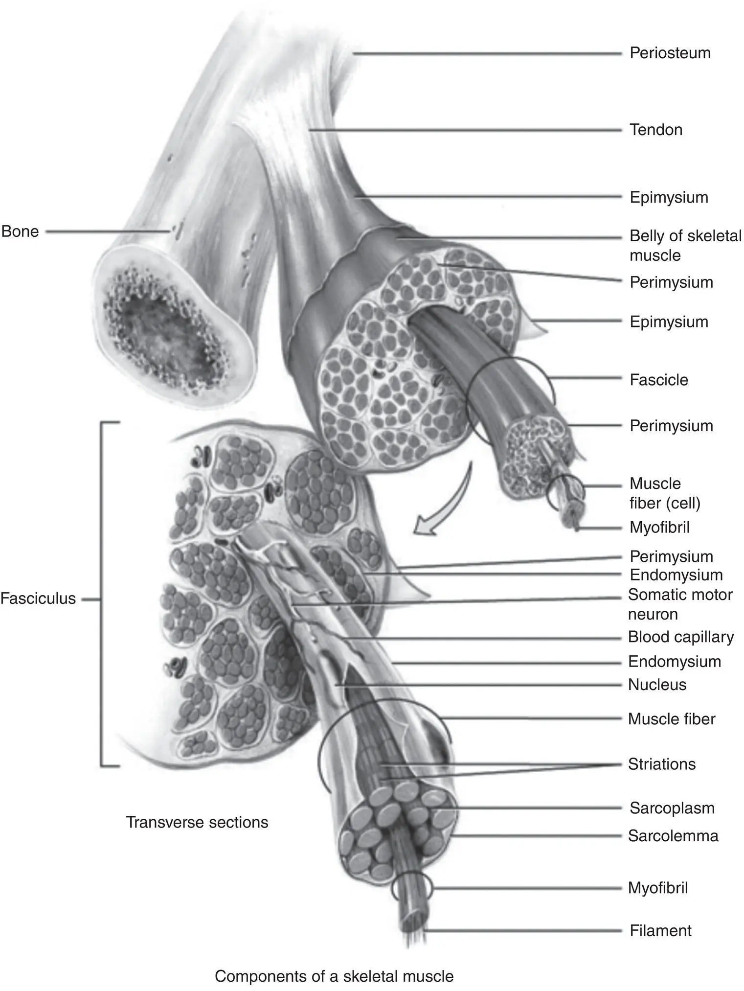
Figure 3.6 Structure of a skeletal muscle.
Tortora, G. J., & Derrickson, B. H. (Eds.), (2010). Muscle. In Introduction to the human body , 11th ed., Wiley.
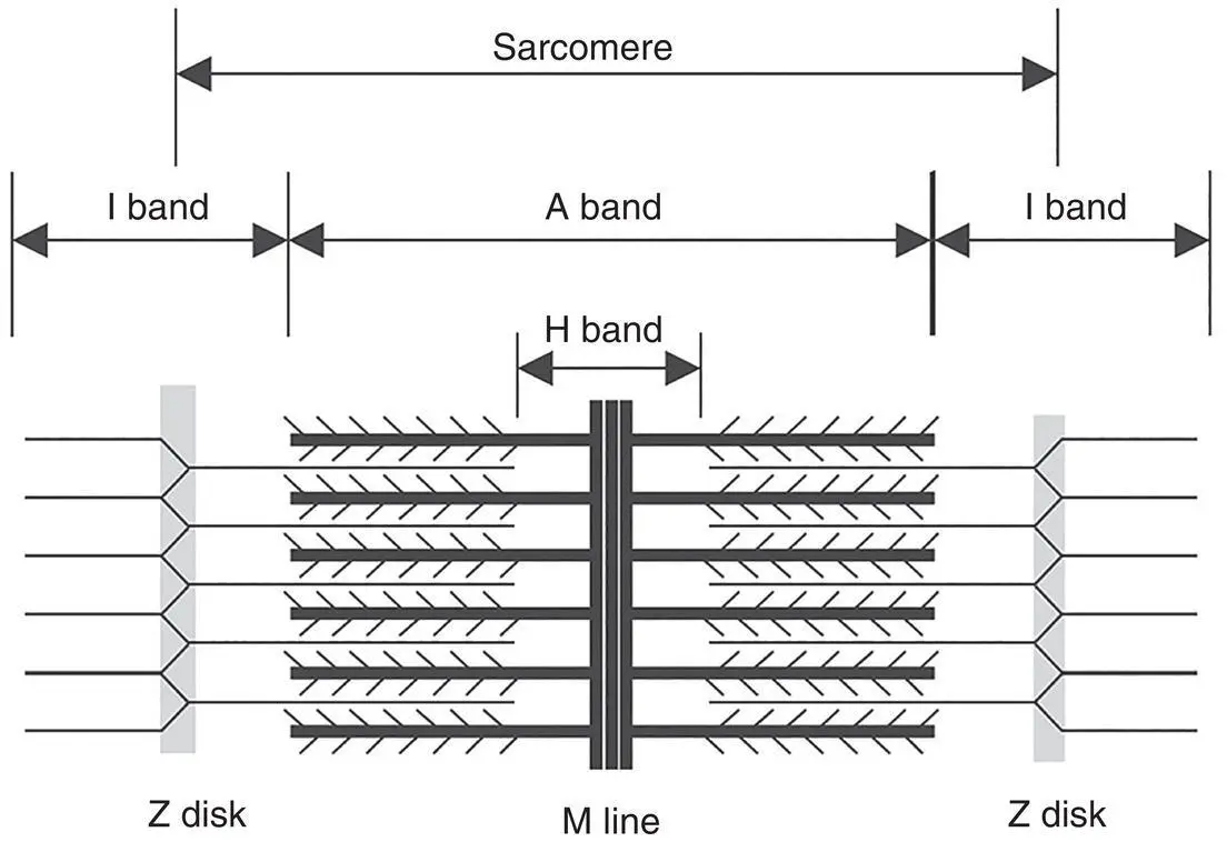
Figure 3.7 A simplified schematic of a sarcomere is shown. A sarcomere is composed of actin and myosin filaments that are organized into a characteristic pattern displaying A‐, I‐, M‐, and Z‐bands.
Modified from Nayak, A. & Amrute‐Nayak, M. (2020). SUMO system – A key regulator in sarcomere organization. FEBS Journal , 287 , 2176–2190. https://doi.org/10.1111/febs.15263.
Sarcoplasmic reticulum and calcium storage and release
The sarcoplasmic reticulum of muscles is a membrane‐bound structure that is similar to the smooth endoplasmic reticulum in other cells (a network of membranous tubules in the cytoplasm of eukaryotic cells that is in essence a transportation system). The sarcoplasmic reticulum is a key structure specialized in calcium ion (Ca 2+) storage ( Figure 3.8). Depolarization of the sarcoplasmic reticulum membrane results in the release of Ca 2+ions. This release is initiated at the specialized neuromuscular junction (to be discussed shortly). For a uniform muscle contraction, the cell membrane of skeletal muscle fibers extends into and penetrates the center of the muscle fiber as a system of transverse (T) tubules that form a complex network encircling the A–I bands of each sarcomere. Adjacent to the opposite sides of each T‐tubule are the expanded terminal cisternae of the sarcoplasmic reticulum. Two small cisternae of the sarcoplasmic reticulum and the T‐tubule form a specialized complex known as a triad. It is this junction where a neurally mediated depolarization of the T‐tubules is transmitted to the sarcoplasmic reticulum, stimulating a quick and widespread release of Ca 2+.
It is important to understand that muscle contraction depends on the availability of Ca 2+ions, while muscle relaxation occurs in the absence of Ca 2+. The sarcoplasmic reticulum ( Figure 3.8) regulates Ca 2+flow necessary for rapid contraction and relaxation cycles. After depolarization of the sarcoplasmic reticulum, Ca 2+ions concentrated within cisterna are passively released into the vicinity of the overlapping thick and thin filaments and bind to troponin. This allows bridging of the actin and myosin molecules. When the membrane depolarization ends, the sarcoplasmic reticulum actively transports the Ca 2+back into the cisternae, ending contractile activity.
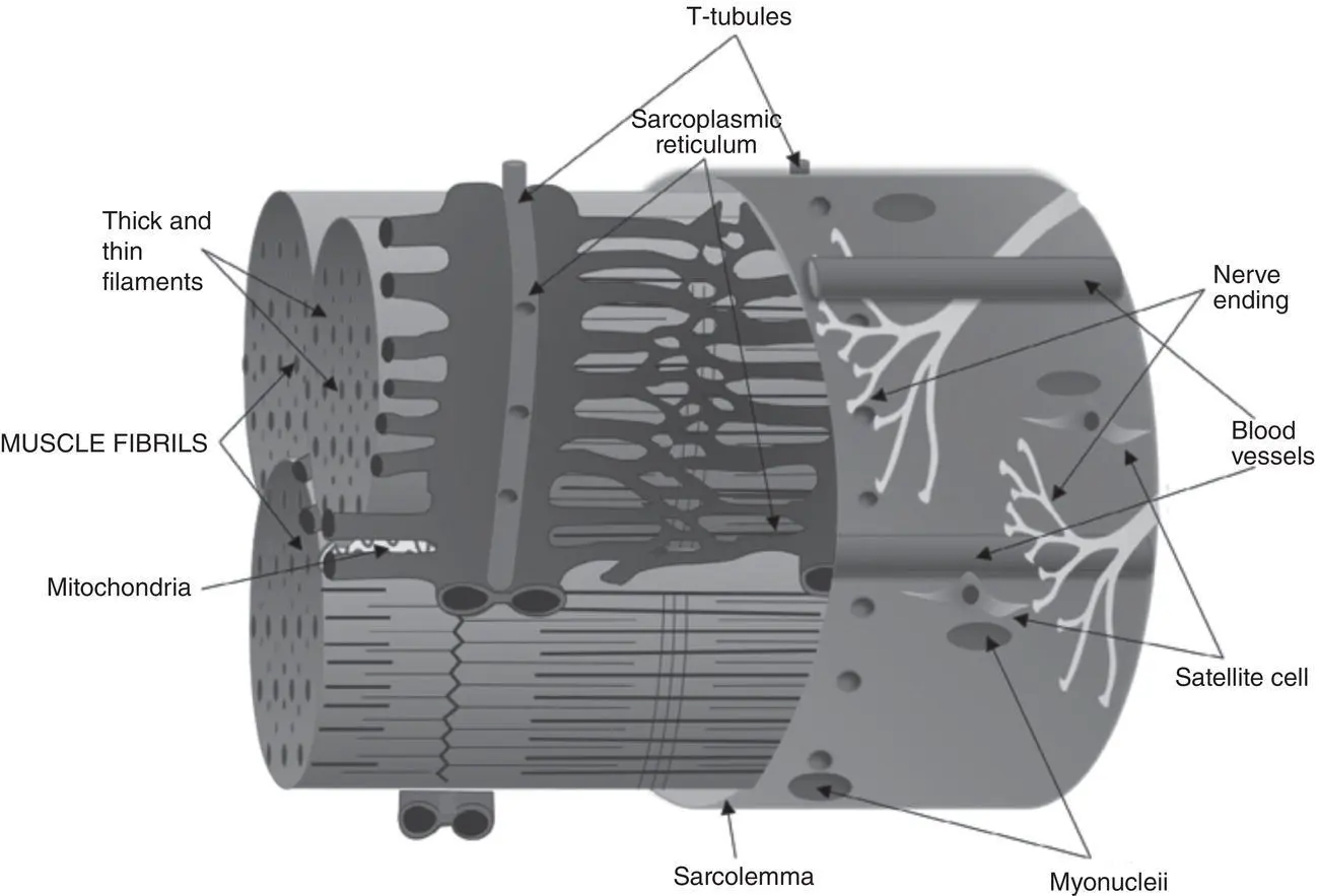
Figure 3.8 Diagram of the sarcoplasmic reticulum and T‐tubule system of a mammalian skeletal muscle fiber. The sarcoplasmic reticulum enmeshes fibrils with transverse (T) tubules intersecting them. Invaginations of the T‐tubules occur at the transition point of A and I bands in every sarcomere. The T‐tubules are associated with terminal cisternae of the sarcoplasmic reticulum, forming triads. The cut surface of myofibrils also shows the thin and thick myofilaments. Satellite cells reside along the host muscle fiber, directly above the sarcolemma under the basal lamina of muscle and in proximity to the nuclei of muscle cells. Nerve endings and local capillaries extend along the length of the muscle fiber. The extracellular matrix, not shown here, successively encases each layer.
Mukund, K. & Subramaniam, S. (2019). Skeletal muscle: A review of molecular structure and function, in health and disease. Wiley Interdisciplinary Reviews: Systems Biology and Medicine, 12 , e1462./John Wiley & Sons/CC BY‐4.0.
Vascularization
Skeletal muscle is highly vascularized and can receive up to 80–85% of the heart’s total cardiac output during heavy exercise ( Figure 3.9) (Joyner & Casey, 2015). Muscle contraction requires a great deal of energy and, therefore, large amounts of nutrients and oxygen. Moreover, waste products (e.g., lactic acid) produced during the muscle’s energy production reactions need to be eliminated. For this purpose, large blood vessels and nerves enter the epimysium and divide into branches that spread throughout the muscle in the perimysium and endomysium. Numerous capillaries run longitudinally through the endomysium as an “endomyseal capillary bed.” There are frequent transverse anastomoses between these capillaries, resulting in a fine elongated capillary network that surrounds each muscle fiber.
Function of Skeletal Muscle Components
Proper function of skeletal muscle also requires careful coordination between muscle fibers and their proteins with connective tissues, blood vessels, and nerves. Overall, muscles, using their ability to contract, transmit their forces through tendons onto the endoskeleton (bones and cartilage), which allows the movement of the skeleton.
Figure 3.9 Vascular anatomy within skeletal muscle. (a) Highly organized vasculature with dense capillary networks run parallel to myofibers forming microvascular units optimized for nutrient transfer. Inset: Capillaries are embedded within myofiber sarcolemma, where mitochondria congregate at the contact site between capillary and myofiber.
From: Gilbert‐Honick, J. & Grayson, W. (2020). Vascularized and innervated skeletal muscle tissue engineering. Advanced Healthcare Materials 9(1): e1900626. Wiley.
Myosin filament sliding
The contraction of muscle tissues provides motion, maintenance of posture, and heat production. The mechanisms of contraction of sarcomeres are key to muscle function. The thin and thick filaments only partially overlap in resting sarcomeres ( Figure 3.7). However, during contraction, the amount of overlap between these filaments increases due to sliding of the filaments past one another ( Figure 3.10), which causes shortening of the sarcomere. This contraction is induced by an action potential produced at the myoneural synapse (the juncture of a somatic motor axon and a muscle fiber). At the sarcomere level, during contraction, a small subset of myosin heads align with available actin‐binding sites. As the bound myosin heads move the actin, they provide for alignment of new actin–myosin cross bridges. The old bridges detach only after the myosin binds a new ATP molecule. This action resets the myosin head and prepares it for another contraction cycle. If no ATP is available, the actin–myosin complex become stable, which accounts for the extreme muscle rigidity associated with death (rigor mortis). A single muscle contraction is the result of hundreds of bridge‐forming and bridge‐breaking cycles. The contraction activity that leads to a complete overlap between thin and thick filaments continues until Ca 2+ions are removed, and the troponin–tropomyosin complex again covers the myosin‐binding site.
Neuromuscular junction and muscle contraction
Skeletal muscle is considered as a voluntary muscle because it can be made to contract by conscious controls. In order for a skeletal muscle fiber to contact, it must first be stimulated by an impulse (an action potential) from a motor nerve cell. The neurons that stimulate muscles to contract are somatic (of the body) motor neurons and have cell bodies that originate in the spinal cord or brainstem and have nerve processes (axons) that extend into the periphery to muscles (see Chapter 4). In skeletal muscle, myelinated somatic motor nerves branch into the perimysium and divide into several terminal smaller branches that innervate the muscle fibers. At the site of innervation, the axon loses its myelin sheath and forms a dilated terminal situated within a trough on the muscle cell surface, the neuromuscular junction (also known as a motor end plate) ( Figure 3.11). Within this axon terminal are numerous synaptic vesicles containing the neurotransmitter acetylcholine. When a somatic neuron fires and the action potential reaches the motor end plate, acetylcholine is released into a synaptic cleft (a space between the axonal terminal and the muscle). The synaptic cleft is a highly folded region of the sarcolemma that allows for more surface area. There are numerous mitochondria, ribosomes, and glycogen granules at this site. When acetylcholine binds to its receptor, Na +channels open and membrane depolarization results. Excess acetylcholine in the synaptic cleft is hydrolyzed by the enzyme cholinesterase to avoid prolonged contact of the neurotransmitter with its receptors. The depolarization is then propagated deep into the myofiber by the previously discussed transverse tubule system. At each triad (a T‐tubule and two cisterna), the depolarization signal is passed to the sarcolemma reticulum. This results in a release of Ca 2+and initiation of the muscle cells’s contraction cycle. When the depolarization ceases, the Ca 2+is actively transported back into the cisterna for storage, and the muscle cell relaxes.
Читать дальшеИнтервал:
Закладка:
Похожие книги на «Musculoskeletal Disorders»
Представляем Вашему вниманию похожие книги на «Musculoskeletal Disorders» списком для выбора. Мы отобрали схожую по названию и смыслу литературу в надежде предоставить читателям больше вариантов отыскать новые, интересные, ещё непрочитанные произведения.


![Ally Carter - [Gallagher Girls 02 ] - Cross My Heart & Hope To Spy](/books/262178/ally-carter-gallagher-girls-02-thumb.webp)


![John Bruce - The Lettsomian Lectures on Diseases and Disorders of the Heart and Arteries in Middle and Advanced Life [1900-1901]](/books/749387/john-bruce-the-lettsomian-lectures-on-diseases-and-disorders-of-the-heart-and-arteries-in-middle-and-advanced-life-1900-1901-thumb.webp)
Обсуждение, отзывы о книге «Musculoskeletal Disorders» и просто собственные мнения читателей. Оставьте ваши комментарии, напишите, что Вы думаете о произведении, его смысле или главных героях. Укажите что конкретно понравилось, а что нет, и почему Вы так считаете.