Charles S. Cockell - Astrobiology
Здесь есть возможность читать онлайн «Charles S. Cockell - Astrobiology» — ознакомительный отрывок электронной книги совершенно бесплатно, а после прочтения отрывка купить полную версию. В некоторых случаях можно слушать аудио, скачать через торрент в формате fb2 и присутствует краткое содержание. Жанр: unrecognised, на английском языке. Описание произведения, (предисловие) а так же отзывы посетителей доступны на портале библиотеки ЛибКат.
- Название:Astrobiology
- Автор:
- Жанр:
- Год:неизвестен
- ISBN:нет данных
- Рейтинг книги:3 / 5. Голосов: 1
-
Избранное:Добавить в избранное
- Отзывы:
-
Ваша оценка:
Astrobiology: краткое содержание, описание и аннотация
Предлагаем к чтению аннотацию, описание, краткое содержание или предисловие (зависит от того, что написал сам автор книги «Astrobiology»). Если вы не нашли необходимую информацию о книге — напишите в комментариях, мы постараемся отыскать её.
offers an introductory text that explores the structure of living things, the formation of the elements required for life in the Universe, the biological and geological history of the Earth, and the habitability of other planets. Written by a noted expert on the topic, the book examines many of the major conceptual foundations in astrobiology, which cover a diversity of traditional fields including chemistry, biology, geosciences, physics, and astronomy.
The book explores many profound questions such as: How did life originate on Earth? How has life persisted on Earth for over three billion years? Is there life elsewhere in the Universe? What is the future of life on Earth?
is centered on investigating the past and future of life on Earth by looking beyond Earth to get the answers. Astrobiology links the diverse scientific fields needed to understand life on our own planet and, potentially, life beyond. This new second edition:
Expands on information about the nature of astrobiology and why it is useful Contains a new chapter “What is Life?” that explores the history of attempts to understand life Contains 20% more material on the astrobiology of Mars, icy moons, the structure of life, and the habitability of planets New ‘Discussion Boxes’ to stimulate debate and thought about key questions in astrobiology New review and reflection questions for each chapter to aid learning New boxes describing the careers of astrobiologists and how they got into the subject Offers revised and updated information throughout to reflect the latest advances in the field Written for students of life sciences, physics, astronomy and related disciplines, the updated edition of
is an essential introductory text that includes recent advances to this dynamic field.
Astrobiology — читать онлайн ознакомительный отрывок
Ниже представлен текст книги, разбитый по страницам. Система сохранения места последней прочитанной страницы, позволяет с удобством читать онлайн бесплатно книгу «Astrobiology», без необходимости каждый раз заново искать на чём Вы остановились. Поставьте закладку, и сможете в любой момент перейти на страницу, на которой закончили чтение.
Интервал:
Закладка:
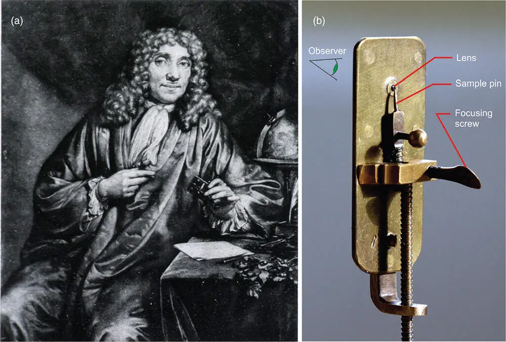
Figure 5.1 Early microbiology. (a) Antonie van Leeuwenhoek, discoverer of microbes, and (b) one of his first microscopes. The microscope (about 10 cm long) is held up to the eye, and objects are observed through the tiny glass lens.
Source: Reproduced with permission of Jeroen Rouwkema.
The creatures he observed had different shapes and he published numerous papers through The Royal Society in which he described these organisms and their appearances. Remarkably, he managed, even with his primitive microscopes, to observe some of the major shapes (morphologies) of microorganisms, including coccoids (spheres), rods, spiral organisms, and microbes with a bent cell shape. He even observed bacterial movement or motility, which you can see in Figure 5.2.
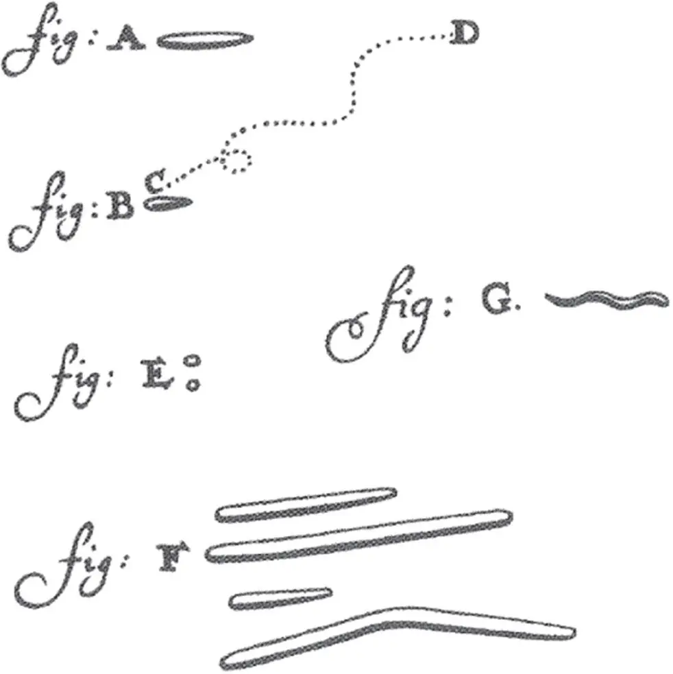
Figure 5.2 Leeuwenhoek's diagrams in the 1670s showing the first drawing of prokaryotes and their diverse morphologies. In the part labeled “fig: B (C–D)” you can see a depiction of bacterial motility.
Leeuwenhoek even showed that he could kill bacteria. In a letter of 1684, he wrote: “I took a very little wine-Vinegar and mixt it with the water in which the scurf [tooth plaque he had scrapped from his teeth] was dissolved, whereupon the Animals dyed presently.”
For the moment, we leave historical accounts. However, I have no compunction here in recommending the now rather classic book The Microbe Hunters , written in 1926 by Paul de Kruif, an amusing and popular account of the early history of microbiology, which remains relevant today.
5.3 Shapes of Cells
Cells take on a diversity of morphologies (shapes) depending on the species, their local environment, and their function. Eukaryotic cells in multicellular organisms can vary from the rectangular cells of plants whose dimensions range from 10 to 100 μm to the long thin cells of neurons that transmit nerve impulses through organs such as your brain, which although thin (from about 4 to 100 μm in width) can be up to 1 m long.
The most consistent patterns of morphology are found in the prokaryotes. Prokaryotic cells fall into a number of major classes of shapes (Figure 5.3).
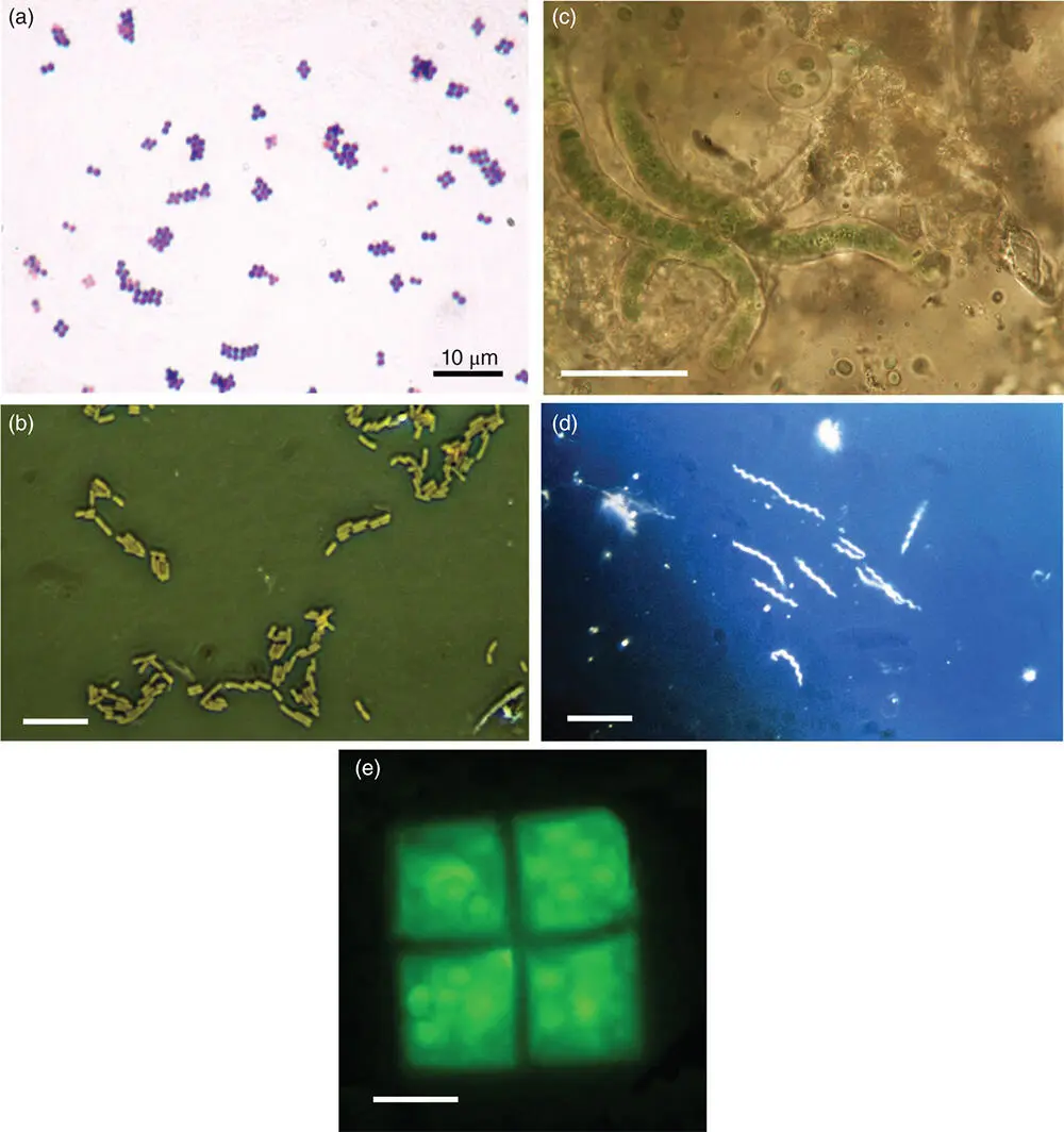
Figure 5.3 The wide variety of prokaryote shapes as seen under a microscope. (a) Staphylococcus (coccoid bacteria forming grape-like clusters). Here stained purple with the Gram stain. Scale bar: 10 μm.
Source: Reproduced with permission of Y. Tambe.
(b) Rod-shaped Escherichia coli . Scale bar: 5 μm.
Source: Reproduced with permission of Josef Reischig.
(c) Filamentous cyanobacteria from the Arctic. Scale bar: 10 μm. (d) Spiral-shaped spirochaete bacteria. Scale bar: 10 μm. (e) The square-shaped bacterium, Haloquadratum walsbyi , here shown stained and observed under fluorescence. Scale bar: 2 μm.
Many microbes have a coccoid (spherical) morphology and are referred to as coccus. An example is Staphylococcus (Figure 5.3a), which is responsible for some infections in humans. In some species, coccoids aggregate to form pairs (a diplococcus) or even collections of cells that look like bunches of grapes, such as observed in Staphylococcus . Many organisms are rod-shaped or bacilli, which are round-ended cylinders. An example of these organisms is bacteria in the genus Bacillus , or the gut bacterium Escherichia coli (Figure 5.3b). To complicate matters, there are even prokaryotes that are something of a mix between a coccus and a rod (coccobacillus) and can be considered as a slightly elongated coccus.
Microorganisms can be found that are filamentous, such as many cyanobacteria (Figure 5.3c), fungi, and some species of soil bacteria such as microorganisms in the phylum Actinobacteria . Filamentous organisms can have branched morphologies, most commonly observed in fungi and cyanobacteria. Yet other organisms have a spiral shape, such as Spirochaetes(Figure 5.3d), which are responsible for some diseases such as Lyme disease ( Borrelia burgdorferi ). There are organisms that are disc-shaped, star-shaped, and tapered.
Perhaps the most unusual shape is found in a member of the archaea, Haloquadratum , which adopts a square shape (Figure 5.3e). The organisms divide into sheets, looking somewhat like a sheet of postage stamps, achieving a layer of microbes up to 40 μm in length. The reason for this shape may be linked to its growth in briny pools and the resulting balance of osmotic stresses inside and outside the cells.
Some microorganisms can change their shape under stress. When exposed to nutrient stress or low temperatures, certain species become filamentous in growth. It is thought that the filamentous shape enhances the surface area for nutrient acquisition. Filamentous shapes may also improve attachment to surfaces and enhance the formation of layers of organisms or biofilms. It is clear that prokaryote shape is not just a serendipitous and ephemeral evolutionary feature, but that there is a whole diversity of environmental pressures that might influence microbial shapes, which are only just beginning to be understood.
5.4 The Structure of Cells
Despite the diversity of cell shapes and types, all cells have three basic components that are essential for an organism to function. These basic components might be considered to be universal. They are: (i) a membrane to hold the cell contents in, (ii) an information storage system to direct molecular synthesis and ultimately reproduction of the cell itself, and (iii) a system for gathering energy from the outside environment to drive chemical reactions, growth, reproduction, and so on. In this chapter, we look at the structure of cells, focusing on the membranes that enclose cells and the information storage system. The ability to gain energy from the external environment dramatically influences whether environments are habitable. There are a very wide diversity of ways to gather energy. For this reason, we spend the whole of the next chapter focusing on how life gets energy to maintain itself, grow, and reproduce.
5.5 The Structure of Cellular Membranes
To create a cell, we require some type of vessel in which to concentrate molecules. Most aqueous environments on Earth, and elsewhere, such as lakes, rivers, and oceans, have a tendency to dilute molecules. A cell enclosure, or membrane, provides a way to keep molecules together at relatively high concentrations. A membrane will also retain water, allowing for chemical reactions to proceed even when outside conditions are desiccating.
In the previous chapter, we discussed the lipids, some of which contain a hydrophilic head (which is attracted to water) and a hydrophobic tail made from a fatty acid (which is repelled from water). One important class of lipids that are involved in membrane formation is the phospholipids in which the charged polar end is a phosphate group (Figure 5.4). It is worth examining these in more detail to see how they form a cellular membrane. The principles are the same for all membrane lipids.
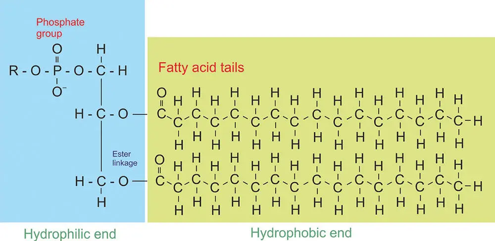
Figure 5.4 Amphiphilic molecules such as phospholipids that make up some cell membranes. They have a hydrophilic end (attracted to water) and a hydrophobic end (repelled from water).
Читать дальшеИнтервал:
Закладка:
Похожие книги на «Astrobiology»
Представляем Вашему вниманию похожие книги на «Astrobiology» списком для выбора. Мы отобрали схожую по названию и смыслу литературу в надежде предоставить читателям больше вариантов отыскать новые, интересные, ещё непрочитанные произведения.


Обсуждение, отзывы о книге «Astrobiology» и просто собственные мнения читателей. Оставьте ваши комментарии, напишите, что Вы думаете о произведении, его смысле или главных героях. Укажите что конкретно понравилось, а что нет, и почему Вы так считаете.