Gastrointestinal Surgical Techniques in Small Animals
Здесь есть возможность читать онлайн «Gastrointestinal Surgical Techniques in Small Animals» — ознакомительный отрывок электронной книги совершенно бесплатно, а после прочтения отрывка купить полную версию. В некоторых случаях можно слушать аудио, скачать через торрент в формате fb2 и присутствует краткое содержание. Жанр: unrecognised, на английском языке. Описание произведения, (предисловие) а так же отзывы посетителей доступны на портале библиотеки ЛибКат.
- Название:Gastrointestinal Surgical Techniques in Small Animals
- Автор:
- Жанр:
- Год:неизвестен
- ISBN:нет данных
- Рейтинг книги:5 / 5. Голосов: 1
-
Избранное:Добавить в избранное
- Отзывы:
-
Ваша оценка:
Gastrointestinal Surgical Techniques in Small Animals: краткое содержание, описание и аннотация
Предлагаем к чтению аннотацию, описание, краткое содержание или предисловие (зависит от того, что написал сам автор книги «Gastrointestinal Surgical Techniques in Small Animals»). Если вы не нашли необходимую информацию о книге — напишите в комментариях, мы постараемся отыскать её.
Describes techniques for canine and feline gastrointestinal surgery in detail Presents the state of the art for GI surgery in dogs and cats Includes access to a companion website with video clips demonstrating techniques
is an essential resource for small animal surgeons and veterinary residents.
Gastrointestinal Surgical Techniques in Small Animals — читать онлайн ознакомительный отрывок
Ниже представлен текст книги, разбитый по страницам. Система сохранения места последней прочитанной страницы, позволяет с удобством читать онлайн бесплатно книгу «Gastrointestinal Surgical Techniques in Small Animals», без необходимости каждый раз заново искать на чём Вы остановились. Поставьте закладку, и сможете в любой момент перейти на страницу, на которой закончили чтение.
Интервал:
Закладка:
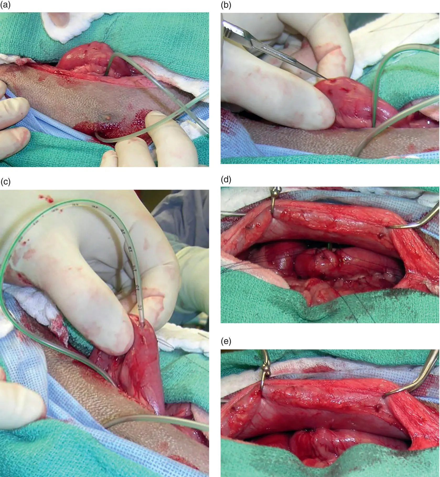
Figure 4.10
4.4.5 Utilization
A jejunostomy feeding tube can be used immediately even if the patient is heavily sedated. A liquid diet is required. After calculating the daily requirement the diet is delivery over 24 hours with a pump. The tube can be removed four days after its implantation.
An esophagojejunostomy tube can be placed also if an abdominal surgery is not indicated or possible (Cummings and Daley 2014).
4.4.6 Complications
Complications with jejunostomy tube are not frequent. The most common problem is obstruction of the tube. Also if the concentration of the liquid diet is too high it might induce diarrhea. Usually it is recommended to start with 1/4 to 1/3 of the daily maintenance and increase by 1/4 or 1/3 every day the amount of feeding.
4.5 Gastrojejunostomy Tube
4.5.1 Indications
The combination of a jejunostomy tube and gastrostomy tube is very common during the surgical treatment to septic peritonitis and during the reconstruction of the upper gastrointestinal tract (Cavanaugh et al. 2008). The jejunostomy tube is used first to support the patient while vomiting is occurring or the patient is lateral recumbent. The gastrostomy tube is used first to keep the stomach decompressed in an attempt to minimize gastroesophageal reflux, vomiting, and aspiration pneumonia. The gastrostomy is used later to provide more long‐term support to the patient when the vomiting episodes have subsided.
Instead of placing two separate tubes, it is possible to surgically place a gastrotomy tube and then advance within the gastrostomy tube a jejunostomy tube that is directed through the pylorus into the duodenum and the proximal jejunum. A gastrojejunostomy tube can be placed percutaneously (Jergens et al. 2007).
4.5.2 Materials and Equipment
A large diameter (28 Fr) gastrostomy (Kangaroo gastrostomy feeding tube, Medtronics, Minneapolis, MN) and a 9 Fr jejunal feeding tube 89 cm long (Kangaroo jejunostomy feeding Tube, Medtronics, Minneapolis, MN) are used. The jejunostomy tube is weighted.
4.5.3 Technique
The gastrostomy tube is placed first as described above. A flexible tip wire is advanced in the jejunostomy tube. The wire‐jejunostomy tube construct is then advanced in the gastrostomy tube and manually directed through the wall of the stomach in the pylorus and the duodenum. The jejunostomy tube is advanced in the proximal jejunum ( Figure 4.11). The wire is removed and the jejunostomy tube is flushed to make sure there is no resistance due to a kink. The jejunostomy tube and the gastrotomy tube connect together with a special adaptor in the hub of the gastrostomy tube.
4.5.4 Tips
To facilitate the placement of the jejunostomy tube it is very helpful to advance the gastrostomy tube first through the pylorus. The gastrostomy tube can be manipulated through the stomach wall to be directed into the pylorus. Then it is easier to feed the jejunostomy tube with its wire in the duodenum. It is also very important not to let the jejunostomy tube make a loop in the stomach. If a loop of the jejunostomy tube is in the stomach, it might kink and occlude the jejunostomy tube. Also, if there is a loop in the stomach the jejunostomy tube might migrate back and coil in the lumen of the stomach.
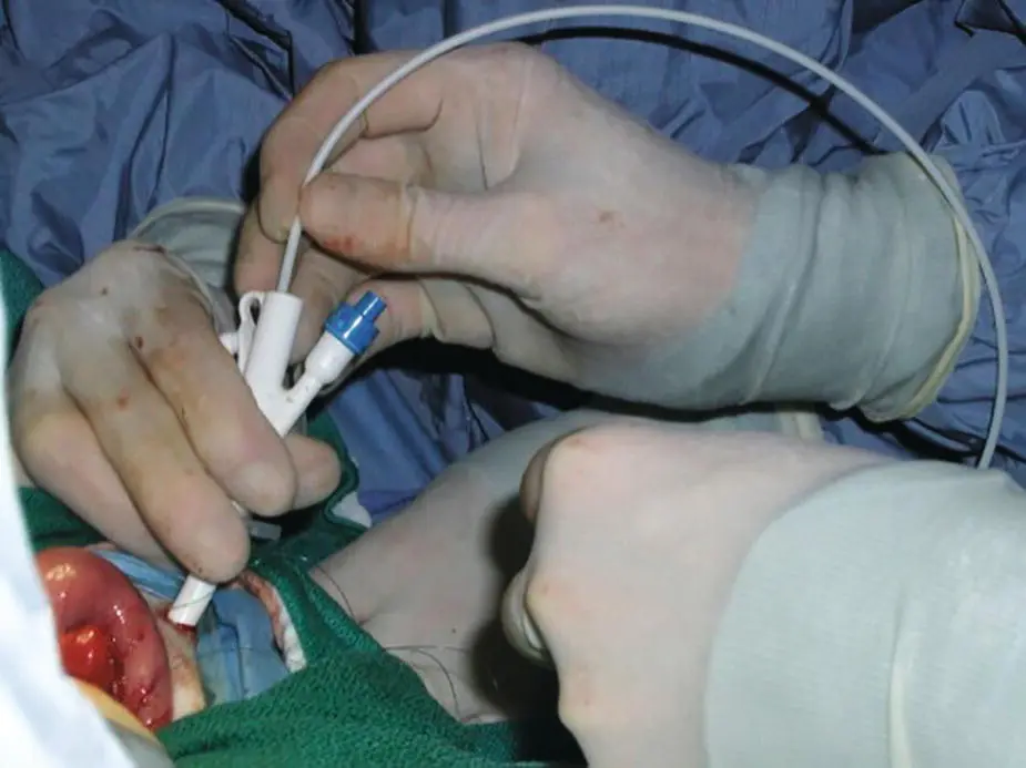
Figure 4.11
References
1 Abood, S.K. and Buffington, C.A. (1991). Improved nasogastric intubation technique for administration of nutritional support in dogs. J. Am. Vet. Med. Assoc. 199 (5): 577–579.
2 Abood, S.K. and Buffington, C.A. (1992). Enteral feeding of dogs and cats: 51 cases (1989–1991). J. Am. Vet. Med. Assoc. 201 (4): 619–622.
3 Armstrong, P.J. and Hardie, E.M. (1990b). Percutaneous endoscopic gastrostomy – a retrospective study of 54 clinical cases in dogs and cats. J. Vet. Intern. Med. 4 (4): 202–206.
4 Armstrong, P.J. et al. (1990a). Enteral nutrition by tube. Vet. Clin. North Am. Small Anim. Pract. 20 (1): 237–275.
5 Bright, R.M. et al. (1991). Percutaneous tube gastrostomy for enteral alimentation in small animals. Compend. Contin. Educ. Pract. Vet. 13: 15–22.
6 Cavanaugh, R.P. et al. (2008). Evaluation of surgically placed gastrojejunostomy feeding tubes in critically ill dogs. J. Am. Vet. Med. Assoc. 232 (3): 380–388.
7 Crowe, D.T. Jr. (1986). Use of a nasogastric tube for gastric and esophageal decompression in the dog and cat. J. Am. Vet. Med. Assoc. 188 (10): 1178–1182.
8 Crowe, D.T. and Devey, J.J. (1997a). Esophagostomy tubes for feeding and decompression: clinical experience in 29 small animal patients. J. Am. Anim. Hosp. Assoc. 33: 393–403.
9 Crowe, D.T. and Devey, J.J. (1997b). Esophagostomy tubes for feeding and decompression: clinical experience in 29 small animal patients. J. Am. Anim. Hosp. Assoc. 33 (5): 393–403.
10 Cummings, F. and Daley, C.A. (2014). Esophagojejunostomy feeding tube placement in 5 dogs with pancreatitis and anorexia. Vet. Med. Int. 2014: 197294.
11 Daye, R.M. et al. (1999). Interlocking box jejunostomy: a new technique for enteral feeding. J. Am. Anim. Hosp. Assoc. 35 (2): 129–134.
12 Devitt, C.M. and Seim, H.B. (1997). Clinical evaluation of tube esophagostomy in small animals. J. Am. Anim. Hosp. Assoc. 33 (1): 55–60.
13 Herring, J.M. (2016). A novel placement technique for nasogastric and nasoesophageal tubes. J. Vet. Emerg. Crit. Care 26 (4): 593–597.
14 Heuter, K. (2004). Placement of jejunal feeding tubes for post‐gastric feeding. Clin. Tech. Small Anim. Pract. 19 (1): 32–42.
15 Hewitt, S.A. et al. (2004). Evaluation of laparoscopic‐assisted placement of jejunostomy feeding tubes in dogs. J. Am. Vet. Med. Assoc. 225 (1): 65–71.
16 Jergens, A.E. et al. (2007). Percutaneous endoscopic gastrojejunostomy tube placement in healthy dogs and cats. J. Vet. Intern. Med. 21 (1): 18–24.
17 Kanemoto, Y. et al. (2017). Long‐term management of a dog with idiopathic megaesophagus and recurrent aspiration pneumonia by use of an indwelling esophagostomy tube for suction of esophageal content and esophagogastric tube feeding. J. Vet. Med. Sci. 79 (1): 188–191.
18 Levine, P.B. et al. (1997). Esophagostomy tubes as a method of nutritional management in cats: a retrospective study. J. Am. Anim. Hosp. Assoc. 33 (5): 405–410.
19 Marks, S.L. (1998). The principles and practical application of enteral nutrition. Vet. Clin. North Am. Small Anim. Pract. 28 (3): 677–708.
20 Song, E.K. et al. (2008). Comparison of different tube materials and use of Chinese finger trap or four friction suture technique for securing gastrostomy, jejunostomy, and thoracostomy tubes in dogs. Vet. Surg. 37 (3): 212–221.
21 Yoshimoto, S.K. et al. (2006). Owner experiences and complications with home use of a replacement low profile gastrostomy device for long‐term enteral feeding in dogs. Can. Vet. J. 47 (2): 144–150.
22 Yu, M.K. et al. (2013). Comparison of complication rates in dogs with nasoesophageal versus nasogastric feeding tubes. J. Vet. Emerg. Crit. Care 23 (3): 300–304.
5 Drainage Techniques for the Peritoneal Space
Интервал:
Закладка:
Похожие книги на «Gastrointestinal Surgical Techniques in Small Animals»
Представляем Вашему вниманию похожие книги на «Gastrointestinal Surgical Techniques in Small Animals» списком для выбора. Мы отобрали схожую по названию и смыслу литературу в надежде предоставить читателям больше вариантов отыскать новые, интересные, ещё непрочитанные произведения.


Обсуждение, отзывы о книге «Gastrointestinal Surgical Techniques in Small Animals» и просто собственные мнения читателей. Оставьте ваши комментарии, напишите, что Вы думаете о произведении, его смысле или главных героях. Укажите что конкретно понравилось, а что нет, и почему Вы так считаете.