Gastrointestinal Surgical Techniques in Small Animals
Здесь есть возможность читать онлайн «Gastrointestinal Surgical Techniques in Small Animals» — ознакомительный отрывок электронной книги совершенно бесплатно, а после прочтения отрывка купить полную версию. В некоторых случаях можно слушать аудио, скачать через торрент в формате fb2 и присутствует краткое содержание. Жанр: unrecognised, на английском языке. Описание произведения, (предисловие) а так же отзывы посетителей доступны на портале библиотеки ЛибКат.
- Название:Gastrointestinal Surgical Techniques in Small Animals
- Автор:
- Жанр:
- Год:неизвестен
- ISBN:нет данных
- Рейтинг книги:5 / 5. Голосов: 1
-
Избранное:Добавить в избранное
- Отзывы:
-
Ваша оценка:
Gastrointestinal Surgical Techniques in Small Animals: краткое содержание, описание и аннотация
Предлагаем к чтению аннотацию, описание, краткое содержание или предисловие (зависит от того, что написал сам автор книги «Gastrointestinal Surgical Techniques in Small Animals»). Если вы не нашли необходимую информацию о книге — напишите в комментариях, мы постараемся отыскать её.
Describes techniques for canine and feline gastrointestinal surgery in detail Presents the state of the art for GI surgery in dogs and cats Includes access to a companion website with video clips demonstrating techniques
is an essential resource for small animal surgeons and veterinary residents.
Gastrointestinal Surgical Techniques in Small Animals — читать онлайн ознакомительный отрывок
Ниже представлен текст книги, разбитый по страницам. Система сохранения места последней прочитанной страницы, позволяет с удобством читать онлайн бесплатно книгу «Gastrointestinal Surgical Techniques in Small Animals», без необходимости каждый раз заново искать на чём Вы остановились. Поставьте закладку, и сможете в любой момент перейти на страницу, на которой закончили чтение.
Интервал:
Закладка:
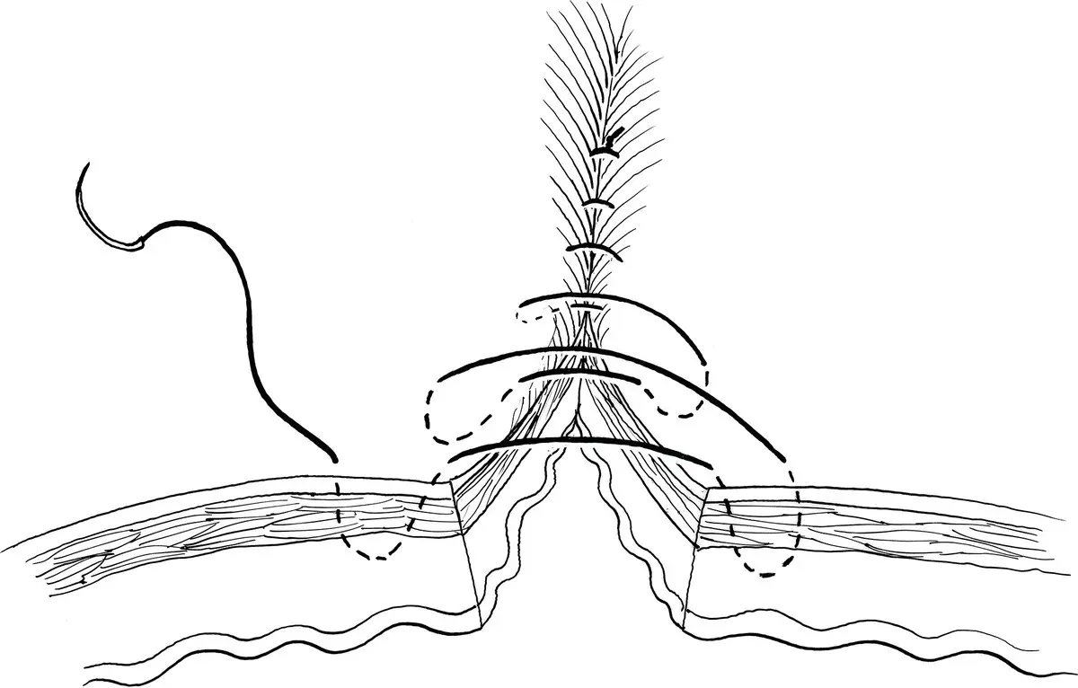
Figure 3.5
3.5.4 Parker–Kerr Oversew
This continuous inverting pattern is used to “blind end” a tubular organ such as bowel or uterine stumps ( Figure 3.6). To reduce contamination and aid in needle purchase, a large hemostatic clamp is first placed perpendicular to the long axis of the tubular organ. Any remaining organ tissue extending past the jaws of the clamp is removed. Starting at either the mesenteric or antimesenteric surface, a loose continuous Cushing suture pattern is placed catching 3–4 mm of bowel wall with each needle bite and running at least 3 mm from the edge of the clamp. Once the loose Cushing pattern is complete, the jaws of the clamp are partially opened releasing the incised bowel edges, while the suture end with the needle is slowly tensioned away from the side closest to the clamp hinge. The clamp is removed and the suture line is tensioned from both sides to completely invert the stump edges. The needle is then reversed and a Lembert pattern is placed and tied to the original free suture end to form a leak‐proof double inverted stump.
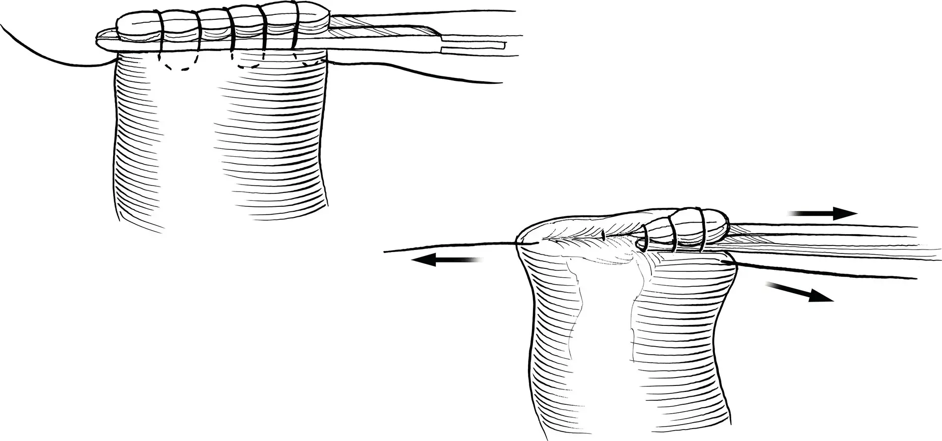
Figure 3.6
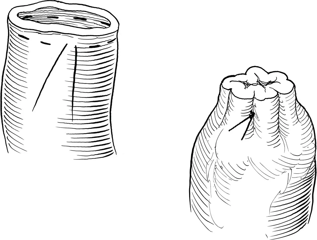
Figure 3.7
3.6 Special Supplementary Patterns: Purse‐String
This suture pattern is intended to close a hollow organ opening or body aperture, such as the anal opening, or around a tube entrance in viscera ( Figure 3.7). It is often utilized around the anal opening to prevent fecal contamination during perineal surgery, and to create a seal around a feeding tube placed in hollow viscera, as in a gastrostomy tube procedure. As the suture is placed around the site, tension on the suture end will tighten the continuous pattern much like a purse being pulled together at its neck with a string. The suture pattern is begun with parallel 3–4 mm bites of tissue about 3–5 mm away from the opening or cut edge. Each successive bite is advanced no more than 2–3 mm from the exit site of the last purchase. This forms a circular pattern around the centrally located opening. At the end of the pattern, both beginning and ending suture strands are in close apposition to each other. The strands are pulled firmly to form a tight cuffed rim of tissue around the tube or orifice, preventing leakage.
Bibliography
1 Bellenger, C. (1982). Comparison of inverting and appositional methods of anastomosis of the small intestines in cats. Vet. Rec. 110: 265–268.
2 Chassin, J.L., Rifkind, K.M., and Tumer, J.W. (1984). Errors and pitfalls in stapling gastrointestinal tract anastomoses. Surg. Clin. North Am. 64: 441–459.
3 Chung, R.S. (1987). Blood flow in colonic anastomoses. Effect of stapling and suturing. Ann. Surg. 206: 335–339.
4 Davis, D.D., Demianiuk, R.M., Musser, J. et al. (2018). Influence of preoperative septic peritonitis and anastomotic technique on the dehiscence of enterectomy sites in dogs. A retrospective review of 210 anastomoses. Vet. Surg. 47: 125–129.
5 Ellison, G. (1989). Healing in the gastrointestinal tract. Semin. Vet. Med. Surg. (Small Anim.) 4: 287–293.
6 Getzen, L.C., Roe, R.D., and Holloway, C.K. (1966). Comparative study of intestinal anastomotic healing in inverted and everted closures. Surg. Gynecol. Obstet. 123: 1219–1227.
7 Goligher, J.C., Lee, P.W., Simpkins, K.C. et al. (1977). A controlled comparison of one‐ and two‐layer techniques of suture for high and low colorectal anastomoses. Br. J. Surg. 64: 609–614.
8 Graham, M.F., Diegelmann, R.F., Elson, C.O. et al. (1988). Collagen content and types in the intestinal strictures of Crohn's disease. Gastroenterology 94: 257–264.
9 Halstead, W.S. (1887). Circular suture of the intestine. An experimental study. Am. J. Med. Sci. 94: 436–461.
10 Hamilton, J.E. (1967). Reappraisal of open intestinal anastomoses. Ann. Surg. 165: 917–924.
11 Hogstrom, H., Haglund, U., and Zederfeldt, B. (1990). Tension leads to increased neutrophil accumulation and decreased laparotomy wound strength. Surgery 107: 215–219.
12 Hunt, T.K., Cederfeldt, B., and Goldstick, T.K. (1969). Oxygen and healing [review]. Am. J. Surg. 118: 521–525.
13 Jansen, A., Becker, A.E., Brummelkamp, W.H. et al. (1981). The importance of apposition of the submucosal intestinal layers for primary wound healing of intestinal anastomoses. Surg. Gynecol. Obstet. 152: 51–58.
14 Kieves N., Thompson D.A. and Krebs A.I. (2014). Comparison of ex vivo leak pressures for single‐layer enterotomy closure between novice and trained participants in a canine model. Scientific Presentation Abstracts: 2014 ACVS Surgery Summit October 16–18, San Diego, CA.
15 McAdams, A.J., Meikle, A.G., and Taylor, J.O. (1970). One layer or two layer colonic anastomoses? Am. J. Surg. 120: 546–550.
16 Moy, R.L., Waldman, B., and Hein, D.W. (1992). A review of sutures and suturing techniques. J. Dermatol. Surg. Oncol. 18: 785–795.
17 Orr, N.W.M. (1969). A single‐layer intestinal anastomosis. Br. J. Surg. 56: 771–774.
18 Sajid, M.S., Siddiqui, M.R.S., and Baig, M.K. (2012). Single layer versus double layer suture anastomosis of the gastrointestinal tract. Cochrane Database Syst. Rev. 18 (1): CD005477. https://doi.org/10.1002/14651858.CD005477.
19 Snowden, K.A., Smeak, D.D., and Chiang, S. (2016). Risk factors for dehiscence of stapled functional end‐to‐end intestinal anastomoses in dogs: 53 cases (2001–2012). Vet. Surg. 45: 91–99.
20 Thornton, F.J. and Barbul, A. (1997). Healing in the gastrointestinal tract. Surg. Clin. N. Am. 77: 549–573.
21 Udenfriend, S. (1966). Formation of hydroxyproline in collagen [review]. Science 152: 1335–1340.
22 Weisman, D.L., Smeak, D.D., Birchard, S.J. et al. (1999). Comparison of a continuous suture pattern with a simple interrupted pattern for enteric closure in dogs and cats: 83 cases (1991–1997). J. Am. Vet. Med. Assoc. 214: 1507–1510.
4 Feeding Tubes
Eric Monnet
Department of Clinical Sciences, College of Veterinary Medicine and Biomedical Sciences, Colorado State University, Fort Collins, CO, USA
Enteral feeding is an important component of the treatment of critically ill patients, and for the support of patients with anorexia related to chronic conditions. Different type of feeding tubes are available to the surgeons to support the patients according to their need and underlying conditions (Armstrong et al. 1990a; Abood and Buffington 1992; Marks 1998).
Each of those feeding tubes has its advantages and disadvantages. The choice of feeding tube is based on the underlying disease, the goal of the enteral nutrition, and the length of time the tube will be needed. Combination of tubes is also possible. It is not rare to combine a gastrostomy tube with a jejunostomy tube to support a patient in the short term and the long term. The jejunostomy tube will be used in the short term to support the patient in the recovery phase of the surgery, especially if the patient is vomiting, while the gastrostomy tube will be used in the long term to support the patient if still anorexic.
Enteral feeding is also very important to support the integrity of the gastrointestinal tract. The enterocytes are getting their nutrient directly from the metabolite present in the lumen of the gastrointestinal tract.
Читать дальшеИнтервал:
Закладка:
Похожие книги на «Gastrointestinal Surgical Techniques in Small Animals»
Представляем Вашему вниманию похожие книги на «Gastrointestinal Surgical Techniques in Small Animals» списком для выбора. Мы отобрали схожую по названию и смыслу литературу в надежде предоставить читателям больше вариантов отыскать новые, интересные, ещё непрочитанные произведения.


Обсуждение, отзывы о книге «Gastrointestinal Surgical Techniques in Small Animals» и просто собственные мнения читателей. Оставьте ваши комментарии, напишите, что Вы думаете о произведении, его смысле или главных героях. Укажите что конкретно понравилось, а что нет, и почему Вы так считаете.