1 ...8 9 10 12 13 14 ...20 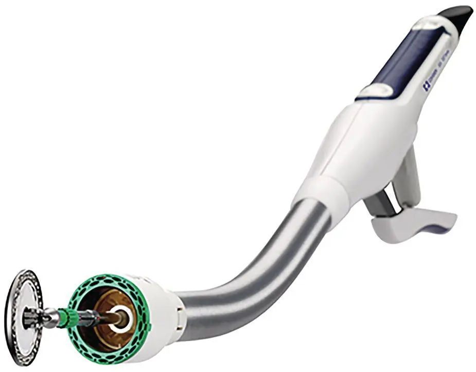
Figure 2.5
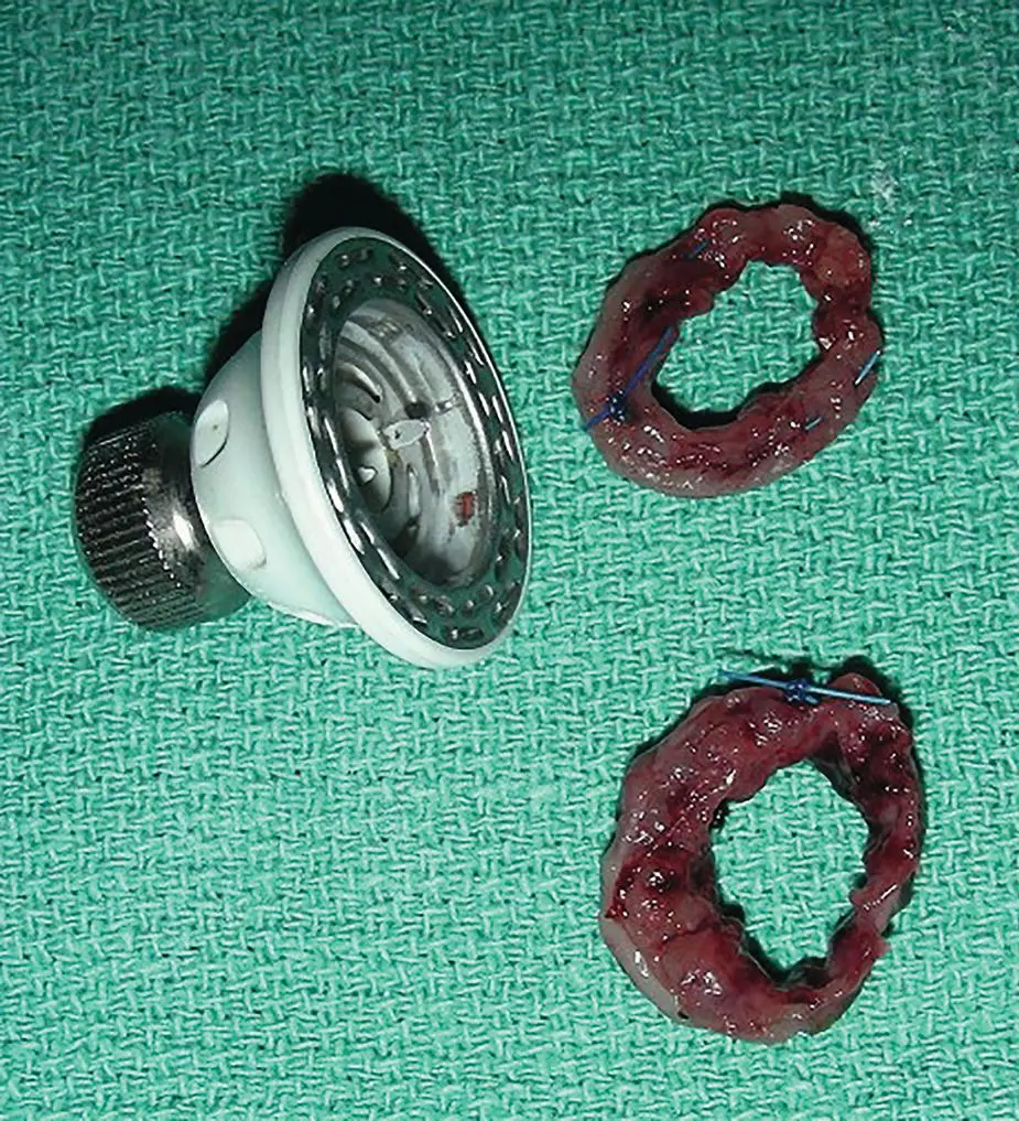
Figure 2.6
1 Arbaugh, M. et al. (2013). Biomechanical comparison of glycomer 631 and glycomer 631 knotless for use in canine incisional gastropexy. Vet. Surg. 42 (2): 205–209.
2 Booth, H.W. (2003). Suture materials, tissue adhesives, staplers, and ligating clips. In: Textbook of Small Animal Surgery, 3e (ed. D. Slatter), 234–244. Philadelphia: Saunders.
3 Capperauld, I. (1989). Suture materials: a review. Clin. Mater. 4 (1): 3–12.
4 Chih‐Chang, C. and Williams, D.F. (1984). Effects of physical configuration and chemical structure of suture materials on bacterial adhesion: A possible link to wound infection. Am. J. Surg. 147 (2): 147–204.
5 Chu, C.C., Von Franhofer, J.A., Greisler, H.P. et al. (eds.) (1997). Wound Closure Biomaterials and Devices. New York: CRC Press.
6 Chu, C.C. and Williams, D.F. (1984). Effects of physical configuration and chemical structure of suture materials on bacterial adhesion. A possible link to wound infection. Am. J. Surg. 147 (2): 197–204.
7 Coolman, B.R. et al. (1999). Evaluation of a skin stapler for belt‐loop gastropexy in dogs. J. Am Anim. Hosp. Assoc. 35 (5): 440–444.
8 Coolman, B.R. et al. (2000). Comparison of skin staples with sutures for anastomosis of the small intestine in dogs. Vet. Surg. 29 (4): 293–302.
9 Corman, M.L. et al. (1989). Comparison of the valtrac biofragmentable anastomosis ring with conventional suture and stapled anastomosis in colon surgery. Results of a prospective, randomized clinical trial. Dis. Colon Rectum 32 (3): 183–187.
10 Denyttenaera, S.V. et al. (2009). Barbed suture for gastrointestinal closure: A randomized control trial. Surgical Innovation. 16 (3): 237–242.
11 Domnick, E.D. (2014). Suture material and needle options in oral and periodontal surgery. J. Vet. Dentistry 31 (3): 204–211.
12 Erhart, N.P. et al. (2013). In vivo assessment of absorbable knotless barbed suture for single layer gastrotomy and enterotomy closure. Vet. Surg. 42 (2): 210–216.
13 Ferrer‐Marquez, M. and Belda‐Lorano, R. (2009). Barbed sutures in general and digestive surgery. Cirugia Espanola. 94 (2): 65–69.
14 Freudenberg, S. et al. (2004). Biodegradation of absorbable sutures in body fluids and pH buffers. Eur. Surg. Res. 36 (6): 376–385.
15 Hansen, L.A. and Monnet, E.L. (2012). Evaluation of a novel suture material for closure of intestinal anastomoses in canine cadavers. Am. J. Vet. Res. 73 (11): 1819–1823.
16 Hardy, T.G. Jr. et al. (1987). Initial clinical experience with a biofragmentable ring for sutureless bowel anastomosis. Dis. Colon Rectum 30 (1): 55–61.
17 Hoer, J., et al. (2001). Influence of suture technique on laparotomy wound healing: An experimental study in the rat. Langenbeck’s archives of surgery/Deutsche Gesellschaft fur Chirurgie 386 (3): 218–223.
18 Masini, B.D. et al. (2011). Bacterial adherence to suture materials. J. Surg. Educ. 68 (2): 101–104.
19 Marturello, D.M. et al. (2014). Knot security and tensile strength of suture materials. Vet. Surg. 43 (1): 73–79.
20 Osterberg, C.A. (1983). Enclosure of bacteria within capillary multifilament sutures as protection against leukocytes. Acta Chir. Scand. 149 (7): 663–668.
21 Ryan, S. et al. (2006). Comparison of biofragmentable anastomosis ring and sutured anastomoses for subtotal colectomy in cats with idiopathic megacolon. Vet. Surg. 35 (8): 740–748.
22 Smeak, D.D. (1998). Selection and use of currently available suture materials and needles. In: Current Techniques in Small Animal Surgery, 4e (eds. M.J. Bojrab et al.), 19–26. Philadelphia: Williams and Wilkins.
23 Thiede, A. et al. (1998). Overview on compression anastomoses: biofragmentable anastomosis ring multicenter prospective trial of 1666 anastomoses. W. J. of Surg. 22 (1): 78–87.
24 Thornton, F.J. and Barbul, A. (2014). Healing in the gastrointestinal tract. Surg. Clin. N. Am. 77 (3): 549–573.
25 Zimmer, C.A. et al. (1991). Influence of knot configuration and tying technique on the mechanical performance of sutures. J. Emerg. Med. 9 (3): 107–113.
3 Suture Patterns for Gastrointestinal Surgery
Daniel D. Smeak
Department of Clinical Sciences, College of Veterinary Medicine and Biomedical Sciences, Colorado State University, Fort Collins, CO, USA
3.1 One‐ or Two‐Layer Closure
Although this is still a controversial topic, both techniques have potential downfalls that could endanger an anastomosis. One would think two‐layer closures are better than one‐layer since they might provide added strength initially. However, they increase the inflammatory response in the early stages of visceral healing owing to the extra tissue handling, suture material, and ischemia of the inverted tissue cuff (Orr 1969; McAdams et al. 1970; Goligher et al. 1977). Excess inflammation at the healing site results in a weaker anastomosis as more collagen is broken down during the inflammatory and debridement phases of healing. Advocates of single‐layer closures argue that this technique results in a larger lumen with less damage to the tissue edges (Thornton and Barbul 1997). Currently, in most conditions, a single‐layer closure for most gastrointestinal repairs is considered adequate (Sajid et al. 2012). Provided the lumen is not compromised, double‐layer repairs are sometimes elected when the surgeon expects higher intraluminal pressures, or when the tissue edge is considered extra friable or when sutures in the first layer tend to cut through the tissue.
3.2 Tissue Inversion, Eversion, or Apposition
Inverting suture patterns cause greater initial narrowing of the intestinal lumen (Bellenger 1982) but their main advantage is that these patterns provide more consistent initial leak‐proof closures (higher leak pressures). Everting patterns elicit greater adhesion formation to exposed mucosal edges, and provide the least leak‐proof closures, and therefore they are not currently recommended for gastrointestinal closure (Hamilton 1967). In theory, approximating (appositional) patterns accurately align tissue layers compared to inverting and everting patterns, and therefore, these patterns are now preferred for gastrointestinal closures. However, in practice, consistent tissue apposition with approximating suture patterns infrequently occurs when anastomoses were evaluated histologically. Nevertheless, direct apposition of all intestinal layers, particularly the submucosal layer, has been found to result in the most rapid direct bridging of the repair (Jansen et al. 1981).
3.3 Stapled or Hand‐Sutured Anastomosis
Stapled anastomoses are technically easier and faster but they do not replace adherence to the principles of good surgical technique for successful intestinal healing (Chassin et al. 1984). Regardless of technique, an adequate blood supply and absence of contamination or tension is paramount to successful repair. Linear staples in thicker visceral tissue may not purchase the strength holding layer on both sides of the staple line leading to a weakened repair (Snowden et al. 2016). It is critical that the correct staple leg length is chosen when visceral edges are edematous or inflamed. In most situations, linear staple cartridges with 3.5 mm staples are acceptable for most intestinal repairs in dogs and cats. Linear staples with 4.8 mm staples are chosen for thicker intestinal edges and for stomach repairs. Stapled anastomoses were not at more risk for dehiscence when performed in the face of septic peritonitis, unlike what has been encountered in hand‐sewn repairs in multiple retrospective studies (Snowden et al., 2016; Davis et al. 2018).
Читать дальше
