Jane Flint - Principles of Virology
Здесь есть возможность читать онлайн «Jane Flint - Principles of Virology» — ознакомительный отрывок электронной книги совершенно бесплатно, а после прочтения отрывка купить полную версию. В некоторых случаях можно слушать аудио, скачать через торрент в формате fb2 и присутствует краткое содержание. Жанр: unrecognised, на английском языке. Описание произведения, (предисловие) а так же отзывы посетителей доступны на портале библиотеки ЛибКат.
- Название:Principles of Virology
- Автор:
- Жанр:
- Год:неизвестен
- ISBN:нет данных
- Рейтинг книги:3 / 5. Голосов: 1
-
Избранное:Добавить в избранное
- Отзывы:
-
Ваша оценка:
Principles of Virology: краткое содержание, описание и аннотация
Предлагаем к чтению аннотацию, описание, краткое содержание или предисловие (зависит от того, что написал сам автор книги «Principles of Virology»). Если вы не нашли необходимую информацию о книге — напишите в комментариях, мы постараемся отыскать её.
Volume I: Molecular Biology
Volume II: Pathogenesis and Control
Principles of Virology, Fifth Edition
Principles of Virology — читать онлайн ознакомительный отрывок
Ниже представлен текст книги, разбитый по страницам. Система сохранения места последней прочитанной страницы, позволяет с удобством читать онлайн бесплатно книгу «Principles of Virology», без необходимости каждый раз заново искать на чём Вы остановились. Поставьте закладку, и сможете в любой момент перейти на страницу, на которой закончили чтение.
Интервал:
Закладка:
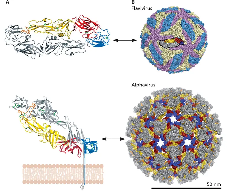
Figure 4.25 Conserved topology and regular packing of envelope proteins of small, (+) strand RNA viruses. (A)Ribbon diagrams of the flavivirus envelope (E) protein dimer (top) and the alphavirus E1/E2 heterodimer (bottom), with one E and the E2 subunit shown in gray. Conserved domains of E and E1 are colored red, yellow, and blue with the fusion loops required for entry in orange. The membrane is below the flavivirus dimer, in the plane of the figure, whereas it is perpendicular to the alphavirus E1/E2 heterodimer as indicated. The parallel and angled orientations to the membrane of the flavivirus and alphavirus envelope proteins, respectively, result in the very different appearances of these particles shown in panel B. (B)Surface renderings on the same scale, showing the regular packing of flavivirus and alphavirus envelope protein dimers. The dimers related by two-, three-, and fivefold axes of icosahedral symmetry are colored blue, pale yellow, and mauve, respectively, except for the central dimer depicted, which is colored as in panel A. In the 80 spikes of the alphavirus envelope, E2 is shown gray and E1 colored by domain as in panel A. Adapted from Vaney MC, Rey FA. 2011. Cell Microbiol 13:1451–1459, with permission. Courtesy of F.A. Rey, Institut Pasteur.
Because the internal capsids or nucleocapsids of these multilayered enveloped viruses are not in direct contact with the envelope, the organization and symmetry of internal structures are not evident from the external appearance of the surface glycoprotein layer. Nor does the organization of these proteins reflect the symmetry of the capsid. For example, the outer surface of all retroviruses appears roughly spherical with an array of projecting knobs or spikes, regardless of whether the internal core is spherical, cylindrical, or cone shaped. Likewise, influenza virus particles, which contain helical nucleocapsids, are generally roughly spherical but are highly pleomorphic with long, filamentous forms common in clinical isolates ( Box 4.10).
Internal proteins that contact the viral envelope are not embedded within the lipid bilayer but rather bind to its inner face. Such viral proteins are targeted to, and interact with, membranes by means of specific signals, which are described in more detail in Chapter 12.
Large Viruses with Multiple Structural Elements
Virus particles that house large DNA genomes are structurally much more intricate than any considered in previous sections. Such particles comprise obviously distinct components with different symmetries and/or multiple layers and in some cases exhibit architectures that do not appear to be based on helical or icosahedral symmetry. In this section, we illustrate various ways in which multiple structural elements can be combined. As we shall see, some of these elements are dedicated to specific functions.
BOX 4.9
DISCUSSION
A virus particle with different structures in different hosts
Throughout this chapter, we describe mature virus particles in terms of a single structure: “the” structure. However, it is important to appreciate that the architectures reported are those of particles isolated and examined under a single set of specific conditions, typically far from physiological. Structural studies of the flavivirus dengue virus, an important human pathogen, illustrate the conformational plasticity of some mature virus particles.
The organization of the single dengue virus envelope glycoprotein, E, described in the text ( Fig. 4.25) is that observed in particles propagated in cells of the mosquito vector maintained at 28°C. As noted previously, the E protein dimers are tightly packed and icosahedrally ordered. However, the epitopes for binding of antibodies that neutralize the virus at 37°C are either partially or entirely buried, suggesting that the virus particle might undergo temperature-dependent conformational transitions. Indeed, when particles are exposed to temperatures encountered in the mammalian host (e.g., 37°C), they do expand significantly, exposing segments of the underlying membrane, and the E protein interactions are altered (compare the left and right panels in the figure). In fact, particles exposed to higher temperatures are heterogeneous, and the example shown in the figure (right) represents but one of multiple forms, identified during selection of particles for three-dimensional reconstruction. Because a heterogeneous population of particles with less well-ordered E protein dimers represents the form of dengue virus recognized by the human immune system, these observations have important implications for the design of dengue virus vaccines.
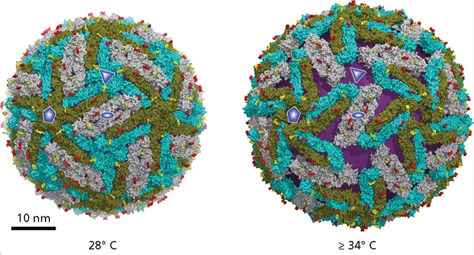
Structures of dengue virus particles at 28°C (left) and at ≥34°C (right), with the axes of five-, three-, and twofold rotational symmetry indicated by a pentagon, triangle, and ellipse, respectively.The E protein dimers that lie at the twofold axes are shown in gray and the other dimers with one subunit in green and one in cyan. The two oligosaccharides attached to each E protein monomer are indicated in red and yellow. The particles exposed to higher temperatures are characterized by exposed patches of membrane (purple) and significant reduction of dimer contacts at the threefold axes of icosahedral symmetry. Reprinted from Rey FA. 2013. Nature 497:443–444, with permission. Courtesy of F.A. Rey, Institut Pasteur.
Fibriansah G, Ng TS, Kostyuchenko VA, Lee J, Lee S, Wang J, Lok SM. 2013. Structural changes in dengue virus when exposed to a temperature of 37°C. J Virol 87:7585–7592.
Zhang X, Sheng J, Plevka P, Kuhn RJ, Diamond MS, Rossmann MG. 2013. Dengue structure differs at the temperatures of its human and mosquito hosts. Proc Natl Acad Sci U S A 110:6795–6799.
Particles with Helical or Icosahedral Parts
Bacteriophage T4
Bacteriophage T4, which has been studied for more than 50 years, is the classic example of an architecturally elaborate virus that contains distinct parts that exhibit icosahedral or helical symmetry. The T4 particle, which is built from ~50 of the proteins encoded in the ~170-kbp double-stranded DNA genome, is a structurally elegant machine tailored for active delivery of the genome to host cells. The most striking feature is the presence of morphologically distinct and functionally specialized structures, notably the head containing the genome and a long tail that terminates in a baseplate from which six long tail fibers protrude ( Fig. 4.26A).
The head of the mature T4 particle, an elongated icosahedron, is built from hexamers of a single viral protein (gp23*). In contrast to the other capsids considered so far, two T numbers are needed to describe the organization of gp23* in the two end structures ( T = 13) and in the elongated midsection ( T = 20). As in adenoviral capsids, the pentamers that occupy the vertices contain a different viral protein, and additional proteins reside on the outer or inner surfaces of the icosahedral shell ( Fig. 4.26B). One of the 12 vertices is occupied by a unique structure termed the connector, which joins the head to the tail. Such structures are derived from a nanomachine termed the portal, which pulls DNA into immature heads. Portals are a characteristic feature of the capsids of other families of DNA-containing bacteriophages, as well as of herpesviruses.
Читать дальшеИнтервал:
Закладка:
Похожие книги на «Principles of Virology»
Представляем Вашему вниманию похожие книги на «Principles of Virology» списком для выбора. Мы отобрали схожую по названию и смыслу литературу в надежде предоставить читателям больше вариантов отыскать новые, интересные, ещё непрочитанные произведения.


Обсуждение, отзывы о книге «Principles of Virology» и просто собственные мнения читателей. Оставьте ваши комментарии, напишите, что Вы думаете о произведении, его смысле или главных героях. Укажите что конкретно понравилось, а что нет, и почему Вы так считаете.