Jane Flint - Principles of Virology
Здесь есть возможность читать онлайн «Jane Flint - Principles of Virology» — ознакомительный отрывок электронной книги совершенно бесплатно, а после прочтения отрывка купить полную версию. В некоторых случаях можно слушать аудио, скачать через торрент в формате fb2 и присутствует краткое содержание. Жанр: unrecognised, на английском языке. Описание произведения, (предисловие) а так же отзывы посетителей доступны на портале библиотеки ЛибКат.
- Название:Principles of Virology
- Автор:
- Жанр:
- Год:неизвестен
- ISBN:нет данных
- Рейтинг книги:3 / 5. Голосов: 1
-
Избранное:Добавить в избранное
- Отзывы:
-
Ваша оценка:
Principles of Virology: краткое содержание, описание и аннотация
Предлагаем к чтению аннотацию, описание, краткое содержание или предисловие (зависит от того, что написал сам автор книги «Principles of Virology»). Если вы не нашли необходимую информацию о книге — напишите в комментариях, мы постараемся отыскать её.
Volume I: Molecular Biology
Volume II: Pathogenesis and Control
Principles of Virology, Fifth Edition
Principles of Virology — читать онлайн ознакомительный отрывок
Ниже представлен текст книги, разбитый по страницам. Система сохранения места последней прочитанной страницы, позволяет с удобством читать онлайн бесплатно книгу «Principles of Virology», без необходимости каждый раз заново искать на чём Вы остановились. Поставьте закладку, и сможете в любой момент перейти на страницу, на которой закончили чтение.
Интервал:
Закладка:
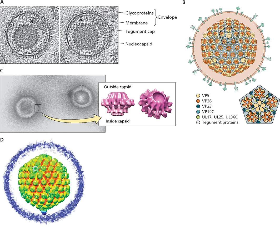
Figure 4.27 Structural features of herpesvirus particles. (A)Two slices through a cryo-electron tomogram of a single herpes simplex virus type 1 particle, showing the eccentric tegument cap (arrowheads). Reprinted from Grunewald K et al. 2003. Science 302:1396–1398, with permission. (B)Diagram of the structure of the herpesviral capsid based on high resolution (3.5 Å or better) cryo-EM structures of herpes simplex virus 1 and 2 and illustrated within a virus particle. Shown below is an enlarged view down a 5-fold axis of icosahedral symmetry. (C)The single portal of herpes simplex virus type 1 nucleocapsids visualized by staining with an antibody specific for the viral UL6 protein conjugated to gold beads is shown to the left. The gold beads are electron dense and appear as dark spots in the electron micrograph. They are present at a single vertex in each nucleocapsid, which therefore contains one portal. A 16-Å reconstruction of the UL6 protein portal based on cryo-EM is shown on the right. Adapted from Trus BL et al. 2004. J Virol 78:12668–12671, with permission. (D)Central slice through a cryo-electron tomographic reconstruction based on symmetryfree averaging is shown radially colored as indicated. Portal vertex-associated density is shown in cyan and purple. Adapted from Schmid MF et al. 2012. PLoS Pathog 8:e1002961, under license CC BY 4.0. © Schmid et al. Courtesy of A.C. Steven, National Institutes of Health (A and C), W. Chiu, Baylor College of Medicine (B), and F.J. Rixon, MRC-University of Glasgow Center for Virus Research, Glasgow, United Kingdom (D).
Alternative Architectures
The particles of other large viruses exhibit regular and sometimes remarkable morphologies that are not obviously based on helical or icosahedral symmetry. One example, Acidianus bottle-shaped virus , is portrayed in Fig. 4.1. We briefly describe two others here to illustrate the structural diversity of such viruses.
Poxviruses
Particles of poxviruses such as vaccinia virus also comprise multiple, distinct structural elements, but none of these exhibit obvious icosahedral or helical symmetry. A second distinctive feature is that two forms of infectious particles are produced in vaccinia virus-infected cells (see Chapter 13), termed mature virions and enveloped extracellular virions, which differ in the number and origin of membranes. Mature virions are large, enveloped structures (~350 to 370 × 250 × 270 nm) comprising at least 75 proteins that appear in the electron microscope as brick or barrel shaped (depending on the orientation) ( Fig. 4.29A). A number of internal structures have been observed by examination of thin sections through purified particles or by cryo-electron tomography ( Fig. 4.29B). These features include the core wall, which surrounds the central core that contains the ~200-kbp DNA genome, and lateral bodies. Remarkably, the core contains some 20 enzymes with many different activities. Although viral proteins that contribute to these various structures have been identified, our understanding of vaccinia virus architecture remains at low resolution.
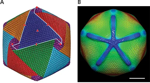
Figure 4.28 Features of mimivirus capsids. (A)Cryo-EM reconstruction of Cafeteria roenbergensis mimivirus at 21-Å resolution. Although not decorated with fibers, the surface of this capsid is characterized by high protrusions formed by surface loops of the double β-barrel jelly roll major capsid protein. These pseudohexagonal structure units are organized in discrete arrays, termed pentasymmetrons (purple) and trisymmetrons (blue, red, green, cyan, and orange), which form the vertices and interact at the threefold axes of symmetry, respectively. One of the 30 edges of the icosahedral particle is indicated in cyan, and the position of five-, three-, and twofold axes of symmetry by red symbols. Adapted from Xiao C et al. 2017. Sci Rep 7:5484, under license CC BY 4.0. Courtesy of C. Xiao, University of Texas at El Paso. (B) Cryo-EM reconstruction of Acanthamoeba polyphaga mimivirus following sequential digestion with lysozyme and the protease bromelain. This treatment was applied to remove (or reduce) the dense array of surface fibers ( Fig. 4.1), which increase ice thickness around the particles and hence signal-to-noise because of random scattering of electrons. The reconstruction (61 Å), which is based on only fivefold averaging, is viewed down the stargate (blue). The size of this assembly with extensions of the arms some 200 Å almost to neighboring vertices is clearly evident. Adapted from Xiao C et al. 2009. PLoS Biol 7:e92 under license CC BY 4.0. © 2009 Xiao et al. Courtesy of M. Rossman, Purdue University.
Pithoviruses
Pithovirus particles are the largest described to date, indeed are visible in the light microscope. They are ovoid or amphora-like in shape and variable in length (most commonly 1.35 to 1.65 nm). The internal nucleoid, which contains the double-stranded DNA genome of some 0.6 Mbp, is enclosed within what is thought to be a lipid membrane, in turn encased in a thick protein layer (the tegument). The apex of pithovirus is closed by a protruding “cork” with a hexagonal, grid-like appearance ( Fig. 4.29Cand D). Following uptake of virus particles into host cells by phagocytosis, this unusual cork structure is expelled to allow fusion of the viral nucleoid membrane with that of the cellular vacuole. Unprecedented assemblies specialized for release of the viral genome in host cells may prove to be a characteristic property of the very large viruses.
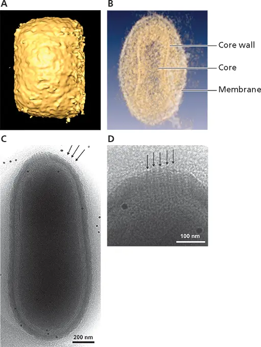
Figure 4.29 Virus particles with alternative architectures. Structural features of the poxvirus vaccinia virus. (A)Surface rendering of intracellular mature particles of vaccinia virus reconstructed from cryo-electron tomograms showing the brick shape and irregular protrusions from the surface. (B)Translucent visualization of the reconstructed particle volume showing the dumbbell-shaped core and external membrane. A and B reprinted from Cyrklaff M et al. 2005. Proc Natl Acad Sci U S A 102:2772–2777, with permission. Courtesy of J.L. Carrascosa, Universida Autonoma de Madrid. The virus Pithovirus sibericum was isolated following culture of a suspension of soil from a sample of permafrost collected in 2000 in Siberia. (C)Morphology of this virus particle, viewed by energy-filtered cryo-EM to facilitate visualization of thick, ice-embedded particles. The internal nucleoid, which appears rather featureless, is likely covered by a membrane, a thick protein layer termed the tegument, and a low-density outer layer. (D)The apical cork, which is made up of vertical fibers and hence appears striated, resides at one end of the particle. C and D reprinted from Okamoto K et al. 2017. Sci Rep 7:13291, under license CC BY 4.0. Courtesy of K. Okamoto, Uppsala University, Sweden.
Other Components of Virions
Интервал:
Закладка:
Похожие книги на «Principles of Virology»
Представляем Вашему вниманию похожие книги на «Principles of Virology» списком для выбора. Мы отобрали схожую по названию и смыслу литературу в надежде предоставить читателям больше вариантов отыскать новые, интересные, ещё непрочитанные произведения.


Обсуждение, отзывы о книге «Principles of Virology» и просто собственные мнения читателей. Оставьте ваши комментарии, напишите, что Вы думаете о произведении, его смысле или главных героях. Укажите что конкретно понравилось, а что нет, и почему Вы так считаете.