An Introduction to Molecular Biotechnology
Здесь есть возможность читать онлайн «An Introduction to Molecular Biotechnology» — ознакомительный отрывок электронной книги совершенно бесплатно, а после прочтения отрывка купить полную версию. В некоторых случаях можно слушать аудио, скачать через торрент в формате fb2 и присутствует краткое содержание. Жанр: unrecognised, на английском языке. Описание произведения, (предисловие) а так же отзывы посетителей доступны на портале библиотеки ЛибКат.
- Название:An Introduction to Molecular Biotechnology
- Автор:
- Жанр:
- Год:неизвестен
- ISBN:нет данных
- Рейтинг книги:3 / 5. Голосов: 1
-
Избранное:Добавить в избранное
- Отзывы:
-
Ваша оценка:
An Introduction to Molecular Biotechnology: краткое содержание, описание и аннотация
Предлагаем к чтению аннотацию, описание, краткое содержание или предисловие (зависит от того, что написал сам автор книги «An Introduction to Molecular Biotechnology»). Если вы не нашли необходимую информацию о книге — напишите в комментариях, мы постараемся отыскать её.
An Introduction to Molecular Biotechnology — читать онлайн ознакомительный отрывок
Ниже представлен текст книги, разбитый по страницам. Система сохранения места последней прочитанной страницы, позволяет с удобством читать онлайн бесплатно книгу «An Introduction to Molecular Biotechnology», без необходимости каждый раз заново искать на чём Вы остановились. Поставьте закладку, и сможете в любой момент перейти на страницу, на которой закончили чтение.
Интервал:
Закладка:
Polar signaling moleculesthat are unable to pass the biomembrane through diffusion are recognized by receptors on the cell surface. There are three categories of such receptors ( Figure 3.8):
Ion channel‐linked receptors are activated by specific ligands. As a reaction, the conformation of the channel protein is modified, leading to the opening or closure of the channel in question. Ions are let in or out accordingly. The changes in ion concentration produce a change in the membrane potential. In this way, the tension in ion channels can be regulated, or new action potentials released. Ion channel‐linked receptors are mainly found in the neuronal system, such as the nicotinic acetylcholine receptor(nAChR), the GABA receptor, the NMDA receptor, and the glycine receptor.
G‐protein‐coupled receptors (GPCRs) communicate with a G‐protein that is bound either to GTP or GDP. The activation of this type of receptor by a ligand causes a conformation change, which is recognized by the G‐protein. The G‐protein (or, to be more precise, its α‐subunit) is activated and can, in turn, interact with a membrane‐bound effector protein. The effector protein is often an enzyme (adenylyl cyclase or phospholipase), which produces second messengers. This mechanism whereby a single signaling molecule activates a multitude of effector proteins, which, in turn, release a host of second messengers, results in an effective amplification of the signal. Adenylyl cyclase turns ATP into cAMP, which acts as second messenger, regulating protein kinase A allosterically. Once protein kinase A has been activated, it may phosphorylate other enzymes or proteins (e.g. transcription factors), which then spring into action ( Figure 3.9). After the dissociation of the α‐subunit, the βγ‐complexes of the activated G‐protein can also be biologically active. In the cardiac muscle, acetylcholine binds to a muscarinic receptor(mAChR), thus activating the βγ‐complex. The βγ‐complex binds to K+ channels and opens them. cAMP is degraded by phosphodiesterase – an enzyme that is considered a target structure for several pharmaceutical products (e.g. caffeine). Table 3.3gives an overview of some essential hormones that are amplified by adenylyl cyclase and cAMP. A few signal system use cGMP instead of cAMP (e.g. in photoreceptors).
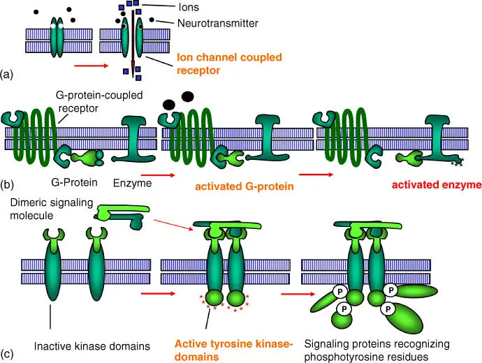
Figure 3.8 Schematic representation of receptor classes on the cell surface. (a) Ion channel‐coupled receptors. (b) G‐protein‐coupled receptors. (c) Enzyme‐coupled receptors (e.g. the tyrosine kinases).
Source: Alberts et al. (2015). Adapted with permission of Garland Science.
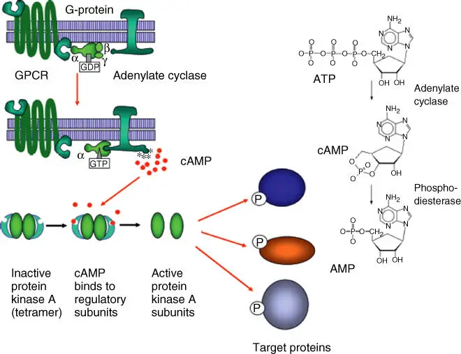
Figure 3.9 Activation of adenylyl cyclase and formation from cAMP as second messenger.
Source: Alberts et al. (2015). Adapted with permission of Garland Science.
Table 3.3 The role of adenylyl cyclase and phospholipase C ‐β in signal transduction.
| Signaling molecule | Target tissue | Main reaction |
|---|---|---|
| Adenylyl cyclase | ||
| Adrenaline | Heart | Raising heart frequency and enhancing contraction, muscles, glycogen degradation |
| Muscle | Breakdown of glycogen | |
| ACTH | Adrenal gland (cortex) | Secretion of cortisone |
| ACTH, adrenaline | Fat tissue | Breakdown of triglycerides |
| Glucagon | Liver | Glycogen degradation, increase of blood glucose levels |
| Parathormone | Bone | Bone resorption |
| Vasopressin | Kidney | Water resorption |
| Luteinizing hormone | Ovary | Progesterone secretion |
| Thyroid‐stimulating hormone (TSH) | Thyroid gland | Synthesis and release of thyroid hormone |
| Phospholipase C‐β | ||
| Vasopressin | Liver | Glycogen degradation |
| Acetylcholine | Pancreas | Secretion of amylase |
| Smooth muscles | Muscle contraction | |
| Thrombin | Platelets | Platelet aggregation |
Four families of trimeric G‐proteins have been discovered, which are active in a diversity of signaling pathways(Table 3.4). Few G‐proteins directly regulate elements of the cytoskeleton or ion channels:
Phospholipase C is another important effector protein, cleaving phosphatidylinositol into inositol‐1,4,5‐triphosphate (IP3) and diacylglycerol(DAG) after activation ( Figure 3.10). IP3 acts as second messenger, binding to ryanodine receptors in the ER and thus activating a calcium channel. Calcium can also act as a signaling substance, activating, for example, protein kinase C(PKC), various calmodulin(CaM)‐dependent kinases, and many other proteins ( Figure 3.11). PKC, a modulator of many target proteins (such as transcription factors), can also be activated by DAG ( Figure 3.11). Table 3.3summarizes the major signaling processes involving phospholipase C‐β. For medical research, G‐protein‐linked signaling pathways are of major interest, as they are used by many currently available pharmaceuticals. There are still many unknown steps in the process, which could prove interesting targets for new drugs to be developed.
Enzyme‐linked receptors can be activated by a signaling molecule (e.g. various growth factors that stimulate cell division) (Figures 3.8and 3.11). In dimeric receptors, two units form an active receptor with enzyme domains on the cytosolic side. The dimerization process activates tyrosine kinases (Table 3.5) that begin to phosphorylate each other. They are termed receptor tyrosine kinase (RTK). The phosphotyrosine residues are recognized by specific adapter proteins that are activated by them and then cause the activation of other signaling proteins ( Figure 3.11). Proteins of the Ras superfamily (monomeric GTPases) mediate signaling in most RTKs. Since such enzyme‐linked receptors are often found in tumor cells where they are overexpressed or permanently activated, their inhibition, especially the inhibition of tyrosine kinase, is a major strategy in the treatment of cancer. The drug Gleevec (STI‐571) binds to the ATP‐binding site and thus inhibits tyrosine kinases effectively.
Nitric oxide is a gaseous signaling mediator with many functions. It is being synthesized from arginine by NO synthases(NOS). In smooth muscles, e.g. those of the endothelium of blood vessels, NO induces a relaxation. NO activates guanylyl cyclase, leading to cGMP formation, which lead to the relaxation of smooth muscles.
Table 3.4 Some functions of trimeric G‐proteins.
| Family | Family members | Effective subunit | Some functions |
|---|---|---|---|
| I | G s | α | Activates adenylyl cyclase and Ca 2+channels |
| G olf | α | Activates adenylyl cyclase in olfactory neurons | |
| II | G i | α | Inhibits adenylyl cyclase |
| βγ | Activates K +channels | ||
| Go | βγ | Activates K +channels, inactivates Ca 2+channels | |
| Gt | α | Activates cGMP phosphodiesterase in photoreceptors | |
| III | Gq | α | Activates phospholipase C‐β |
| IV | G12/13 | α | Activates Rho GTPases to regulate the actin cytoskeleton |
Source: Alberts et al. (2015). Reproduced with permission of Garland Science.
Читать дальшеИнтервал:
Закладка:
Похожие книги на «An Introduction to Molecular Biotechnology»
Представляем Вашему вниманию похожие книги на «An Introduction to Molecular Biotechnology» списком для выбора. Мы отобрали схожую по названию и смыслу литературу в надежде предоставить читателям больше вариантов отыскать новые, интересные, ещё непрочитанные произведения.


![Andrew Radford - Linguistics An Introduction [Second Edition]](/books/397851/andrew-radford-linguistics-an-introduction-second-thumb.webp)


Обсуждение, отзывы о книге «An Introduction to Molecular Biotechnology» и просто собственные мнения читателей. Оставьте ваши комментарии, напишите, что Вы думаете о произведении, его смысле или главных героях. Укажите что конкретно понравилось, а что нет, и почему Вы так считаете.