An Introduction to Molecular Biotechnology
Здесь есть возможность читать онлайн «An Introduction to Molecular Biotechnology» — ознакомительный отрывок электронной книги совершенно бесплатно, а после прочтения отрывка купить полную версию. В некоторых случаях можно слушать аудио, скачать через торрент в формате fb2 и присутствует краткое содержание. Жанр: unrecognised, на английском языке. Описание произведения, (предисловие) а так же отзывы посетителей доступны на портале библиотеки ЛибКат.
- Название:An Introduction to Molecular Biotechnology
- Автор:
- Жанр:
- Год:неизвестен
- ISBN:нет данных
- Рейтинг книги:3 / 5. Голосов: 1
-
Избранное:Добавить в избранное
- Отзывы:
-
Ваша оценка:
An Introduction to Molecular Biotechnology: краткое содержание, описание и аннотация
Предлагаем к чтению аннотацию, описание, краткое содержание или предисловие (зависит от того, что написал сам автор книги «An Introduction to Molecular Biotechnology»). Если вы не нашли необходимую информацию о книге — напишите в комментариях, мы постараемся отыскать её.
An Introduction to Molecular Biotechnology — читать онлайн ознакомительный отрывок
Ниже представлен текст книги, разбитый по страницам. Система сохранения места последней прочитанной страницы, позволяет с удобством читать онлайн бесплатно книгу «An Introduction to Molecular Biotechnology», без необходимости каждый раз заново искать на чём Вы остановились. Поставьте закладку, и сможете в любой момент перейти на страницу, на которой закончили чтение.
Интервал:
Закладка:
Peroxisomesare small membrane‐enclosed, mostly rounded vesicles in which H 2O 2is produced or degraded (e.g. by the enzyme catalase).
3.1.3 Mitochondria and Chloroplasts
Mitochondriaare very striking organelles that are found in nearly all eukaryotic cells ( Figure 3.14). They look like worms or sausages and are between 1 μm and several micrometers long and 0.5 μm thick. Mitochondria have two separate membrane systems. The inner membrane forms a series of infoldings (cristae) that extend the surface considerably. The large surface area is needed because proteins and enzymes of the respiratory chainneed to find space in or on the inner mitochondrial membrane ( Figure 3.15). In a liver cell, mitochondria occupy about 22% of the cell volume.
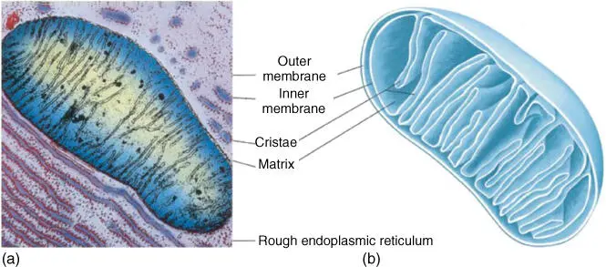
Figure 3.14 Composition of a mitochondrion. (a) Electron microscope photograph.
Source: Courtesy of K.R. Porter/Photo Researchers, Inc.
(b) Schematic representation.
Source: Voet et al. (2016). Adapted with permission of John Wiley and Sons.

Figure 3.15 Function of mitochondrion: metabolism and respiratory chain. (a) Metabolism and respiration in mitochondrion. (b) Schematic representation of the respiratory chain with the complexes I–IV; the proton gradient is used by ATP synthase to produce ATP. Rotenone, malonate, antimycin A, and KCN are the inhibitors of complexes I–IV. FeS, iron–sulfur cluster; Cyt, cytochrome; CoQ, ubiquinone; FMN, flavin mononucleotide.
The respiratory chainproduces ATP from the reduction equivalents NADHand FADH 2. During this process, electrons are transported through several intermediate stages, and a proton gradientis built up to provide energy for ATP synthase. This is also referred to as cellular respirationbecause the respiratory chain uses up oxygen. Without mitochondria, aerobic organisms such as animals, fungi, and plants would not be able to use oxygen from the air for the oxidation of organic matter – in other words, to produce energy. There are, however, some bacteria and a few eukaryotes that are anaerobic (i.e. they do not need oxygen). These organisms do not contain mitochondria.
During the citric acid cycle (Krebs cycle ), which takes place in the mitochondria, acetyl CoA is introduced, and in each run of the cycle, CO 2and reduction equivalents are generated. The acetyl CoA is derived from pyruvate, a product of glycolysis, which has been taken up by the mitochondria through a pyruvate transporter. It is then converted into acetyl CoA by a pyruvate decarboxylase complex. Another way of generating acetyl CoA is by β‐oxidationof fatty acids – a process that also takes place in mitochondria ( Figure 3.15).
Mitochondria contain their own ring‐shaped DNA ( Figure 3.16). In animals, the mitochondrial genome (mtDNA) is significantly smaller (16–19 kb) than in plants. It contains 13 genes coding for enzymes or other proteins involved in electron transport, 22 genes for tRNAs, and two genes for rRNAs. As every animal cell contains several hundred or even thousand mitochondria, each of which contains 5–10 mtDNA copies, the total of mtDNA copies amounts to several thousand per cell. mtDNA makes up about 1% of the total amount of DNA contained in a cell. The analysis of nucleotide sequences from mitochondrial genes has become an important tool in systematics to establish phylogenies and to define species.
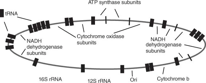
Figure 3.16 Schematic overview of the arrangement of genes in the mtDNA of mammals.
Plant mitochondria, by contrast, have large genomes (150–2500 kb). Some of their genes even have an intron/exon structure.
Mitochondria contain functional ribosomesequivalent to the prokaryotic 70S type, and the nucleotide sequences in mitochondrial genes and the amino acid sequences of the respective proteins are more closely related to the corresponding prokaryotic genes than to equivalents coded in the nucleus. The genetic code of mitochondria shows a few differences to the universal code: UGA (stop codon) codes in animals and fungi for tryptophan, AUA (for isoleucine) codes in animals and fungi for methionine, and AGG (arginine) codes in mammals for stop and in invertebrates for serine.
These findings as well as other mitochondrial characteristics led to the endosymbiont hypothesis, which states that mitochondria are derived from α‐purple bacteriathat were ingested by an ancestral eucyte 1.2 billion years ago and lived on as endosymbionts. The cell provides nutrients for the endosymbionts and receives ATP in return. Figure 3.17shows a likely ingestion path for the α‐purple bacteria into the ancestral eucyte. It is assumed that the ancestral eucyte came into being by the infolding of a bacterial cytoplasmic membrane to form an ER. The membrane then began to surround the chromosome, thus forming a nucleus.
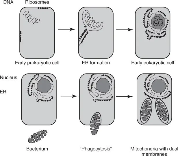
Figure 3.17 Development of an early eucyte and origin of mitochondria. α‐Purple bacteria were ingested by the early eucyte in a kind of phagocytosis. Hence, the outer mitochondrial membrane is derived from the host cell, whereas the inner mitochondrial membrane is the original bacterial cytoplasmic membrane.
Green plantsand algaecontain an additional organelle, the conspicuous chloroplasts, which are significantly larger and structurally more complex than mitochondria ( Figure 3.18). Apart from the surrounding inner and outer biomembranes, the chloroplast contains an extensively folded membrane system, known as thylakoids. These contain chlorophyll, as well as the proteins and enzymes required for photosynthesis, to enable the plants to turn sunlight into energy in the form of ATP and NADPH ( Figure 3.19). The electron transport between photosystem II and I and the production of NADPH are explained in Figure 3.19b. The light reaction leads to the buildup of a proton gradient, which is then used by ATP synthaseto produce ATP. During the subsequent CO 2 fixation process( Calvin cycle), CO 2is first bound to ribulose‐1,5‐biphosphate, which is then cleaved into two C3 units (3‐phosphoglycerate). 3‐Phosphoglycerate is transformed into glycerol aldehyde‐3‐phosphate, which is used for the regeneration of ribulose‐1,5‐biphosphate and for building glucose, fatty acids, and amino acids. A plant cell can generate additional ATP from glucose for the energy supply of the cell. This makes plants autotrophic and a suitable basic nutrient for heterotrophic animals that live on organic matter.
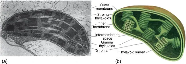
Figure 3.18 Structure of a chloroplast. (a) Electron microscope photo of a chloroplast.
Читать дальшеИнтервал:
Закладка:
Похожие книги на «An Introduction to Molecular Biotechnology»
Представляем Вашему вниманию похожие книги на «An Introduction to Molecular Biotechnology» списком для выбора. Мы отобрали схожую по названию и смыслу литературу в надежде предоставить читателям больше вариантов отыскать новые, интересные, ещё непрочитанные произведения.


![Andrew Radford - Linguistics An Introduction [Second Edition]](/books/397851/andrew-radford-linguistics-an-introduction-second-thumb.webp)


Обсуждение, отзывы о книге «An Introduction to Molecular Biotechnology» и просто собственные мнения читателей. Оставьте ваши комментарии, напишите, что Вы думаете о произведении, его смысле или главных героях. Укажите что конкретно понравилось, а что нет, и почему Вы так считаете.