Biological Mechanisms of Tooth Movement
Здесь есть возможность читать онлайн «Biological Mechanisms of Tooth Movement» — ознакомительный отрывок электронной книги совершенно бесплатно, а после прочтения отрывка купить полную версию. В некоторых случаях можно слушать аудио, скачать через торрент в формате fb2 и присутствует краткое содержание. Жанр: unrecognised, на английском языке. Описание произведения, (предисловие) а так же отзывы посетителей доступны на портале библиотеки ЛибКат.
- Название:Biological Mechanisms of Tooth Movement
- Автор:
- Жанр:
- Год:неизвестен
- ISBN:нет данных
- Рейтинг книги:5 / 5. Голосов: 1
-
Избранное:Добавить в избранное
- Отзывы:
-
Ваша оценка:
Biological Mechanisms of Tooth Movement: краткое содержание, описание и аннотация
Предлагаем к чтению аннотацию, описание, краткое содержание или предисловие (зависит от того, что написал сам автор книги «Biological Mechanisms of Tooth Movement»). Если вы не нашли необходимую информацию о книге — напишите в комментариях, мы постараемся отыскать её.
delivers a comprehensive reference for orthodontic trainees and specialists.
It is fully updated to include new chapters on personalized orthodontics as well as the inflammatory process occurring in the dental and paradental tissues. It is heavily illustrated throughout, making it easier for readers to understand and retain the information discussed within. The topics covered range from bone biology, the effects of mechanical loading on tissues and cells, genetics, tissue remodeling, and the effects of diet, drugs, and systemic diseases.
The Third Edition of
features seven sections that cover subjects such as:
The development of biological concepts in orthodontics, including the cellular and molecular biology behind orthodontic tooth movement Mechanics meets biology, including the effects of mechanical loading on hard and soft tissues and cells, and biological reactions to temporary anchorage devices Inflammation and orthodontics, including markers for tissue remodeling in the gingival crevicular fluid and saliva Personalized diagnosis and treatment based on genomic criteria, including the genetic influences on orthodontic tooth movement Rapid orthodontics, including methods to accelerate or decelerate orthodontic tooth movement Perfect for residents and PhD students of orthodontic and periodontal programs,
is also useful to academics, clinicians, bone biologists, and researchers with an interest in the mechanics and biology of tooth movement.
Biological Mechanisms of Tooth Movement — читать онлайн ознакомительный отрывок
Ниже представлен текст книги, разбитый по страницам. Система сохранения места последней прочитанной страницы, позволяет с удобством читать онлайн бесплатно книгу «Biological Mechanisms of Tooth Movement», без необходимости каждый раз заново искать на чём Вы остановились. Поставьте закладку, и сможете в любой момент перейти на страницу, на которой закончили чтение.
Интервал:
Закладка:
Cell–matrix interactions
The ECM provides a scaffold for cell adhesion, which can occur in two ways: by hemidesmosomes, connecting the ECM to intermediate filaments such as keratin, and by focal adhesions, connecting the ECM to actin filaments within the cell. The latter is the most important for OTM. In both types of adhesion, specific cellular adhesion molecules known as integrins are essential (Barczyk et al., 2013; Walko et al., 2015).
Integrins are transmembrane proteins formed as heterodimers of specific α and β transmembrane proteins that bind cells to ECM structures ( Figure 3.5). Integrins can contain many combinations of 18 different α subunits and eight β subunits. For example, osteoblasts have mainly α2β1 integrins, and osteoclasts have mainly αVβ3 integrins (Duong et al ., 2000).
Integrins act as receptors for ECM glycoproteins such as fibronectin, vitronectin, and proteins such as collagen and laminin, which contain the amino acid sequence arginine, glycine, aspartate (RGD motif). They can also bind to integrins on the surface of other cells (Duong et al ., 2000; Kechagia et al., 2019)
Intracellularly, integrins induce the formation of focal adhesion complexes which consist of the intracellular part of the integrins, focal adhesion kinase, which triggers intracellular mechanotransduction, by activating downstream mechanotransducers and many cytoplasmic proteins, such as tallin, vinculin, paxillin, and alpha‐actinin ( Figure 3.6)
The focal adhesion complex binds the integrin to actin filaments, the most important constituent of the cytoskeleton (Meikle, 2006; Martino et al ., 2018). Focal adhesions as well as the cytoskeleton are constantly remodeled under the influence of the ECM: proteins associate and disassociate with it continually as signals are transmitted to other parts of the cell. Furthermore, the cytoskeleton can contract by F‐actin sliding on the motor protein myosin II. These processes are responsible for cell deformation and cell migration (Burridge and Chrzanowska‐Wodnicka, 1996). Gradients of different environmental cues, such as diffusible ligands (chemotaxis), substrate‐bound ligands in the ECM (haptotaxis), or ECM rigidity (durotaxis) dictate the direction of migration (Kechagia et al ., 2019).

Figure 3.5 Integrin and its subunits.
(Source: Jaap Maltha.)
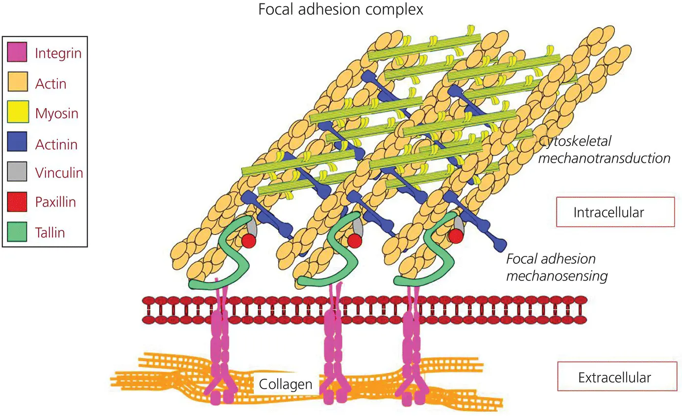
Figure 3.6 The focal adhesion complex.
(Source: Jaap Maltha.)
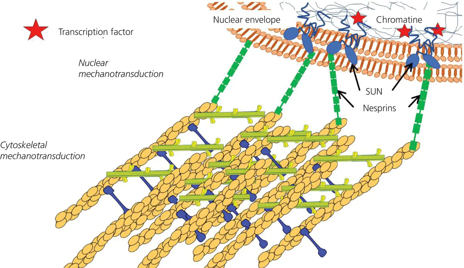
Figure 3.7 The nucleus‐related part of the cytoskeleton.
(Source: Jaap Maltha.)
It is essential that the composition and the distribution of the focal adhesions within a cell change to allow its migration. Initially, new focal adhesion complexes and cytoskeletal structures are formed at cellular protrusions, the lamellipodia. They mature and remain stationary with respect to the ECM through integrins. The cell uses this as an anchor on which it can push or pull itself over the ECM. At the same time, focal adhesion complexes at the trailing edge are disassembled, together with the cytoskeletal structures, allowing cell migration along the ECM (Martino et al ., 2018; Kechagia et al ., 2019).
Furthermore, the cytoskeleton is linked to the nuclear envelope by SUN and nesprin proteins. They transfer mechanical stimuli from the cytoskeleton to the nucleus where mechanosensitive transcription factors activate mechanosensitive genes (Feller et al ., 2015; Martino et al ., 2018) ( Figure 3.7).
Effects of orthodontic force application
Phases of OTM
In 1962, Burstone suggested that, if the rates of OTM were plotted against time, there would be three phases of OTM: the initial phase, a lag phase, and a post‐lag phase. The initial phase is characterized by a period of very rapid movement, which occurs immediately after application of force to the tooth. This rate is attributed to the displacement of the tooth within the PDL space and bending of the alveolar bone. This phase is followed by a lag period, when no or low rates of tooth displacement occur. This lag results from hyalinization of the PDL in areas of compression. No further tooth movement will occur until cells complete the removal of all necrotic tissues. During the third phase, the rate of movement gradually or suddenly increases. Experiments by Hixon and co‐workers (Hixon et al ., 1969, 1970) revealed two phases in OTM: an initial mechanical displacement, and a delayed metabolic response.

Figure 3.8 General time–displacement curve of OTM.
(Source: Jaap Maltha.)
More recently, a new time–displacement model for OTM was proposed (van Leeuwen et al., 1999; Von Böhl, et al ., 2004b) ( Figure 3.8). These studies, performed on beagle dogs, divided the curve of tooth movement into four phases.
The first phase lasts 24 hours to 2 days and represents the initial movement of the tooth inside its bony socket, causing structural changes in the ECM. In this initial phase, the ECM is compressed in the direction of the tooth movement, leading to a temporal increase in tissue pressure, constriction of blood vessels, and deformation of nerves. In many cases this results in an anoxic situation leading to local tissue necrosis, called hyalinization ( Figure 3.9).
At the trailing side of the tooth (formerly incorrectly called the tension side), the periodontal space is widened, which leads to a temporal decrease in tissue pressure, and a widening of the blood vessels (von Böhl and Kuijpers‐Jagtman, 2009).
The initial phase is, in most cases, followed by a second phase, where there is an arrest in tooth movement lasting for approximately 20–30 days. As long as the hyalinized tissue remains, tooth movement is prevented, as direct bone resorption is not possible, because osteoclasts cannot differentiate within the necrotic areas of the PDL. Actual OTM, the third phase, begins with an increasing rate only after the complete removal of the hyalinized tissue.
In the fourth phase, tooth movement takes place at a constant rate, as long as the force is exerted and no obstacles are encountered. This is the linear phase. In this phase, alveolar bone is resorbed at the leading side of the root (formerly incorrectly called the pressure side) ( Figure 3.10), and bone deposition is found at the trailing side (Pilon, Kuijpers‐Jagtman, and Maltha, 1996; van Leeuwen et al ., 1999) ( Figure 3.11).
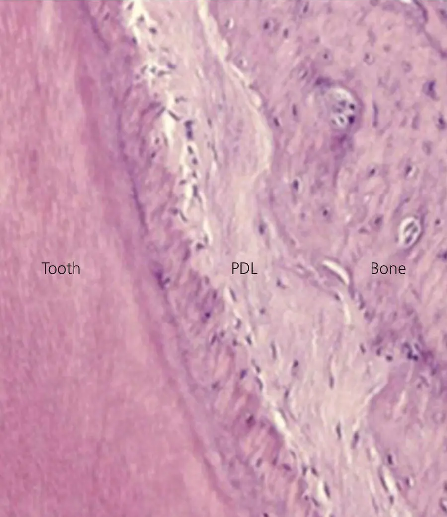
Figure 3.9 Photomicrograph of the PDL after orthodontic force application for 36 hours on a rat molar. The internal structure of the PDL is almost completely lost due to hyalinization.
(Source: Jaap Maltha.)
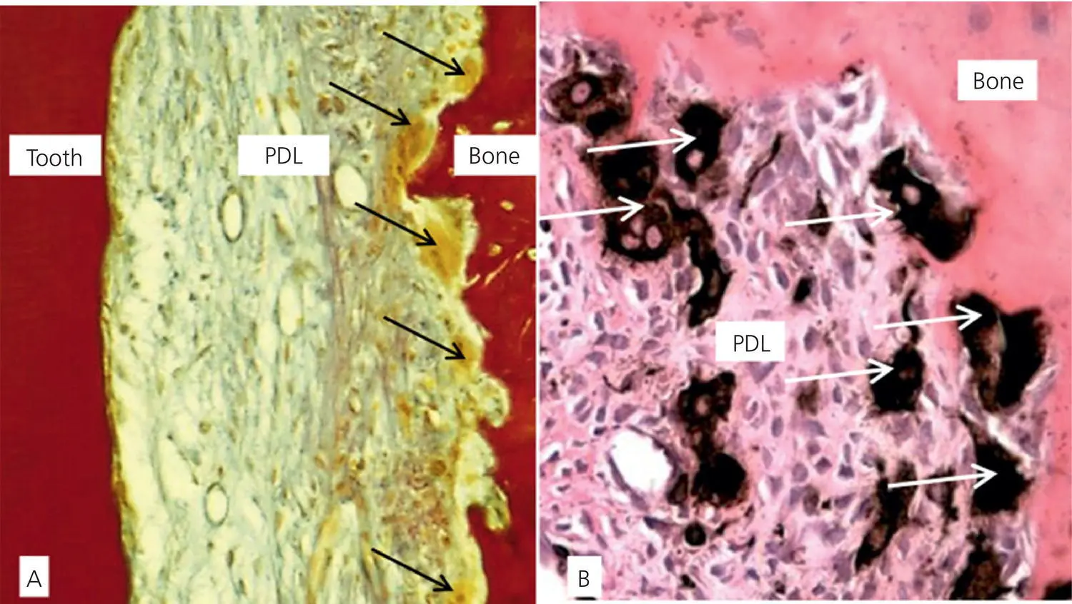
Figure 3.10 Photomicrographs of the leading side of orthodontically moving premolar of a dog. A. Herovici staining showing the absence of type I collagen fibers and their replacement by type III collagen. B. ED1 staining, specific for osteoclast cytoplasm. Arrows indicate osteoclasts.
Читать дальшеИнтервал:
Закладка:
Похожие книги на «Biological Mechanisms of Tooth Movement»
Представляем Вашему вниманию похожие книги на «Biological Mechanisms of Tooth Movement» списком для выбора. Мы отобрали схожую по названию и смыслу литературу в надежде предоставить читателям больше вариантов отыскать новые, интересные, ещё непрочитанные произведения.


Обсуждение, отзывы о книге «Biological Mechanisms of Tooth Movement» и просто собственные мнения читателей. Оставьте ваши комментарии, напишите, что Вы думаете о произведении, его смысле или главных героях. Укажите что конкретно понравилось, а что нет, и почему Вы так считаете.