Biological Mechanisms of Tooth Movement
Здесь есть возможность читать онлайн «Biological Mechanisms of Tooth Movement» — ознакомительный отрывок электронной книги совершенно бесплатно, а после прочтения отрывка купить полную версию. В некоторых случаях можно слушать аудио, скачать через торрент в формате fb2 и присутствует краткое содержание. Жанр: unrecognised, на английском языке. Описание произведения, (предисловие) а так же отзывы посетителей доступны на портале библиотеки ЛибКат.
- Название:Biological Mechanisms of Tooth Movement
- Автор:
- Жанр:
- Год:неизвестен
- ISBN:нет данных
- Рейтинг книги:5 / 5. Голосов: 1
-
Избранное:Добавить в избранное
- Отзывы:
-
Ваша оценка:
Biological Mechanisms of Tooth Movement: краткое содержание, описание и аннотация
Предлагаем к чтению аннотацию, описание, краткое содержание или предисловие (зависит от того, что написал сам автор книги «Biological Mechanisms of Tooth Movement»). Если вы не нашли необходимую информацию о книге — напишите в комментариях, мы постараемся отыскать её.
delivers a comprehensive reference for orthodontic trainees and specialists.
It is fully updated to include new chapters on personalized orthodontics as well as the inflammatory process occurring in the dental and paradental tissues. It is heavily illustrated throughout, making it easier for readers to understand and retain the information discussed within. The topics covered range from bone biology, the effects of mechanical loading on tissues and cells, genetics, tissue remodeling, and the effects of diet, drugs, and systemic diseases.
The Third Edition of
features seven sections that cover subjects such as:
The development of biological concepts in orthodontics, including the cellular and molecular biology behind orthodontic tooth movement Mechanics meets biology, including the effects of mechanical loading on hard and soft tissues and cells, and biological reactions to temporary anchorage devices Inflammation and orthodontics, including markers for tissue remodeling in the gingival crevicular fluid and saliva Personalized diagnosis and treatment based on genomic criteria, including the genetic influences on orthodontic tooth movement Rapid orthodontics, including methods to accelerate or decelerate orthodontic tooth movement Perfect for residents and PhD students of orthodontic and periodontal programs,
is also useful to academics, clinicians, bone biologists, and researchers with an interest in the mechanics and biology of tooth movement.
Biological Mechanisms of Tooth Movement — читать онлайн ознакомительный отрывок
Ниже представлен текст книги, разбитый по страницам. Система сохранения места последней прочитанной страницы, позволяет с удобством читать онлайн бесплатно книгу «Biological Mechanisms of Tooth Movement», без необходимости каждый раз заново искать на чём Вы остановились. Поставьте закладку, и сможете в любой момент перейти на страницу, на которой закончили чтение.
Интервал:
Закладка:
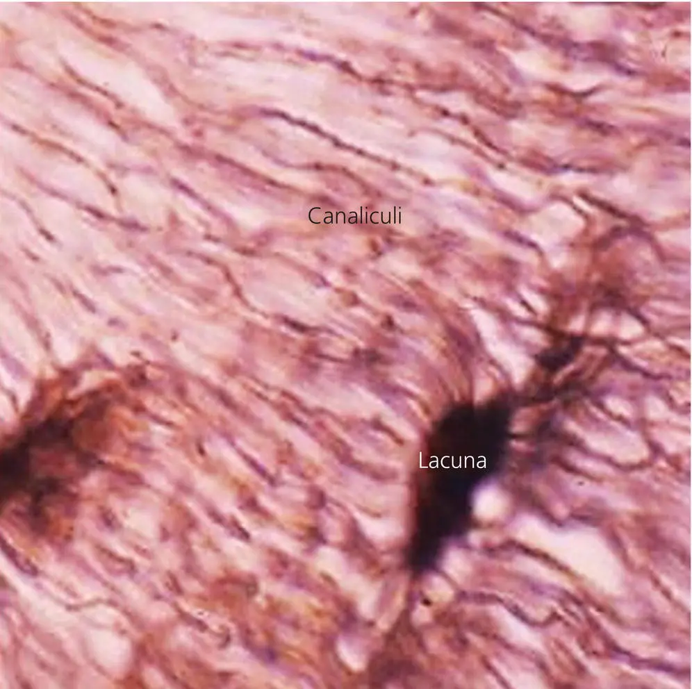
Figure 3.2 Photomicrograph of the bone matrix, showing osteocyte lacuna and lacuna–canalicular network.
(Source: Jaap Maltha.)
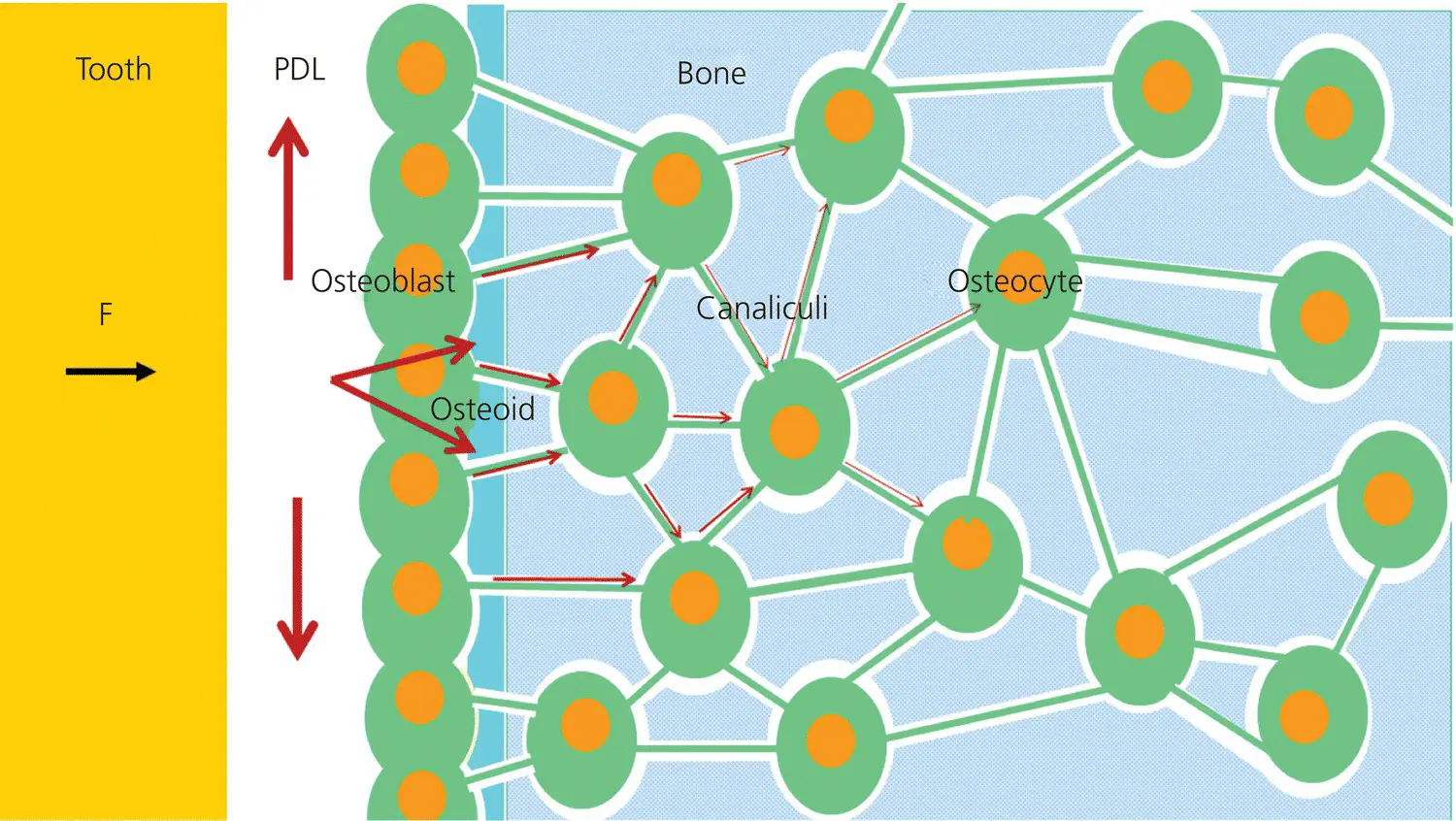
Figure 3.3 Fluid flow (arrows) after the application of an orthodontic force (F). Apart from a very rapid redistribution of fluid within the PDL, a limited fluid flow in the lacuna‐canalicular system leads to shear stress as a message for the mechanosensory system.
(Source: Jaap Maltha.)

Figure 3.4 The transposition of a dog premolar during the first 5 hours after the application of an orthodontic force. Two phases can be recognized: an initial phase lasting only a few seconds with a very rapid tooth movement, and a second phase in which the rate of tooth movement gradually decreases until it stabilizes.
(Source: Jaap Maltha.)
In addition, cementoblasts are part of the PDL and are derived from the mesenchymal dental follicle. They cover the root surface and are responsible for cementogenesis, through processes more or less comparable with osteogenesis. Some cementoblasts are buried in the cementum matrix to form cementocytes, especially in the more apical areas of the root. Similarly to osteocytes, they show thin cell processes in the ECM. However, they are far less extensive than in bone and do not form an intercellular network (Nanci and Bosshardt, 2006; Yamamoto et al ., 2016).
Macrophages are a type of white blood cells that have the same origin as osteoclasts, namely extravasated monocytes. They are essential as phagocytes in defense against pathogenic microorganisms, in clearance of dead or senescent cells, and in removal of cell debris. Furthermore, they promote homeostasis through their trophic, regulatory, and repair functions (Gordon and Martinez‐Pomares, 2017; Gordon and Plüddemann, 2017)
Biomechanical characteristics of the PDL
Orthodontic forces are applied in an environment where the mechanical properties depend on the material properties, sizes, and shapes of the constituent entities within the dento‐alveolar units (Viecilli et al ., 2008, 2013). The initial effect of the application of an orthodontic force is deformation or strain in the PDL and, to a lesser extent, the alveolar bone. This deformation depends on the biomechanical characteristics of the PDL, which in turn are dependent on the constituents of the extracellular compartment, i.e. fluid, fibers, and ground substance. This complicates mechanical behavior. In most biomechanical literature on the PDL a linear elastic behavior is assumed, which is an oversimplification of the real situation.
Experimental studies have described the time‐dependent behavior of the PDL in the first period after orthodontic force application (van Driel et al ., 2000; Jónsdóttir et al., 2006; Bergomi et al ., 2010). Two subperiods can be recognized: the first lasts for only a few seconds when a rapid transposition within the socket indicates a redistribution of free‐floating fluid; the second lasts for about 5 hours and shows a decelerating transposition, indicating viscoelastic behavior of the ECM of the PDL, with the collagen fibers leading to fiber‐reinforced properties (Jónsdóttir et al ., 2006; Jonsdottir et al ., 2012) ( Figure 3.4).
Therefore, the most likely model for the biomechanical properties of the normal PDL would be a biphasic poroviscoelastic fiber‐reinforced material (van Driel et al., 2000; Wang et al ., 2012; Ortún‐Terrazas et al ., 2018; Uhlir et al ., 2017; Wu et al ., 2019).
However, a serious drawback of all the literature pertaining to the mechanical properties of the PDL is that it is exclusively based on the normal state. The dramatic changes in the structure and composition of the PDL during OTM have never been taken into account.
General regulatory mechanisms
Cell–cell interactions
Cell–cell interactions allow cells to communicate with each other in response to changes in their microenvironment. These interactions can be stable through intercellular junctions, such as tight junctions, desmosomes, and gap junctions. They can also be variable, through the binding of soluble proteins secreted by one cell to receptor proteins on another cell. Such interactions allow cells to communicate with adjacent cells (autocrine actions), nearby cells (paracrine actions) (Tse and Wong, 2019), and even distant cells via the vascular system (endocrine actions). The latter actions are dealing with hormonal regulation.
For OTM, autocrine and paracrine interactions are essential. Secreted regulatory factors can bind to specific receptors on the target cell. These receptors are transmembrane structures containing proteins, carbohydrates, and lipids that project into the extracellular compartment. The binding of the signaling molecule to the receptor induces conformational changes in the receptor which, in turn, elicits a response in the corresponding cell. These responses include changes in cytoskeletal structure and subsequently change gene expression (McGeachie and Tennant, 1997; Meikle, 2006; Jiang et al ., 2015).
Two groups of local regulatory proteins are distinguished, namely growth factors and cytokines. The growth factors mainly affect cellular growth, proliferation, differentiation, and maturation, while the cytokines are primarily associated with hematopoietic and immunological processes. The latter can act as proinflammatory or as anti‐inflammatory mediators, and they can enhance cellular immune and antibody responses. The distinction between growth factors and cytokines is not very strict, since some cytokines can also act as growth factors and stimulate or inhibit cell growth and differentiation.
Growth factors and cytokines are grouped in several families. The most important factors involved in bone remodeling and thus in OTM belong to the transforming growth factor‐β (TGFβ) super family including TGFβs and bone morphogenetic proteins (BMPs), epidermal growth factors (EGF) including EGF and transforming growth factor‐α (TGFα), fibroblast growth factors (FGFs), insulin‐like growth factors (IGFs), vascular endothelial growth factors (VEGFs), tumor necrosis factors (TNFs), and colony‐stimulating factors (CSFs) (Roodman, 1993; McGeachie and Tennant, 1997; Hadjidakis and Androulakis, 2006; Jiang et al ., 2015).
Another group of local regulatory molecules consists of the eicosanoids. They synthetize from arachidonic acid that is released from cell membranes through phospholipase A2. Free arachidonic acid can be converted to bioactive eicosanoids through the so‐called arachidonic cascade, chains of enzymatic reactions, resulting in different subgroups of signaling molecules such as prostacyclines, thromboxanes, lipoxanes, and leukotrienes. For OTM, the most important subgroup is formed by the prostaglandins, which are formed through the activity of cyclooxygenases (COX1 and COX2) and prostaglandin synthase (Harizi et al., 2008; Xia et al ., 2016; Vansant et al ., 2018). Prostaglandins are synthetized within a large variety of cells. After exocytose they can bind to prostaglandin receptors on different target cells, in which a wide variety of effects can be induced (Binderman et al ., 1988; Mundy, 1993; Hadjidakis and Androulakis, 2006)
Читать дальшеИнтервал:
Закладка:
Похожие книги на «Biological Mechanisms of Tooth Movement»
Представляем Вашему вниманию похожие книги на «Biological Mechanisms of Tooth Movement» списком для выбора. Мы отобрали схожую по названию и смыслу литературу в надежде предоставить читателям больше вариантов отыскать новые, интересные, ещё непрочитанные произведения.


Обсуждение, отзывы о книге «Biological Mechanisms of Tooth Movement» и просто собственные мнения читателей. Оставьте ваши комментарии, напишите, что Вы думаете о произведении, его смысле или главных героях. Укажите что конкретно понравилось, а что нет, и почему Вы так считаете.