Robin J. M. Gray - Temporomandibular Disorders
Здесь есть возможность читать онлайн «Robin J. M. Gray - Temporomandibular Disorders» — ознакомительный отрывок электронной книги совершенно бесплатно, а после прочтения отрывка купить полную версию. В некоторых случаях можно слушать аудио, скачать через торрент в формате fb2 и присутствует краткое содержание. Жанр: unrecognised, на английском языке. Описание произведения, (предисловие) а так же отзывы посетителей доступны на портале библиотеки ЛибКат.
- Название:Temporomandibular Disorders
- Автор:
- Жанр:
- Год:неизвестен
- ISBN:нет данных
- Рейтинг книги:4 / 5. Голосов: 1
-
Избранное:Добавить в избранное
- Отзывы:
-
Ваша оценка:
Temporomandibular Disorders: краткое содержание, описание и аннотация
Предлагаем к чтению аннотацию, описание, краткое содержание или предисловие (зависит от того, что написал сам автор книги «Temporomandibular Disorders»). Если вы не нашли необходимую информацию о книге — напишите в комментариях, мы постараемся отыскать её.
delivers a systematic and logical approach to diagnosing and treating temporomandibular disorders.
Using a case-based approach to assist readers with understanding and retention, the book discusses the practical realities of managing patients and promoting effective treatment of temporomandibular disorders. Containing full colour clinical images and diagrams throughout, the chapters include practical guides on how to make splints and samples of patient information sheets which can be used as templates. Readers will get access to topics such as:
The clinical aspects of anatomy, function, pathology, and classification Differential diagnosis of temporomandibular joint problems Clicking joint problems and the use of preliminary investigation in disc displacement Temporomandibular joint locking diagnosis and treatment, including final treatment plans Facial pain examinations, differential diagnosis, and questions to ask patients regarding pain in general Headaches, worn teeth, dislocated jaws, and more issues that arise in the treatment of temporomandibular joint problems Perfect for undergraduate dental students and general dental practitioners, the new edition of
is also useful to postgraduate dental students, academics, and researchers.
Temporomandibular Disorders — читать онлайн ознакомительный отрывок
Ниже представлен текст книги, разбитый по страницам. Система сохранения места последней прочитанной страницы, позволяет с удобством читать онлайн бесплатно книгу «Temporomandibular Disorders», без необходимости каждый раз заново искать на чём Вы остановились. Поставьте закладку, и сможете в любой момент перейти на страницу, на которой закончили чтение.
Интервал:
Закладка:
Temporalis muscle
This muscle can be examined by palpating its origin extraorally. Ask the patient to clench the teeth together and the outline of the muscle origin can be identified, especially the anterior fibres. Digital palpation can be preformed between the superior and inferior temporal lines extending posteriorly ( Figure 3.9).
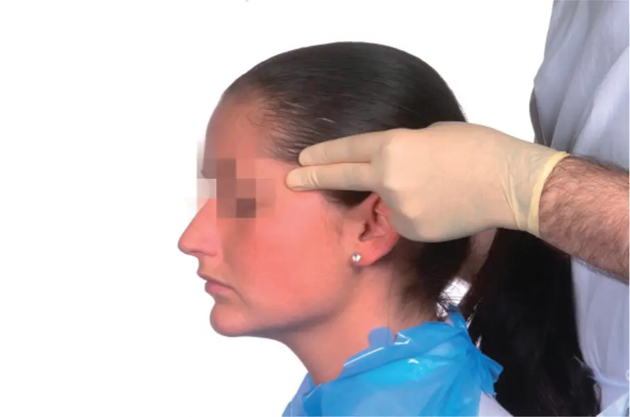
Figure 3.9Palpation of the anterior vertical fibres of the temporalis.
(M. Ziad Al‐Ani, Robin J.M. Gray.)
The anterior, more vertical fibres comprise the main elevator muscle of the jaw and are most commonly tender on palpation. The posterior fibres are almost horizontal in orientation and less frequently tender because their main function is to retrude the mandible.
It is suggested that the insertion of the temporalis muscle into the anterior margin of the coronoid process can be palpated intraorally by placing the little finger on the anterior border of the ramus and running it upwards, but this is not a reliable test because this is an uncomfortable and inaccessible area to try to access even in those who do not have muscle tenderness.
Lateral pterygoid muscle
This muscle is inaccessible to manual palpation so palpation for tenderness lacks validity and reliability and is difficult if not impossible to perform.
A more reliable technique is to examine the response to resistance. The patient is asked to open the mouth. The examiner's hand is placed under the patient's chin and pressure is applied to try to close the mouth while the patient tries to resist ( Figure 3.10a). This results in a more reliable test because the muscle is fixed. If there is tenderness in the lateral pterygoid muscle, this test will produce pain in the preauricular region. The same can be done by resisting lateral mandibular movement ( Figure 3.10b).
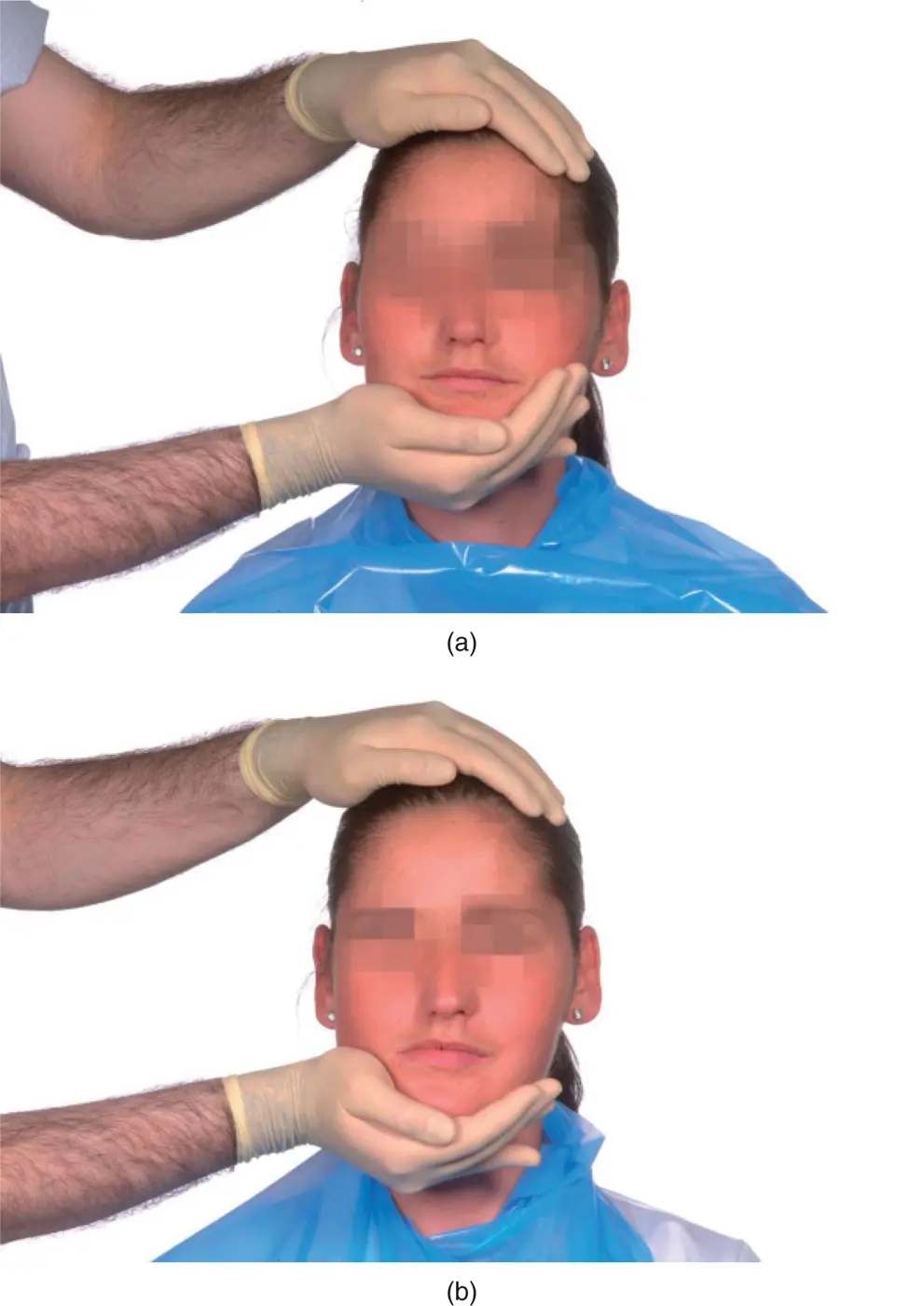
Figure 3.10(a) Examination of lateral pterygoid muscle against vertical resisted movement; (b) Examination of lateral pterygoid muscle against lateral resistance.
(M. Ziad Al‐Ani, Robin J.M. Gray.)
If the patient were, for instance to move the mandible to the right and this movement were resisted, left preauricular pain would arise if there was lateral pterygoid tenderness on the left.
Joint sounds 
 4
4
Clicking
Clicking from the TMJ is often felt by the patient but can be inaudible to the examiner. A click can occasionally be felt by palpating the TMJ in the preauricular region but is more often detected on intra‐auricular palpation.
If joint sounds are to be listened for, a reliable method is use of a stereo‐stethoscope. This consists of a standard earpiece with two outlets, rather than one, and two tubes, each of which is connected to a separate diaphragm ( Figure 3.11).
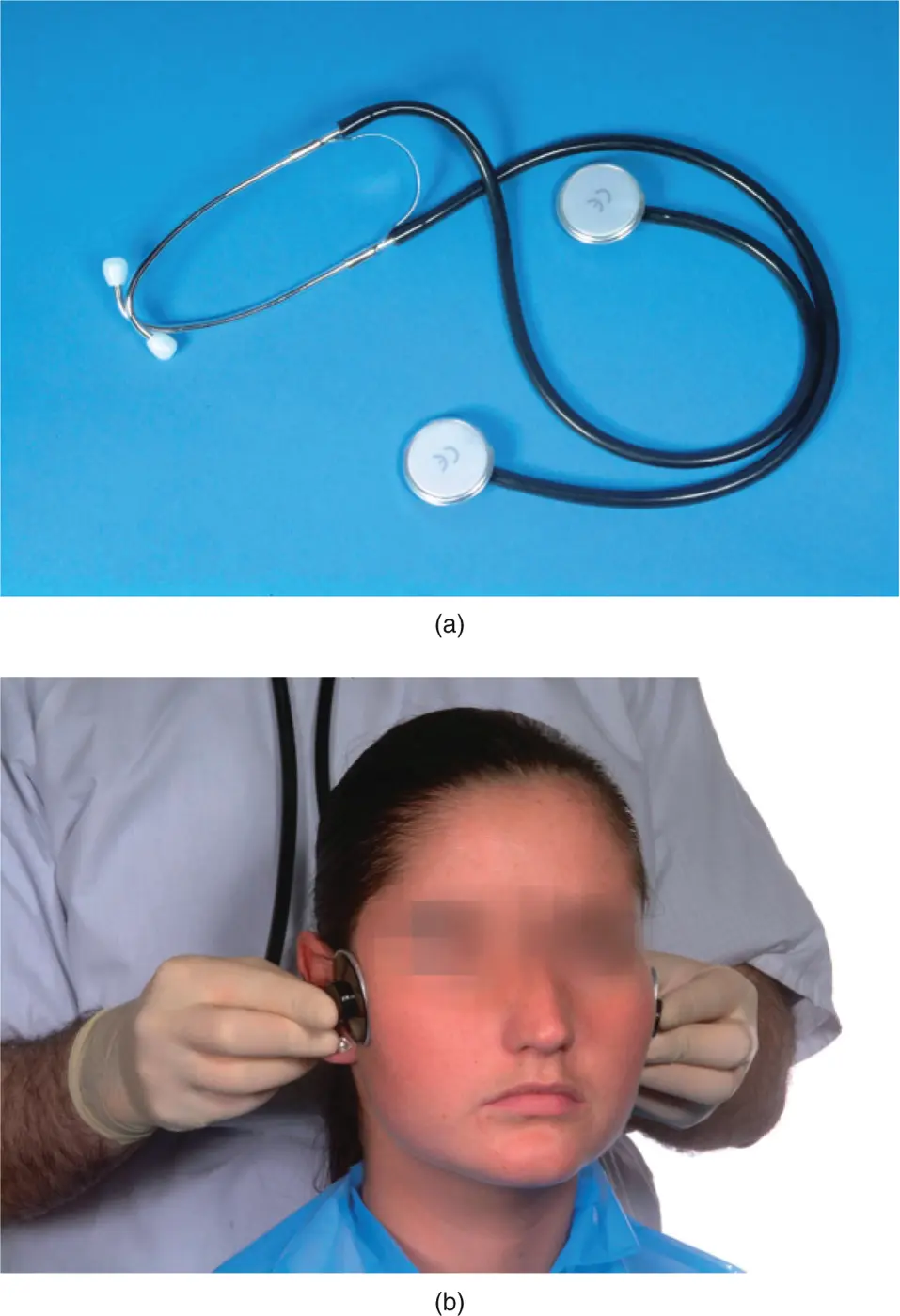
Figure 3.11(a) A stereo‐stethoscope used for listening to the temporomandibular joint. (b) Stereo‐stethoscope in use allowing auscultation and comparison of one TMJ with the other.
(M. Ziad Al‐Ani, Robin J.M. Gray.)
The apparatus provides a method of detecting TMJ sounds and determining whether they emanate from the right or left side or are bilateral. It should be remembered that it is sometimes extremely difficult to determine which side a click is coming from by listening with a stethoscope because of the ‘echo’ and reverberation across the bones of the skull from the contralateral side. In addition, auscultation permits the clinician to detect the frequently softer closing click that is sometimes difficult to detect on joint palpation alone.
For the diagnosis of disc displacement with reduction and to assist in determining a suitable treatment plan, it is important to determine whether the click can be eliminated by protrusion of the mandible. At the chair side, the patient is asked to protrude the mandible and then perform a series of opening and closing mouth movements, usually with the upper and lower incisors in an ‘edge‐to‐edge’ relationship. The click will be present during the first movement but, if the click is eliminated in subsequent movements in this protrusive mandibular position, the diagnosis of disc displacement with reduction is highly probable and it is likely that provision of a suitable splint design will reduce or eliminate the symptoms.
Crepitus
Crepitus is a crunching or grating sound that indicates degenerative joint disease. It can be heard with a stethoscope or, if severe, without when it may be readily audible to others. It can be present throughout the movement cycle or at any point in the cycle.
Signs of bruxism
 Tooth surface loss or tooth wear cannot be taken as a sign that the patient is an activebruxist. Even if bruxism is the cause of tooth surface loss, the patient may no longer be performing this parafunctional activity ( Figure 3.12a).
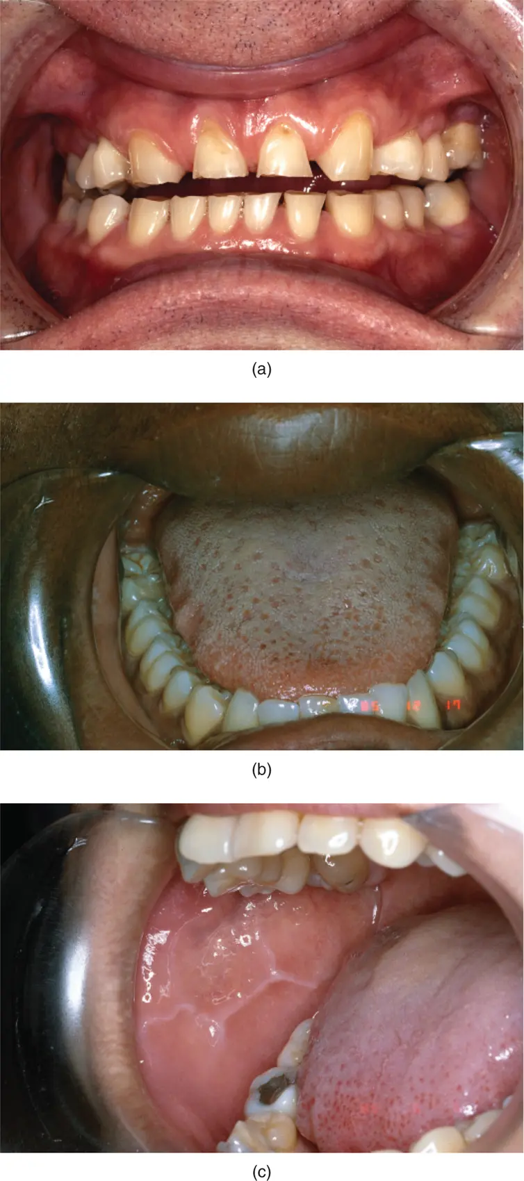
Figure 3.12(a) Attrition, (b) tongue scalloping, and (c) cheek ridging seen in patients who parafunction.
(M. Ziad Al‐Ani, Robin J.M. Gray.)
Dental sensitivity is a common symptom of active bruxism. The anterior teeth are often affected and this is frequently noticed on waking from sleep. Repeated tooth and/or restoration fracture is often also reported.
The two most reliable signs of activebruxism are, however, scalloping of the lateral border of the tongue ( Figure 3.12b) and ridging of the buccal cheek mucosa along the occlusal line ( Figure 3.12c). These features are due to the soft tissues being thrust against the surfaces of the teeth during parafunction. Ridging of the cheek mucosa is occasionally severe enough to present clinically as frictional hyperkeratosis. Both scalloping of the tongue and ridging of the cheek mucosa usually disappear when the parafunction ceases.
Occlusal examination 1
For the purposes of occlusal examination of a patient with a TMD, a straightforward examination technique can be employed. Further and more detailed examination will be necessary if it is determined that the occlusion is a major aetiological factor in the TMD or if restorative treatment is planned.
Читать дальшеИнтервал:
Закладка:
Похожие книги на «Temporomandibular Disorders»
Представляем Вашему вниманию похожие книги на «Temporomandibular Disorders» списком для выбора. Мы отобрали схожую по названию и смыслу литературу в надежде предоставить читателям больше вариантов отыскать новые, интересные, ещё непрочитанные произведения.


![John Bruce - The Lettsomian Lectures on Diseases and Disorders of the Heart and Arteries in Middle and Advanced Life [1900-1901]](/books/749387/john-bruce-the-lettsomian-lectures-on-diseases-and-disorders-of-the-heart-and-arteries-in-middle-and-advanced-life-1900-1901-thumb.webp)
Обсуждение, отзывы о книге «Temporomandibular Disorders» и просто собственные мнения читателей. Оставьте ваши комментарии, напишите, что Вы думаете о произведении, его смысле или главных героях. Укажите что конкретно понравилось, а что нет, и почему Вы так считаете.