Naoki Sugimoto - Chemistry and Biology of Non-canonical Nucleic Acids
Здесь есть возможность читать онлайн «Naoki Sugimoto - Chemistry and Biology of Non-canonical Nucleic Acids» — ознакомительный отрывок электронной книги совершенно бесплатно, а после прочтения отрывка купить полную версию. В некоторых случаях можно слушать аудио, скачать через торрент в формате fb2 и присутствует краткое содержание. Жанр: unrecognised, на английском языке. Описание произведения, (предисловие) а так же отзывы посетителей доступны на портале библиотеки ЛибКат.
- Название:Chemistry and Biology of Non-canonical Nucleic Acids
- Автор:
- Жанр:
- Год:неизвестен
- ISBN:нет данных
- Рейтинг книги:3 / 5. Голосов: 1
-
Избранное:Добавить в избранное
- Отзывы:
-
Ваша оценка:
Chemistry and Biology of Non-canonical Nucleic Acids: краткое содержание, описание и аннотация
Предлагаем к чтению аннотацию, описание, краткое содержание или предисловие (зависит от того, что написал сам автор книги «Chemistry and Biology of Non-canonical Nucleic Acids»). Если вы не нашли необходимую информацию о книге — напишите в комментариях, мы постараемся отыскать её.
Chemistry and Biology of Non-canonical Nucleic Acids — читать онлайн ознакомительный отрывок
Ниже представлен текст книги, разбитый по страницам. Система сохранения места последней прочитанной страницы, позволяет с удобством читать онлайн бесплатно книгу «Chemistry and Biology of Non-canonical Nucleic Acids», без необходимости каждый раз заново искать на чём Вы остановились. Поставьте закладку, и сможете в любой момент перейти на страницу, на которой закончили чтение.
Интервал:
Закладка:
1.5 Perspective of the Research for Non-canonical Nucleic Acid Structures
As the regulation of gene expression by the specific structure of nucleic acids has been clarified, the next important issue is knowing what specific structures are formed where and when in cells. For example, Hoogsteen base pairs are affected by the molecular environments such as ions, pH, and water activity. Cells are in an environment crowded with molecules, so-called molecular crowding (see Chapters 3and 4), and the molecular environment changes depending on the cell cycle [14]. For example, the nucleolus causes a change in the molecular density in the nucleus by repeating formation and dissociation according to the cell cycle. This regulates the timing of activation of rRNA transcription in each cell cycle, because the transcription of rRNA specifically occurs in nucleolus. In addition, the environment of mitochondria is particularly crowded (up to 500 mg ml −1) but heterogeneous due to locally increased proton concentration by the proton gradient required for ATP synthesis. Therefore, it is desirable to develop a technology that can predict physicochemical property of specific structures due to Hoogsteen base pairs in each characteristic molecular environment [15]. In addition, there is a possibility to make a new approach of drug development that treats diseases by changing the molecular environments of cells, rather than targeting genes and proteins.
1.6 Conclusion and Perspective
According to Pauling's personal communication revealed by the Nobel Foundation's disclosure, he considered Watson and Crick's Nobel award to be premature. In spite of his opinion, the Nobel Foundation decided to award the Prize to Watson and Crick. This might suggest that Watson–Crick base pairs were very common and meaningful at that time but non-Watson–Crick base pairs were artifact and meaningless. Nowadays, non-Watson–Crick base pairs are becoming common and significant as Pauling perhaps predicted. Now, the day when the essence of nucleic acids becomes beyond the concept of Watson and Crick is coming closer.
References
1 1 Schrödinger, E. (1944). What Is Life? The Physical Aspect of the Living Cell and Mind. Cambridge: Cambridge University Press.
2 2 Tamm, C., Hodes, M., and Chargaff, E. (1952). J. Biol. Chem. 195: 49–63.
3 3 Watson, J.D. and Crick, F.H. (1953). Nature 171: 737–738.
4 4 Pauling, L. and Corey, R.B. (1953). Nature 171: 346–346.
5 5 (a) Hoogsteen, K. (1959). Acta Crystallogr. 12: 822–823.(b) Hoogsteen, K.R. (1963). Acta Crystallogr. 16: 907–916.
6 6 (a) Day, R.O., Seeman, N.C., Rosenberg, J.M., and Rich, A. (1973). Proc. Natl. Acad. Sci. U. S. A. 70: 849–853.(b) Rosenberg, J.M., Seeman, N.C., Kim, J.J. et al. (1973). Nature 243: 150–154.
7 7 Wing, R., Drew, H., Takano, T. et al. (1980). Nature 287: 755–758.
8 8 Felsenfeld, G. and Rich, A. (1957). Biochim. Biophys. Acta 26: 457–468.
9 9 Kallenbach, N.R., Daniel, W.E. Jr. and Kaminker, M.A. (1976). Biochemistry 15: 1218–1224.
10 10 Gellert, M., Lipsett, M.N., and Davies, D.R. (1962). Proc. Natl. Acad. Sci. U. S. A. 48: 2013–2018.
11 11 Robertus, J.D., Ladner, J.E., Finch, J. et al. (1974). Nature 250: 546.
12 12 (a) Kruger, K., Grabowski, P.J., Zaug, A.J. et al. (1982). Cell 31: 147–157.(b) Zaug, A.J., Grabowski, P.J., and Cech, T.R. (1983). Nature 301: 578–583.
13 13 Gehring, K., Leroy, J.L., and Gueron, M. (1993). Nature 363: 561–565.
14 14 Nakano, S., Miyoshi, D., and Sugimoto, N. (2014). Chem. Rev. 114: 2733–2758.
15 15 (a) Takahashi, S. and Sugimoto, N. (2020). Chem. Soc. Rev. 49: 8439–8468.(b) Takahashi, S. and Sugimoto, N. (2021). Acc. Chem. Res. 54. In press.
2 Structures of Nucleic Acids Now
The main points of the learning:
1 Learn interactions in nucleic acid structures.
2 Understand structure polymorphisms of nucleic acids.
3 Study differences in conformational properties between DNA and RNA.
2.1 Introduction
Nucleic acids are basically molecules with a high degree of structural flexibility and polymorphic property. Phosphates in nucleic acids are negatively charged and cause electrostatic repulsion in each phosphate moiety. This electrostatic repulsion is disadvantageous for nucleic acids to form a compact and ordered structure. Nucleic acids form the higher-order structures by offsetting unfavorable entropy changes and electrostatic repulsion by internal interactions such as hydrogen bonding and stacking interactions and external factors such as interactions of nucleic acids with cations and cosolutes. In other words, the canonical nucleic acid structure consisting of double helix with Watson–Crick base pairs is a part of the possible structural forms, and nucleic acids form various non-canonical structures depending on the internal and external factors. This chapter shows basic elements that form non-canonical nucleic acid structures including unusual base pairing, whose existence has been revealed by structural analyses, and their properties of thermodynamic stabilities. Detailed analyses of the stabilities of nucleic acid structures and factors that affect them are explained in Chapter 3.
2.2 Unusual Base Pairs in a Duplex
As in Chapter 1, the canonical structure of nucleic acids is double-stranded helix with Watson–Crick base pairs ( Figure 2.1), which are almost identical in their geometric and dimensional arrangement in the helix. The sugar groups are both attached to the bases on the same side of the base pair. The distance between C1′ atoms of the sugars on opposite strands is essentially the same. These geometric features enable any sequence of Watson–Crick base pairs fit into the duplex. On the other hand, a large number of other arrangements and hydrogen bonding patterns of base pairs are possible, and many have been observed experimentally such as using X-ray and NMR analyses. X-ray analysis can define the duplex structures, which incorporate the non-Watson–Crick base pairs and provide details of their hydrogen bonding scheme [1]. On the other hand, NMR analysis provides information regarding the dynamics of the nucleic acid conformation such as mismatched base pairs, in which transient tautomeric and anionic species form Watson–Crick-type hydrogen bonds [2].
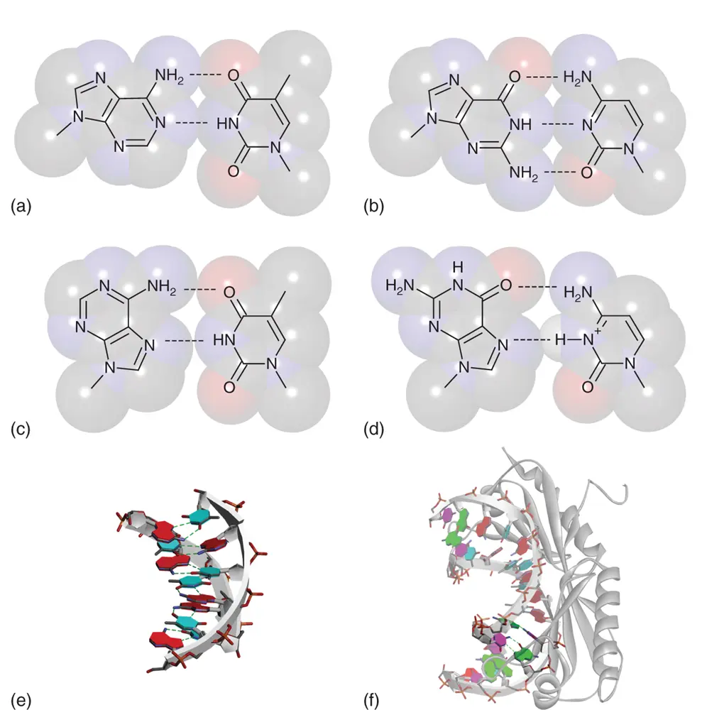
Figure 2.1 Watson–Crick and Hoogsteen base pairs in double helix. Chemical structures of A-T (a) and G-C (b) Watson–Crick base pairs. Chemical structures of A-T (c) and G-C +(d) Hoogsteen base pairs. N3 atom of cytosine nucleobase is protonated. (e) Structure of DNA duplex consisting of all A-T Hoogsteen base pairs (PDBID: 1RSB). (f) Structure of DNA duplex containing two consecutive G-C +Hoogsteen base pairs (PDB ID: 1QN3). The DNA duplex is bending due to interaction of TATA-box binding protein. Nucleobases forming the Hoogsteen base pairs are emphasized dark. In (e) and (f), hydrogen bonds between the Hoogsteen base pairs are shown in dashed lines.
2.2.1 Hoogsteen Base Pair
Hoogsteen base pair ( Figure 2.1) is one of the major non-Watson–Crick base pairs that can be seen in several crystal structures of duplexes containing A·T base pairs. For example, the crystal structure of AT-rich sequences adopted parallel and antiparallel stranded duplexes with all Hoogsteen-type hydrogen bonding in their A·T base pairs ( Figure 2.1) [3]. In the case of antiparallel duplex with the Hoogsteen base pairs, the overall structure features of the duplex such as diameter of the duplex, number of base pairs per turn, and sugar pucker conformation are similar to the canonical B-type DNA duplex. A unique characteristic is that the adenine nucleobases in the duplex have syn conformation in their glycosidic bond angles. Although the same pattern of hydrogen bonding is possible, A-type RNA duplexes disfavor the A·U Hoogsteen base pair because the A-form geometry disfavors the syn conformation in the adenine nucleobase due to sugar-backbone rearrangements needed to sterically accommodate the adenine [4]. Formation of Hoogsteen-type hydrogen bonding is also possible between guanine (G) and cytosine (C +), in which N3 atom is protonated. Formation of transient Hoogsteen base pairs including the G·C +in diverse sequence composition has been demonstrated by relaxation dispersion assay using NMR ( Figure 2.1) [5]. It is considered that the Hoogsteen base pairs play roles in modulating interaction of proteins and biological reactions such as induction or repair of DNA damage and replication of DNA by altering the structural and chemical properties of the duplex [5].
Читать дальшеИнтервал:
Закладка:
Похожие книги на «Chemistry and Biology of Non-canonical Nucleic Acids»
Представляем Вашему вниманию похожие книги на «Chemistry and Biology of Non-canonical Nucleic Acids» списком для выбора. Мы отобрали схожую по названию и смыслу литературу в надежде предоставить читателям больше вариантов отыскать новые, интересные, ещё непрочитанные произведения.


Обсуждение, отзывы о книге «Chemistry and Biology of Non-canonical Nucleic Acids» и просто собственные мнения читателей. Оставьте ваши комментарии, напишите, что Вы думаете о произведении, его смысле или главных героях. Укажите что конкретно понравилось, а что нет, и почему Вы так считаете.