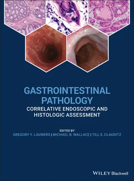
Gastrointestinal Pathology

Здесь есть возможность читать онлайн «Gastrointestinal Pathology» — ознакомительный отрывок электронной книги совершенно бесплатно, а после прочтения отрывка купить полную версию. В некоторых случаях можно слушать аудио, скачать через торрент в формате fb2 и присутствует краткое содержание. Жанр: unrecognised, на английском языке. Описание произведения, (предисловие) а так же отзывы посетителей доступны на портале библиотеки ЛибКат.

Gastrointestinal Pathology: краткое содержание, описание и аннотация

Предлагаем к чтению аннотацию, описание, краткое содержание или предисловие (зависит от того, что написал сам автор книги «Gastrointestinal Pathology»). Если вы не нашли необходимую информацию о книге — напишите в комментариях, мы постараемся отыскать её.

An illustrated guide to best practices when performing
 and assessing biopsies for GI conditions of all kinds
Accurate diagnosis of GI conditions necessarily entails both the careful taking of biopsies and the informed analysis of tissue material. With that being so, gastroenterologists and GI pathologists alike must have a solid understanding of the techniques, handling requirements, and diagnostic characteristics involved if they are to collaborate effectively. 
 has been designed to provide a clinically focussed and richly illustrated guide to real-world scenarios faced by practicing GI specialists, offering step-by-step instruction and professional advice on the correct diagnosis of all major GI conditions. This essential new book includes: 
Full-color illustrations throughout Complete details of biopsy samples required to diagnose specific conditions Reviews of differential diagnoses Clinical management clues based on pathologic findings Featuring information to improve the practice of all gastroenterologists and GI pathologists, 
 is a practical and every-day resource for the precise diagnosis of a wide range of GI conditions.
Gastrointestinal Pathology — читать онлайн ознакомительный отрывок

Ниже представлен текст книги, разбитый по страницам. Система сохранения места последней прочитанной страницы, позволяет с удобством читать онлайн бесплатно книгу «Gastrointestinal Pathology», без необходимости каждый раз заново искать на чём Вы остановились. Поставьте закладку, и сможете в любой момент перейти на страницу, на которой закончили чтение.

Тёмная тема
Сбросить

Интервал:

Закладка:

Сделать

2 Chapter 2 Figure 2.1 Endoscopic appearance of distal esophagus with normal squamous mu... Figure 2.2 Normal esophagus squamous mucosa with normal stratified non‐kerat... Figure 2.3 Endoscopic appearance of white exudate typical of Candida albican ... Figure 2.4 Debris of slough off superficial squamous epithelium with fungal ... Figure 2.5 PAS stain of Figure 2.4 illustrating the fungal elements of Candi ... Figure 2.6 Multinucleated basal keratinocytes typical of HSV esophagitis. Figure 2.7 Erosive esophagitis with prominent granulation and atypical cellu... Figure 2.8 High‐magnification evaluation of Figure 2.7 demonstrated (a) endo... Figure 2.9 Endoscopic appearance of erosive esophagitis in a patient with ga... Figure 2.10 Characteristic appearance of low‐power view of reflux esophagiti... Figure 2.11 High‐power magnification of reflux esophagitis with basal cell h... Figure 2.12 Endoscopic appearance of eosinophilic esophagitis (a) with so‐ca... Figure 2.13 Characteristic histology of eosinophilic esophagitis with basal ... Figure 2.14 Characteristic histology of lymphocytic esophagitis. Figure 2.15 Endoscopic appearance of inflammatory stricture of the esophagus... Figure 2.16 (a) Iron tablet erosive esophagitis with fibrinopurulent debris.... Figure 2.17 Endoscopic appearance of necrotizing esophagitis with appearance... Figure 2.18 Characteristic appearance of sloughing esophagitis with two‐tone... Figure 2.19 Endoscopic appearance of acute GVHD of esophagus with diffuse mu... Figure 2.20 Endoscopic appearance of inflammatory stricture with persistent ... Figure 2.21 Microscopic appearance of acute GVHD of esophagus with basal apo... Figure 2.22 Endoscopic appearance of esophageal lichen planus with diffuse m... Figure 2.23 Esophageal granuloma in a previously diagnosed patient with Croh...

3 Chapter 3 Figure 3.1 Endoscopic view of Barrett's esophagus. From upper left and clock... Figure 3.2 (a) Typical appearance of Barrett's esophagus with numerous goble... Figure 3.3 Barrett's esophagus indefinite for dysplasia. Nuclear hyperchroma... Figure 3.4 Low‐grade dysplasia. Note the sharp transition between nuclear hy... Figure 3.5 High‐grade dysplasia. The architectural features with back to bac... Figure 3.6 Crypt dysplasia. This lesion is characterized by neoplastic featu... Figure 3.7 (a) Foveolar dysplasia, low grade. This lesion is characterized b... Figure 3.8 Serrated dysplasia—note the serrated profile of the glands with l... Figure 3.9 (a) Intramucosal adenocarcionma—the lesion is characterized by di... Figure 3.10 Duplicated muscularis mucosae—note the inner and outer (duplicat... Figure 3.11 (a) Barrett's esophagus with high‐grade dysplasia in the 8–12 o'... Figure 3.12 Buried BE glands with low‐grade dysplasia. This phenomenon can b... Figure 3.13 Barrett's esophagus with high‐grade dysplasia showing p53 (left)... Figure 3.14 Squamous low‐grade dysplasia/intraepithelial neoplasia. Note the... Figure 3.15 (a) Squamous high‐grade dysplasia/intraepithelial neoplasia with... Figure 3.16 Basal layer dysplasia/intraepithelial neoplasia—note the basal a... Figure 3.17 (a) Schema of endoscopic and macroscopic classification of super... Figure 3.18 Schematic presentation of early and superficial esophageal cance... Figure 3.19 Well‐differentiated squamous cell carcinoma showing nests of aty... Figure 3.20 Basaloid squamous cell carcinoma characteristically showing pleo... Figure 3.21 Verrucous squamous cell carcinoma showing hyper‐ and parakeratos... Figure 3.22 Sarcomatoid squamous cell carcinoma. This picture shows atypical... Figure 3.23 Lugol's iodine staining in vivo of an early squamous cell carcin... Figure 3.24 Endoscopic appearance of an esophageal papilloma appearing as a ... Figure 3.25 Exophytic squamous papilloma showing delicate fibro‐vascular cor... Figure 3.26 Gastric heterotopia “inlet patch.” The salmon‐colored mucosa is ... Figure 3.27 Typical endoscopic image of heterotopic sebaceous glands in mid‐... Figure 3.28 Common histomorphological appearance of “inlet patch.” Figure 3.29 Heterotopic sebaceous glands. Note the typical vacuolated cytopl... Figure 3.30 Glycogenic acanthosis characterized by thickening of the epithel... Figure 3.31 Submucosal granular cell tumor showing a sharp demarcation to th... Figure 3.32 Granular cell tumors are typically immunohistochemically positiv... Figure 3.33 Esophageal leiomyoma (arrow) arising from the muscularis propria... Figure 3.34 Well‐defined submucosal leiomyoma with typical whorled fascicula... Figure 3.35 Leiomyosarcoma—fascicles of high cellularity with marked nuclear... Figure 3.36 Kaposi sarcoma with characteristic slit‐like spaces filled with ...

4 Chapter 4Figure 4.1 Endoscopic appearance of acute gastritis with superficial erosion...Figure 4.2 HP. Gastric antral biopsy with a severe population of Helicobacte ...Figure 4.3 HP inflammation. H.E. stain of a punch biopsy with typical inflam...Figure 4.4 Acute erosive changes. Antral biopsy with superficial erosion and...Figure 4.5 Reactive gastropathy. Antral mucosa showing foveolar hyperplasia ...Figure 4.6 (a) Focally enhanced gastritis. Gastric antral mucosa with a smal...Figure 4.7 Autoimmune gastritis. Typical changes of mucosa of the body in th...Figure 4.8 Lymphocytic gastritis. Superficial high magnification of a gastri...Figure 4.9 Syphilitic gastritis. Higher magnification of a corpus biopsy sho...Figure 4.10 High magnification of CMV gastritis with typical cherry red nucl...Figure 4.11 Eosinophilic gastritis. Superficial biopsy of the antrum with a ...Figure 4.12 Collagenous gastritis. Antral mucosa showing a prominent submuco...Figure 4.13 Iron pill induced gastritis. Superficial antral mucosa with appe...Figure 4.14 Calcifying gastritis. Antral punch biopsy with erosive and react...Figure 4.15 Doxycycline erosive gastritis. The classic appearance is compose...Figure 4.16 (a) GvHD. Typical changes in longstanding GvHD showing multiple ...Figure 4.17 Chemoradiation. High magnification of a gastric mucosal biopsy w...Figure 4.18 Kayaxylate. Example of a chronic gastric ulcer caused by Kayaxal...Figure 4.19 GAVE. Antral mucosa with dilation changes of the capillaries and...Figure 4.20 Amyloidosis. Superficial gastric mucosa showing dense homogenous...

5 Chapter 5Figure 5.1 Single (a) or multiple (b) small translucent lesions covered by b...Figure 5.2 Fundic gland polyposis in familial adenomatous polyposis. There a...Figure 5.3 Low‐power photomicrograph of fundic gland polyp with multiple cys...Figure 5.4 Higher magnification of Figure 5.3 showing cystically dilated gla...Figure 5.5 Low‐grade (foveolar type) dysplasia in a sporadic fundic gland po...Figure 5.6 Endoscopic appearance of a large hyperplastic polyp covered by er...Figure 5.7 Low magnification of a hyperplastic polyp (a), showing gastric fo...Figure 5.8 Small pyloric type glands beneath the proliferating foveolar epit...Figure 5.9 Markedly proliferative serrated foveolar epithelium mimicking car...Figure 5.10 Adenomatous low‐grade dysplasia found in hyperplastic polyp show...Figure 5.11 Gastric biopsy from polypoid mass showing both hyperplastic poly...Figure 5.12 A small polypoid foveolar hyperplasia composed solely of prolife...Figure 5.13 Low‐power view of gastritis cystica polyposa.Figure 5.14 Gastric biopsy specimen with bland glands admixed with edematous...Figure 5.15 Adenocarcinoma observed in the herniated gastric mucosa in the s...Figure 5.16 Endoscopic view of a pedunculated gastric juvenile polypFigure 5.17 Typical microscopic features of intestinal type adenoma with low...Figure 5.18 Gastric foveolar type adenoma with low‐grade dysplasia (left) an...Figure 5.19 Gastric foveolar type adenoma with pale clear cytoplasm and high...Figure 5.20 Closely packed pyloric type glands lined by cuboidal/columnar ce...Figure 5.21 Endoscopic appearance of a pedunculated gastric juvenile polyp w...Figure 5.22 Cystically dilated glandular and foveolar elements in juvenile p...Figure 5.23 Endoscopically many sessile polyps are found in patients with Pe...Figure 5.24 Histomorphologically arborizing bundles of smooth muscles emerge...Figure 5.25 Adenocarcinoma developed in patients with hamartomatous polyposi...Figure 5.26 Gastric polyp in patient with Cowden's disease with foveolar hyp...Figure 5.27 Histomorphological appearance of a biopsy from a patient diagnos...Figure 5.28 A recently described finding in GAPPS are polyps showing hyperpr...Figure 5.29 Inflammatory fibroid polyp presenting as broad‐based submucosal ...Figure 5.30 Gastric biopsy specimen with proliferation of loose spindle cell...Figure 5.31 Immunohistochemical staining of the inflammatory fibroid polyp (...Figure 5.32 Submucosal aggregates of xanthoma cells with a distinct cell mem...Figure 5.33 Heterotopic pancreas on the posterior wall of the mid‐body, an s...Figure 5.34 In gastric pinch biopsies, observation of pancreatic acini in th...Figure 5.35 Gastric pinch biopsy of a patient with endoscopical appearance o...

Читать дальше
Тёмная тема
Сбросить

Интервал:

Закладка:

Сделать

Похожие книги на «Gastrointestinal Pathology»

Представляем Вашему вниманию похожие книги на «Gastrointestinal Pathology» списком для выбора. Мы отобрали схожую по названию и смыслу литературу в надежде предоставить читателям больше вариантов отыскать новые, интересные, ещё непрочитанные произведения.


Отзывы о книге «Gastrointestinal Pathology»

Обсуждение, отзывы о книге «Gastrointestinal Pathology» и просто собственные мнения читателей. Оставьте ваши комментарии, напишите, что Вы думаете о произведении, его смысле или главных героях. Укажите что конкретно понравилось, а что нет, и почему Вы так считаете.

x