Gastrointestinal Pathology
Здесь есть возможность читать онлайн «Gastrointestinal Pathology» — ознакомительный отрывок электронной книги совершенно бесплатно, а после прочтения отрывка купить полную версию. В некоторых случаях можно слушать аудио, скачать через торрент в формате fb2 и присутствует краткое содержание. Жанр: unrecognised, на английском языке. Описание произведения, (предисловие) а так же отзывы посетителей доступны на портале библиотеки ЛибКат.

- Название:Gastrointestinal Pathology
- Автор:
- Жанр:
- Год:неизвестен
- ISBN:нет данных
- Рейтинг книги:3 / 5. Голосов: 1
-
Избранное:Добавить в избранное
- Отзывы:
-
Ваша оценка:
Gastrointestinal Pathology: краткое содержание, описание и аннотация
Предлагаем к чтению аннотацию, описание, краткое содержание или предисловие (зависит от того, что написал сам автор книги «Gastrointestinal Pathology»). Если вы не нашли необходимую информацию о книге — напишите в комментариях, мы постараемся отыскать её.
and assessing biopsies for GI conditions of all kinds
Accurate diagnosis of GI conditions necessarily entails both the careful taking of biopsies and the informed analysis of tissue material. With that being so, gastroenterologists and GI pathologists alike must have a solid understanding of the techniques, handling requirements, and diagnostic characteristics involved if they are to collaborate effectively.
has been designed to provide a clinically focussed and richly illustrated guide to real-world scenarios faced by practicing GI specialists, offering step-by-step instruction and professional advice on the correct diagnosis of all major GI conditions. This essential new book includes:
Full-color illustrations throughout Complete details of biopsy samples required to diagnose specific conditions Reviews of differential diagnoses Clinical management clues based on pathologic findings Featuring information to improve the practice of all gastroenterologists and GI pathologists,
is a practical and every-day resource for the precise diagnosis of a wide range of GI conditions.
Gastrointestinal Pathology — читать онлайн ознакомительный отрывок
Ниже представлен текст книги, разбитый по страницам. Система сохранения места последней прочитанной страницы, позволяет с удобством читать онлайн бесплатно книгу «Gastrointestinal Pathology», без необходимости каждый раз заново искать на чём Вы остановились. Поставьте закладку, и сможете в любой момент перейти на страницу, на которой закончили чтение.
Интервал:
Закладка:
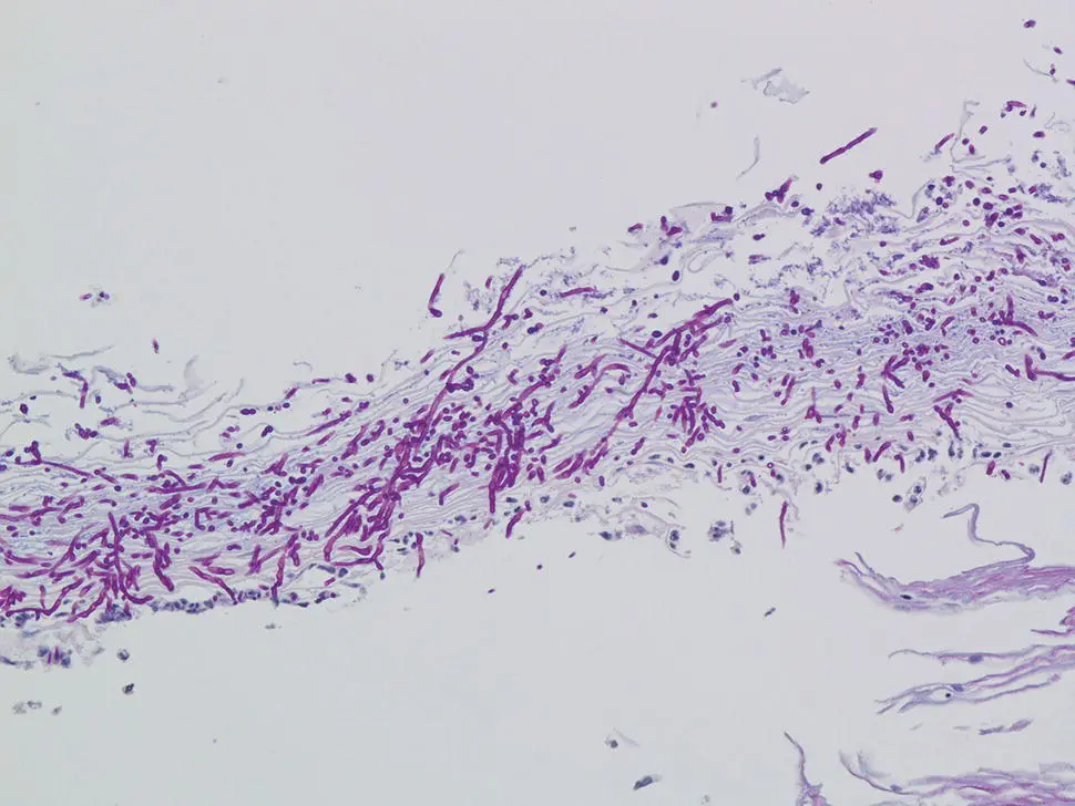
Figure 2.5 PAS stain of Figure 2.4illustrating the fungal elements of Candida albicans .
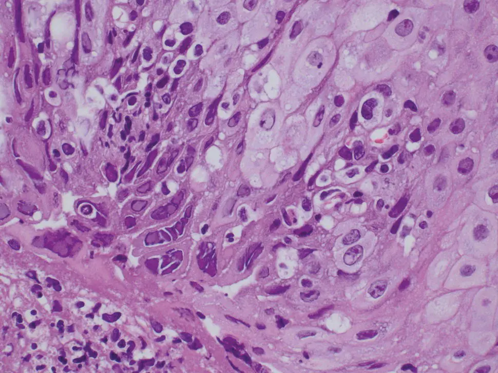
Figure 2.6 Multinucleated basal keratinocytes typical of HSV esophagitis.
Candida can colonize preexisting ulcers or damaged mucosa of any etiology, and in such cases the possibility of dual infection or pathology should be considered. Also, when Candida yeast forms are present only in desquamated keratin debris unassociated with inflammation or endoscopic findings, carry over from an oral infection should be suspected. Cytologic brushings are also very useful in the diagnosis of Candida esophagitis, as the organisms are readily identified on Papanicolaou stained smears.
Herpes Simplex (HSV)
If HSV infection is suspected, in addition to biopsies for histology, fresh tissue can be sent for viral culture to confirm the diagnosis and identify strains that may be resistant to acyclovir. HSV infection is associated with ulceration and a mixed inflammatory infiltrate of intraepithelial neutrophils, eosinophils, and lymphocytes, as well as aggregates of macrophages that can be a diagnostic clue. HSV infects squamous cells of intact or denuded epithelium and is best identified at the edge of an ulcer and in cells within the ulcer slough. Therefore, optimal histologic diagnosis requires sampling of the ulcer edge rather than the ulcer bed. The typical viral cytopathic effects include multinucleation, ground glass nuclei, and dense intranuclear eosinophilic inclusions with a thickened nuclear membrane and a clear halo (Cowdry type A inclusion bodies) ( Figure 2.6). Viral inclusions and multinucleated cells are not always identifiable in biopsies, and ancillary immunohistochemistry (IHC) staining for HSV may be required. Concomitant infection of Candida , cytomegalovirus, or bacteria can exist, particularly in immunocompromised patients. Varicella Zoster Virus has been associated with esophagobronchial fistula formation.
Cytomegalovirus (CMV)
Culture is not used routinely, and does not distinguish the simple presence of CMV from active infection; however, culture may be useful for identifying drug resistance.
Ulceration and granulation tissue formation with associated acute inflammation is common. In contrast to HSV esophagitis, CMV rarely infects the squamous epithelium, and preferentially involves endothelial cells, stromal cells, and glandular epithelium. Therefore, biopsies of the ulcer bed should preferentially be performed when CMV esophagitis is suspected ( Figure 2.7). CMV cytopathic effect is characterized by nuclear enlargement with classic “owl eye” large intranuclear inclusions, and granular, eosinophilic cytoplasmic inclusions ( Figure 2.8a). Smaller, atypical intranuclear inclusions may be present and are more subtle, simulating activated fibroblasts. These infected cells are best identified by immunohistochemical staining ( Figure 2.8b). CMV may coexist with HSV and Candida infection in some cases.
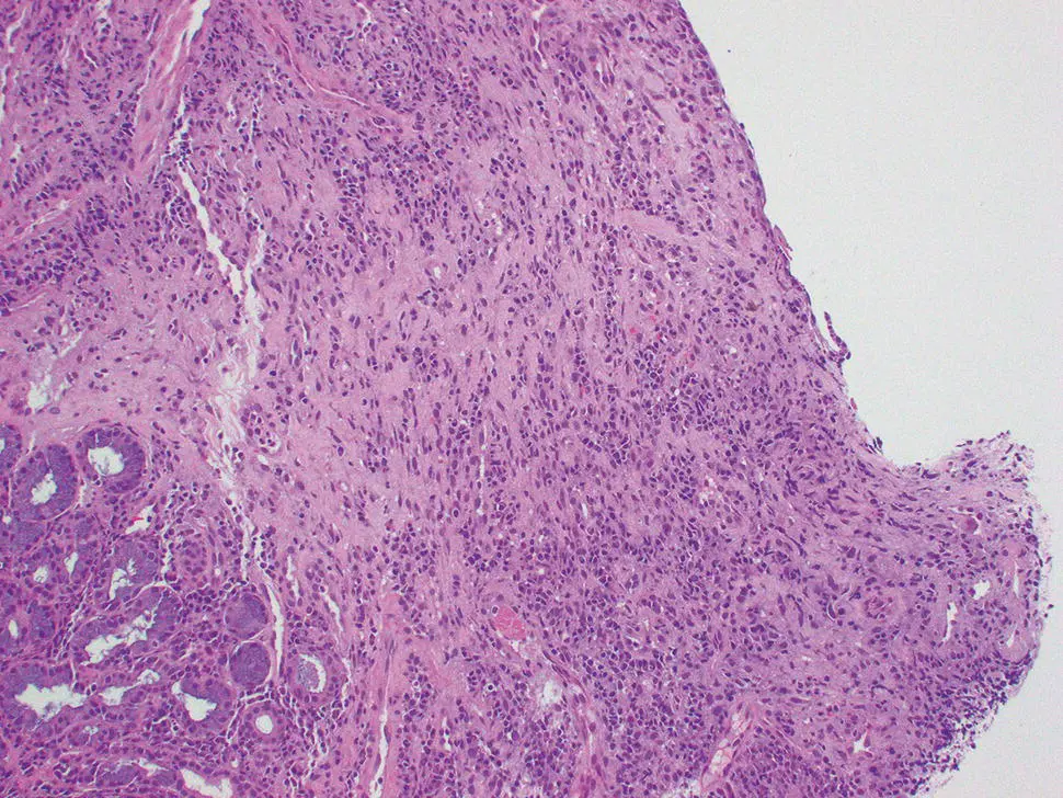
Figure 2.7 Erosive esophagitis with prominent granulation and atypical cellular elements.
Bacterial Esophagitis
Sheets of bacteria are typically present with associated necrosis and mucosal erosion, highlighted by a tissue Gram stain. Inflammation may be scant or absent in neutropenic patients.
Mycobacterial Esophagitis
Histologic features include subepithelial and mural necrotic and non‐necrotic granulomas with fibrosis and chronic inflammation. Acid fast bacilli can be recognized on Ziehl–Neelsen stain in approximately 2/3 of cases.
Immunohistochemical Studies and Molecular Features
IHC staining for HSV (HSV 1 and 2 cocktail stain) may be useful for suspected infections if diagnostic inclusions are not readily identified on H&E sections. Similarly, IHC for CMV is useful to highlight infected cells without typical CMV morphology, and may be more sensitive than light microscopy.
PCR for M. tuberculosis may be useful in selected cases.
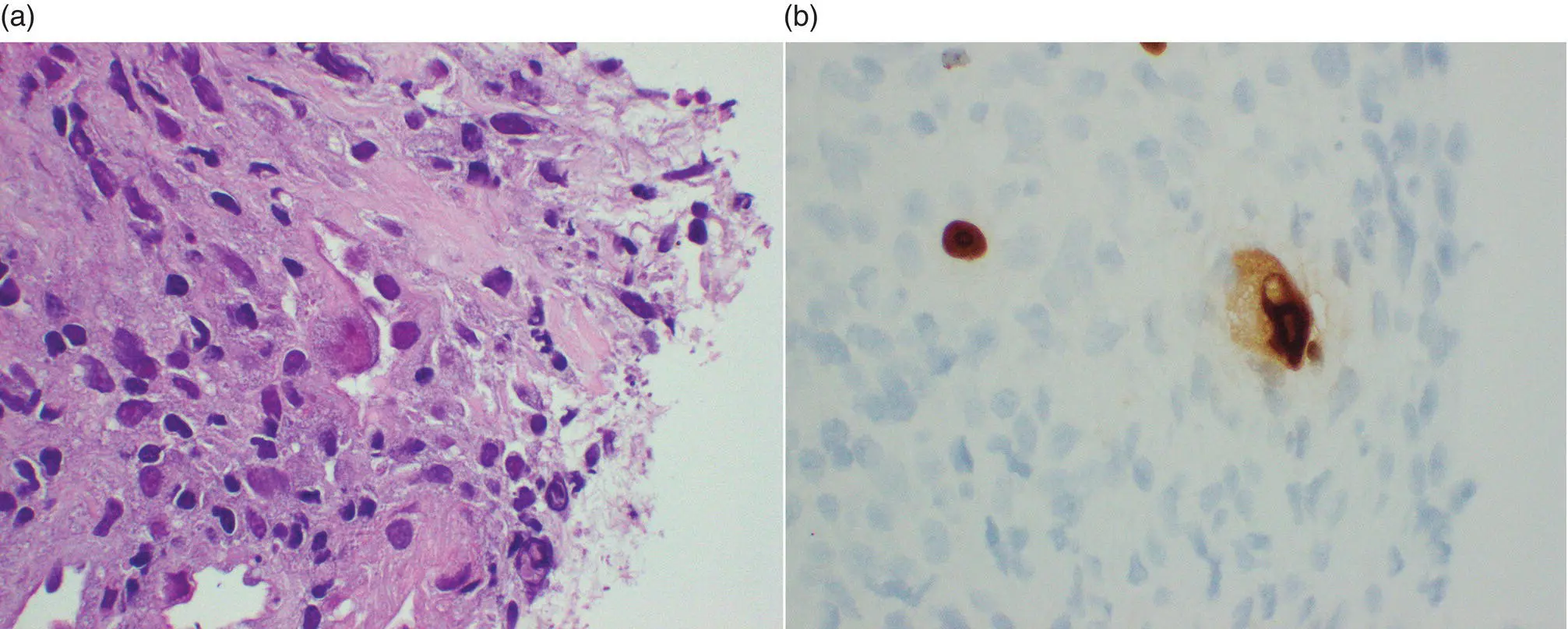
Figure 2.8 High‐magnification evaluation of Figure 2.7demonstrated (a) endothelial CMV infection (b) confirmed by immunohistochemistry.
Differential Diagnosis
Clinical
Inflammatory changes of the esophagus can be seen in many conditions, most commonly acid reflux disease. Behcet's disease may present with focal ulcers, as can pill‐induced (associated with an impacted pill, which may or may not be present during exam) ulcers.
Microscopic
The differential diagnosis of Candida esophagitis includes reflux esophagitis, drug injury, glycogenic acanthosis, ectopic sebaceous glands, and other infections.
The differential diagnosis of HSV esophagitis includes other etiologies of mucosal ulceration, including other infections, Crohn's disease, HIV‐associated ulceration, and drug injury. In some cases, the cytologic atypia induced in the squamous epithelium by the viral cytopathic effects of HSV may be difficult to distinguish from squamous dysplasia/carcinoma, as well as radiation esophagitis.
Prognosis, Evolution, and Clinical Management
Most patients present with dysphagia or odynophagia and undergo upper endoscopy with biopsy. Infectious etiologies are treated with appropriate antimicrobials (e.g. fluconazole for candida), typically with high response rates. In immunosuppressed patients, including those on chronic inhaled steroids, frequent retreatment, or chronic suppressive treatment, may be necessary after full efforts to restore immune function.
Gastroesophageal Reflux Disease
Definition, General Features, Predisposing Factors
The chronic regurgitation of gastroduodenal juice into the esophagus causes gastroesophageal reflux disease (GERD). The mucosal injury results from the effects of refluxed gastric acid, bile, pepsin, and duodenal contents that overwhelm the normal protective antireflux barriers, such as esophageal acid clearance and mucosal resistance. Risk factors and predisposing conditions include obesity, diet, sedentary lifestyle, tobacco and alcohol use, hiatus hernia, reduced lower esophageal sphincter tone, loss of esophageal peristaltic function, gastric hypersecretory states, and delayed gastric emptying. Symptomatic GERD is prevalent worldwide, with considerable geographic variation. Prevalence estimates are approximately 20% in North America and Europe, with only East Asia showing estimates <10%.
Clinical Features and Endoscopic Characteristics
There are several endoscopic classifications of gastroesophageal reflux disease ( Figure 2.9). The Los Angeles Classification divides esophagitis into four grades:
Grade A: one or more mucosal breaks, each no longer than 5 mm.
Grade B: at least one mucosal break more than 5 mm long, but not continuous between the tops of 2 mucosal folds.
Читать дальшеИнтервал:
Закладка:
Похожие книги на «Gastrointestinal Pathology»
Представляем Вашему вниманию похожие книги на «Gastrointestinal Pathology» списком для выбора. Мы отобрали схожую по названию и смыслу литературу в надежде предоставить читателям больше вариантов отыскать новые, интересные, ещё непрочитанные произведения.


Обсуждение, отзывы о книге «Gastrointestinal Pathology» и просто собственные мнения читателей. Оставьте ваши комментарии, напишите, что Вы думаете о произведении, его смысле или главных героях. Укажите что конкретно понравилось, а что нет, и почему Вы так считаете.