Practical Procedures in Implant Dentistry
Здесь есть возможность читать онлайн «Practical Procedures in Implant Dentistry» — ознакомительный отрывок электронной книги совершенно бесплатно, а после прочтения отрывка купить полную версию. В некоторых случаях можно слушать аудио, скачать через торрент в формате fb2 и присутствует краткое содержание. Жанр: unrecognised, на английском языке. Описание произведения, (предисловие) а так же отзывы посетителей доступны на портале библиотеки ЛибКат.
- Название:Practical Procedures in Implant Dentistry
- Автор:
- Жанр:
- Год:неизвестен
- ISBN:нет данных
- Рейтинг книги:3 / 5. Голосов: 1
-
Избранное:Добавить в избранное
- Отзывы:
-
Ваша оценка:
Practical Procedures in Implant Dentistry: краткое содержание, описание и аннотация
Предлагаем к чтению аннотацию, описание, краткое содержание или предисловие (зависит от того, что написал сам автор книги «Practical Procedures in Implant Dentistry»). Если вы не нашли необходимую информацию о книге — напишите в комментариях, мы постараемся отыскать её.
covers core topics such as:
Rationale and assessment for implant placement and restoration, including the diagnostic records and surgical considerations required for optimal planning and risk management Incision design considerations and flap management, with an essential knowledge of regional neuro-vascular structures Implant placement, encompassing the timing of the placement, bone requirements and understanding the importance of the peri-implant interface for soft tissue stability Impression techniques, loading protocols, digital workflows and the aesthetic considerations of implants Prosthetic rehabilitation of single tooth implants to fully edentulous workflows, including discussions of soft tissue support, biomechanics and occlusal verification Perfect for both general dental practitioners and specialists in implant dentistry,
is also a valuable reference to senior undergraduate and postgraduate dental students.
Practical Procedures in Implant Dentistry — читать онлайн ознакомительный отрывок
Ниже представлен текст книги, разбитый по страницам. Система сохранения места последней прочитанной страницы, позволяет с удобством читать онлайн бесплатно книгу «Practical Procedures in Implant Dentistry», без необходимости каждый раз заново искать на чём Вы остановились. Поставьте закладку, и сможете в любой момент перейти на страницу, на которой закончили чтение.
Интервал:
Закладка:
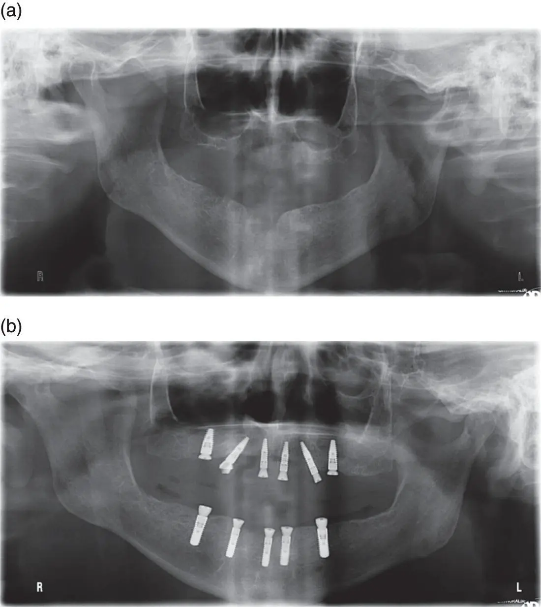
Figure 7.4 Implant placement following maxillary sinus augmentation in a staged approach. Sinus floor elevation (SFE) via a lateral window approach was performed on both the right and left posterior edentulous segments, as the bone quality and residual bone height as shown in the panoramic radiograph (a) were not favourable for a simultaneous implant placement in conjunction with the grafting procedure. Following sinus graft consolidation over a period of six months, implants were placed in the posterior segments uneventfully as shown in the panoramic radiograph (b).
7.6 Greater Palatine Artery and Nerve
Upon exiting the greater palatine foramen located medial and slightly distal to the maxillary third molar, the greater palatine artery and nerve run anterior along the hard palate to the incisive foramen as described earlier. The greater palatine neurovascular bundle is typically located at the junction of the vertical and horizontal palatal walls of the palatal vault ( Figure 7.5).
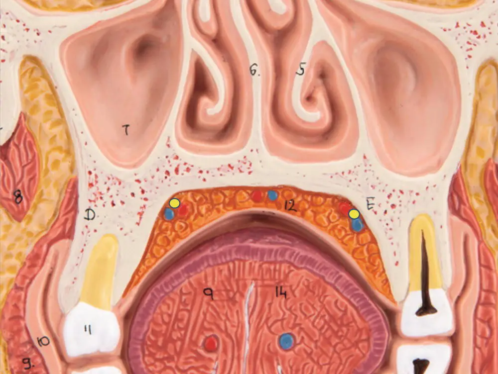
Figure 7.5 Position of the greater palatine artery and nerve. The greater palatine neurovascular bundle is outlined in purple in the frontal section of the skull at the level of the premolars. Individuals with higher palatal vaults exhibit greater distances from the lingual gingival margin to the neurovascular bundle than lower palatal vaults.
7.6.1 Importance in Oral Implantology
When creating an incision in the region of the greater palatine artery a zone of safety should be maintained to avoid injury to the artery with potential resulting bleeding and soft tissue necrosis. This zone of safety will often depend on the anatomical variation of the individual patient. Although the greater palatine neurovascular bundle is typically located at the junction of the vertical and horizontal palatal walls, significant variations in palatal vault depth mean the neurovascular bundle could be as close as 7 mm from the lingual gingival margin at the first molar site in low palatal vault phenotypes to as much as 17 mm in high palatal vault phenotypes [28]. The harvest of connective tissue grafts and free gingival grafts is routinely performed in the maxillary posterior palatal tissues from the first molar site forward to the canine. In most dentate patients without significant periodontal disease it is possible to harvest connective tissue and free gingival grafts up to 8 mm in height without injury to the neurovascular bundle [29].
References
1 1 Song, W., Jo, D.I., Lee, J.Y. et al. (2009). Microanatomy of the incisive canal using three‐dimensional reconstruction of microCT images: an ex vivo; study. Oral Surg. Oral Med. Oral Pathol. Oral Radiol. Endod. 108 (4): 583–590.
2 2 Friedrich, R., Laumann, F., Zrnc, T., and Assaf, A. (2015). The nasopalatine canal in adults on cone beam computed tomograms – a clinical study and review of the literature. in vivo; 29 (4): 467–486.
3 3 Mraiwa, N., Jacobs, R., and Van Cleynenbreugel, J. (2004). The nasopalatine canal revisited using 2D and 3D CT imaging. Dentomaxillofac. Radiol. 33: 396–402.
4 4 Marcantonio, E.J. (2009). Incisive canal deflation for correct implant placement: case report. Implant Dent. 18: 473–479.
5 5 Rosenquist, J. and Nystrom, E. (1992). Occlusion of the incisal canal with bone chips. A procedure to facilitate insertion of implants in the anterior maxilla. Int. J. Oral Maxillofac. Surg. 21: 210–211.
6 6 Garg, A. (1997). Nasal sinus lift: an innovative technique for implant insertions. Dent. Implantol. Update 8: 49.
7 7 Garg, A. (2008). Subnasal elevation and bone augmentation in dental implantology. Dent. Implantol. Update 19: 17.
8 8 Hising, P., Bolin, A., and Branting, C. (2001). Reconstruction of the severely resorbed alveolar ridge crests with dental implants using bovine bone mineral for augmentation. Int. J. Oral Maxillofac. Implants 16: 90.
9 9 Mazor, Z., Lorean, A., and Mijiritsky, E. (2012). Nasal floor elevation combined with dental implant placement. Clin. Implant Dent. Relat. Res. 14 (5): 768–771.
10 10 El‐Ghareeb, M., Pi‐Anfruns, J., Khosousi, M. et al. (2012). Nasal floor augmentation for the reconstruction of the atrophic maxilla: a case series. J. Oral Maxillofac. Surg. 70 (3): 235–241.
11 11 Sicher, H. and DuBrul, E. (1975). The viscera of the head and neck. In: Oral Anatomy, 7e, 418–424. St. Louis: Mosby.
12 12 Mehra, P. and Murad, H. (2004). Maxillary sinus disease of odontogenic origin. Otolaryngol. Clin. North Am. 37: 347–364.
13 13 Sahlstrand‐Johnson, P., Jannert, M., Strombeck, A., and Abul‐Kasim, K. (2011). Computed tomography measurements of different dimensions of maxillary and frontal sinuses. BMC Med. Imaging 11: 8.
14 14 Sharma, S., Jehan, M., and Kumar, A. (2014). Measurements of maxillary sinus volume and dimensions by computed tomography scan for gender determination. J. Anat. Soc. India 63: 36–42.
15 15 El‐Anwar, M., Raafat, A., Mostafa, R. et al. (2018). Maxillary sinus ostium assessment: a CT study. Egypt. J. Radiol. Nucl. Med. 49 (4): 1009–1013.
16 16 Kim, M., Jung, U., and Kim, C. (2006). Maxillary sinus septa: prevalence, height, location, and morphology. A reformatted computed tomography scan analysis. J. Periodontol. 77: 903–908.
17 17 Velasquez‐Plata, D., Hover, L., Peach, C., and Adler, M. (2002). Maxillary sinus septa: a 3‐dimensional computerized tomographic scan analysis. Int. J. Oral Maxillofac. Implants 17: 854–860.
18 18 McGowan, D., Baxter, P., and James, J. (1993). The Maxillary Sinus and its Dental Implications. Oxford: Butterworth‐Heinemann.
19 19 Mogensen, C. and Tos, M. (1977). Quantitative histology of the maxillary sinus. Rhinology 15: 129–140.
20 20 Cawood, J. and Howell, R. (1988). A classification of the edentulous jaws. Int. J. Oral Maxillofac. Surg. 17: 232–236.
21 21 Garg, A. (1999). Augmentation grafting of the maxillary sinus for placement of dental implants: anatomy, physiology, and procedures. Implant Dent. 8: 36–46.
22 22 Del Fabbro, M., Wallace, S., and Testori, T. (2013). Long‐term implant survival in the grafted maxillary sinus: a systematic review. Int. J. Periodontics Restorative Dent. 33: 773–783.
23 23 Jensen, O., Shulman, L., Block, M., and Iacono, V. (1998). Report of the sinus consensus conference of 1996. Int. J. Oral Maxillofac. Implants 13 (Suppl): 11–45.
24 24 Seong, W.J., Barczak, M., Jung, J. et al. (2013). Prevalence of sinus augmentation associated with maxillary posterior implants. J. Oral Implantol. 39: 680–688.
25 25 Esposito, M., Felice, P., and Worthington, H.V. (2010). Interventions for replacing missing teeth: augmentation procedures for the maxillary sinus. Cochrane Database Syst. Rev. 3.
26 26 Felice, P., Pistilli, R., Piattelli, M. et al. (2014). 1‐stage versus 2‐stage lateral sinus lift procedures: 1‐year post‐loading results of a multicentre randomised controlled trial. Eur. J. Oral Implantol. 7 (1): 65–75.
27 27 Esposito, M., Felice, P., and Worthington, H. (2014). Interventions for replacing missing teeth: augmentation procedures of the maxillary sinus. Cochrane Database Syst. Rev. 13 (5).
28 28 Reiser, G., Bruno, J., Mahan, P., and Larkin, L. (1996). The subepithelial connective tissue graft palatal donor site: anatomic considerations for surgeons. Int. J. Periodontics Restorative Dent. 16: 130–137.
Читать дальшеИнтервал:
Закладка:
Похожие книги на «Practical Procedures in Implant Dentistry»
Представляем Вашему вниманию похожие книги на «Practical Procedures in Implant Dentistry» списком для выбора. Мы отобрали схожую по названию и смыслу литературу в надежде предоставить читателям больше вариантов отыскать новые, интересные, ещё непрочитанные произведения.


Обсуждение, отзывы о книге «Practical Procedures in Implant Dentistry» и просто собственные мнения читателей. Оставьте ваши комментарии, напишите, что Вы думаете о произведении, его смысле или главных героях. Укажите что конкретно понравилось, а что нет, и почему Вы так считаете.