Practical Procedures in Implant Dentistry
Здесь есть возможность читать онлайн «Practical Procedures in Implant Dentistry» — ознакомительный отрывок электронной книги совершенно бесплатно, а после прочтения отрывка купить полную версию. В некоторых случаях можно слушать аудио, скачать через торрент в формате fb2 и присутствует краткое содержание. Жанр: unrecognised, на английском языке. Описание произведения, (предисловие) а так же отзывы посетителей доступны на портале библиотеки ЛибКат.
- Название:Practical Procedures in Implant Dentistry
- Автор:
- Жанр:
- Год:неизвестен
- ISBN:нет данных
- Рейтинг книги:3 / 5. Голосов: 1
-
Избранное:Добавить в избранное
- Отзывы:
-
Ваша оценка:
Practical Procedures in Implant Dentistry: краткое содержание, описание и аннотация
Предлагаем к чтению аннотацию, описание, краткое содержание или предисловие (зависит от того, что написал сам автор книги «Practical Procedures in Implant Dentistry»). Если вы не нашли необходимую информацию о книге — напишите в комментариях, мы постараемся отыскать её.
covers core topics such as:
Rationale and assessment for implant placement and restoration, including the diagnostic records and surgical considerations required for optimal planning and risk management Incision design considerations and flap management, with an essential knowledge of regional neuro-vascular structures Implant placement, encompassing the timing of the placement, bone requirements and understanding the importance of the peri-implant interface for soft tissue stability Impression techniques, loading protocols, digital workflows and the aesthetic considerations of implants Prosthetic rehabilitation of single tooth implants to fully edentulous workflows, including discussions of soft tissue support, biomechanics and occlusal verification Perfect for both general dental practitioners and specialists in implant dentistry,
is also a valuable reference to senior undergraduate and postgraduate dental students.
Practical Procedures in Implant Dentistry — читать онлайн ознакомительный отрывок
Ниже представлен текст книги, разбитый по страницам. Система сохранения места последней прочитанной страницы, позволяет с удобством читать онлайн бесплатно книгу «Practical Procedures in Implant Dentistry», без необходимости каждый раз заново искать на чём Вы остановились. Поставьте закладку, и сможете в любой момент перейти на страницу, на которой закончили чтение.
Интервал:
Закладка:
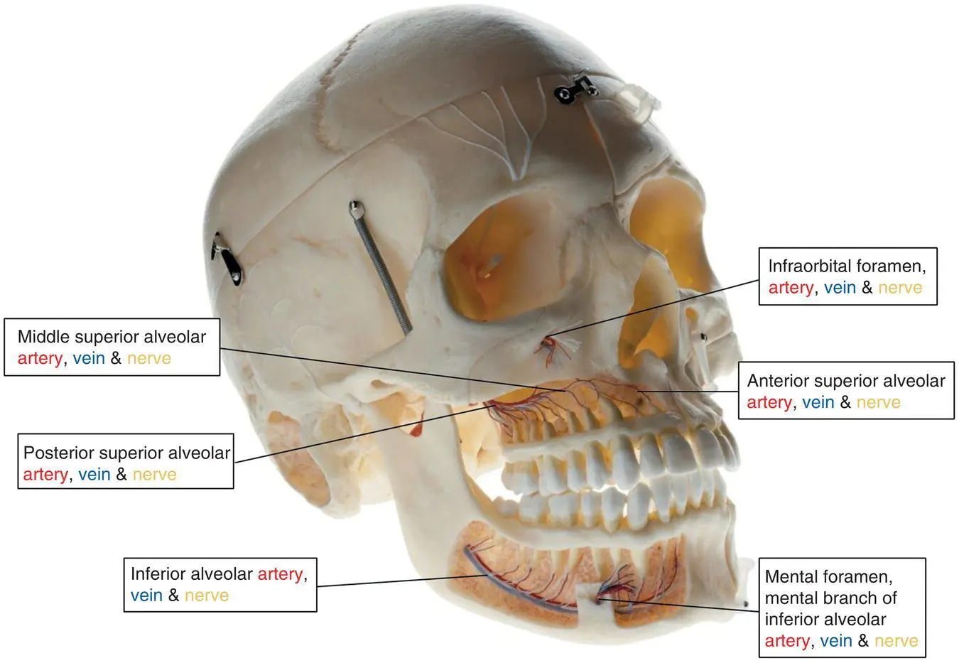
Figure 6.4 Innervation and vascular supply of the maxillary and mandibular dentition.
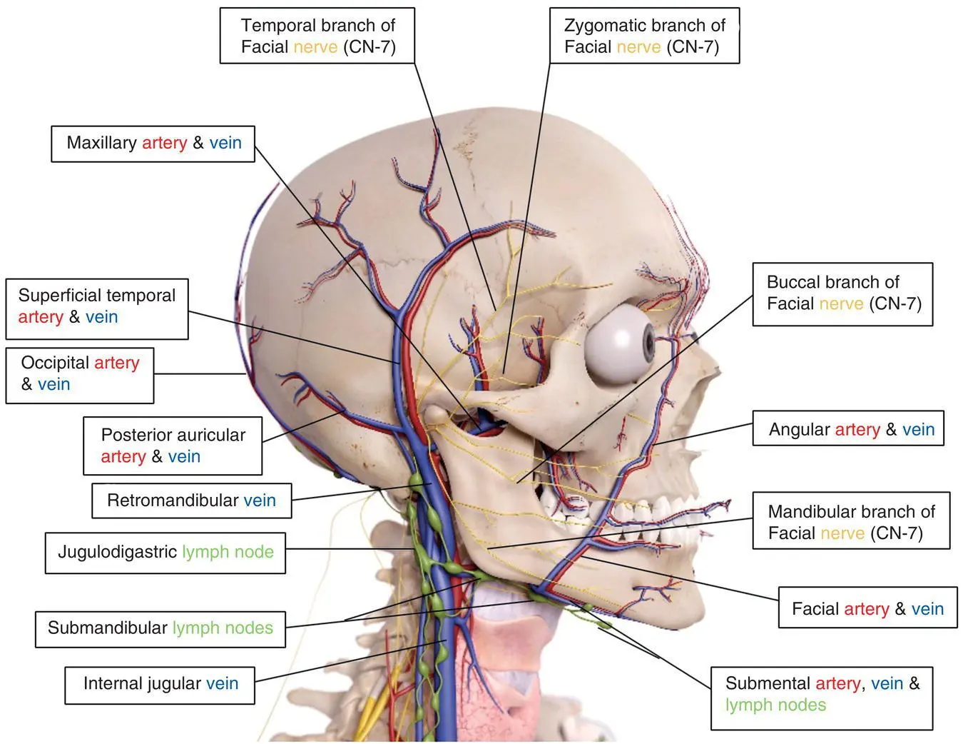
Figure 6.5Anatomical rendering of head and neck – lateral view.
Source : SciePro/Shutterstock.com.
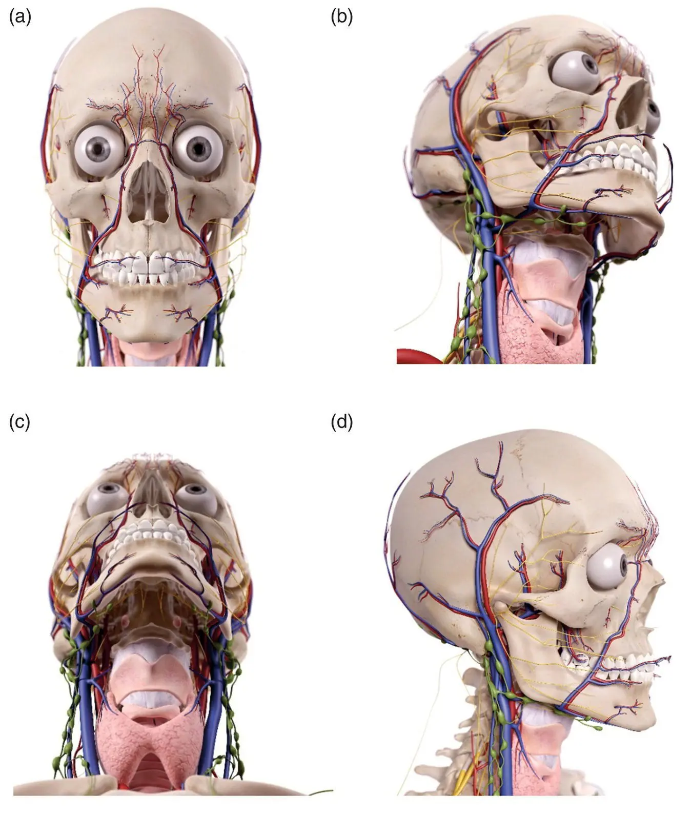
Figure 6.6 Anatomical rendering of head and neck (structures unlabeled). (a) Frontal view. (b) Oblique view. (c) Inferior view. (d) Lateral view.
Source : SciePro/Shutterstock.com.
6.1.3 Musculature
The muscles of the head and neck relevant to oral implantology can be categorised as muscles of mastication, which are paired muscles that aid in the process of grinding and chewing food and turning it into a bolus, and muscles of facial expression, which are generally flat paired muscles that enable movements of the face and facial expression. All muscles of mastication are innervated by branches of the mandibular division of the trigeminal nerve (CN V3), while all muscles of facial expression are innervated by branches of the facial nerve (CN VII). Tables 6.2and 6.3give summaries of key facts [1, 2] and Figure 6.7shows an anatomical rendering.
Table 6.2 Muscles of mastication summary.
Sources: Al‐Faraje, L. (2013). Surgical and Radiologic Anatomy for Oral Implantology . Chicago: Quintessence Publishing Co.; Norton, N. (2007). Netter's Head and Neck Anatomy for Dentistry . Philadelphia: Saunders Elsevier.
| Muscle | Origin | Insertion | Action | Innervation |
|---|---|---|---|---|
| Masseter | Zygomatic arch and maxillary process of zygomatic bone | Lateral surface of ramus of mandible | Elevation and retraction of mandible | Masseteric nerve (CN V3) |
| Temporalis | Temporal fossa | Coronoid process and anterior margin of the ramus of the mandible | Elevation and retraction of the mandible | Deep temporal nerve (CN V3) |
| Medial pterygoid | Medial surface of lateral plate of pterygoid process, pyramidal process of palatine bone (deep head); maxillary tuberosity, pyramidal process of palatine bone (superficial head) | Medial surface of mandible | Elevation and side‐to‐side movements of mandible | Medial pterygoid nerve (CN V3) |
| Lateral pterygoid | Roof of infratemporal fossa (superior head), lateral surface of the lateral pterygoid plate (inferior head) | Pterygoid fovea of mandible and temporomandibular joint articular disc (superior head) and condylar process (inferior head) | Protrusion and side‐to‐side movements of mandible | Lateral pterygoid nerve (CN V3) |
Table 6.3 Muscles of facial expression summary.
Sources: Al‐Faraje, L. (2013). Surgical and Radiologic Anatomy for Oral Implantology . Chicago: Quintessence Publishing Co.; Norton, N. (2007). Netter's Head and Neck Anatomy for Dentistry . Philadelphia: Saunders Elsevier.
| Muscle | Origin | Insertion | Action | Innervation |
|---|---|---|---|---|
| Orbicularis oris | Deep surface of the skin of the maxilla and mandible | Mucous membranes of the lips | Closes or purses the lips | Buccal and mandibular branches of CN VII |
| Buccinator | Molar areas of the alveolar processes of maxilla and mandible | Orbicularis oris, lips, and submucosal surfaces of lips and cheeks | Keeps bolus out of vestibule, expels air from oral cavity | Buccal branch of CN VII |
| Levator labii superiorus | Frontal process of the maxilla and the infraorbital margin | Skin of the upper lip | Elevates upper lip | Buccal and zygomatic branches of CN VII |
| Depressor labii inferioris | Anterior area of oblique line of mandible | Middle of lower lip | Pulls lower lip inferiorly and laterally | Mandibular branch of CN VII |
| Levator labii superioris alaeque nasi | Frontal process of maxilla | Alar cartilage and upper lip muscles (levator labii superioris and orbicularis oris) | Elevates the upper lip and dilates nostrils | Buccal and zygomatic branches of CN VII |
| Mentalis | Frenulum of lower lip | Skin of the chin | Elevates and protrudes the lower lip | Mandibular branch of CN VII |
| Risorius | Fascia superficial to the masseter muscle | Skin of the angle of the mouth | Retracts the corners of the mouth during smiling broadly and laughing | Buccal branch of CN VII |
| Depressor anguli oris | Mandible below canines, premolars, and first molars | Skin of corner of mouth and orbicularis oris | Pulls the angle of the mouth inferiorly and laterally | Buccal and mandibular branches of CN VII |
| Levator anguili oris | Canine fossa of maxilla inferior to infraorbital foramen | Angle of the mouth | Elevates the angle of the mouth | Zygomatic and buccal branches of CN VII |
| Zygomaticus major | Zygomatic bone (lateral and posterior surfaces) | Muscles of the angle of the mouth | Pulls corner of the mouth laterally and superiorly | Zygomatic branch of CN VII |
| Zygomaticus minor | Zygomatic bone (lateral and posterior surfaces) | Corner of the upper lip | Pulls upper lip superiorly | Zygomatic branch of CN VII |
| Nasalis | Transverse part on maxilla Alar part on maxilla | Transverse on aponeurosis at bridge of nose Ala nasi | Compress nostrils Open nostrils | Buccal and zygomatic branches of CN VII |
| Procerus | Facial aponeurosis of the lower nasal bone | Skin between eyebrows | Pulls eyebrows medially and inferiorly | Temporal and zygomatic branches of CN VII |
| Orbicularis oculi | Medial orbital margin, medial palpebral ligament, and lacrimal crest | Close muscles occipitofrontalis, corrugator supercilia, eyelids | Closes eyelid | Temporal and zygomatic branches of CN VII |
| Corrugator supercili | Frontal bone supraorbital ridge | Middle of the eyebrow | Draws the eyebrows medially and inferiorly | Temporal branch of CN VII |
| Platysma | Skin over lower neck and upper lateral thorax | Inferior border of mandible, skin over lower face, angle of mouth | Wrinkles skin of lower face and neck | Cervical branch of CN VII |
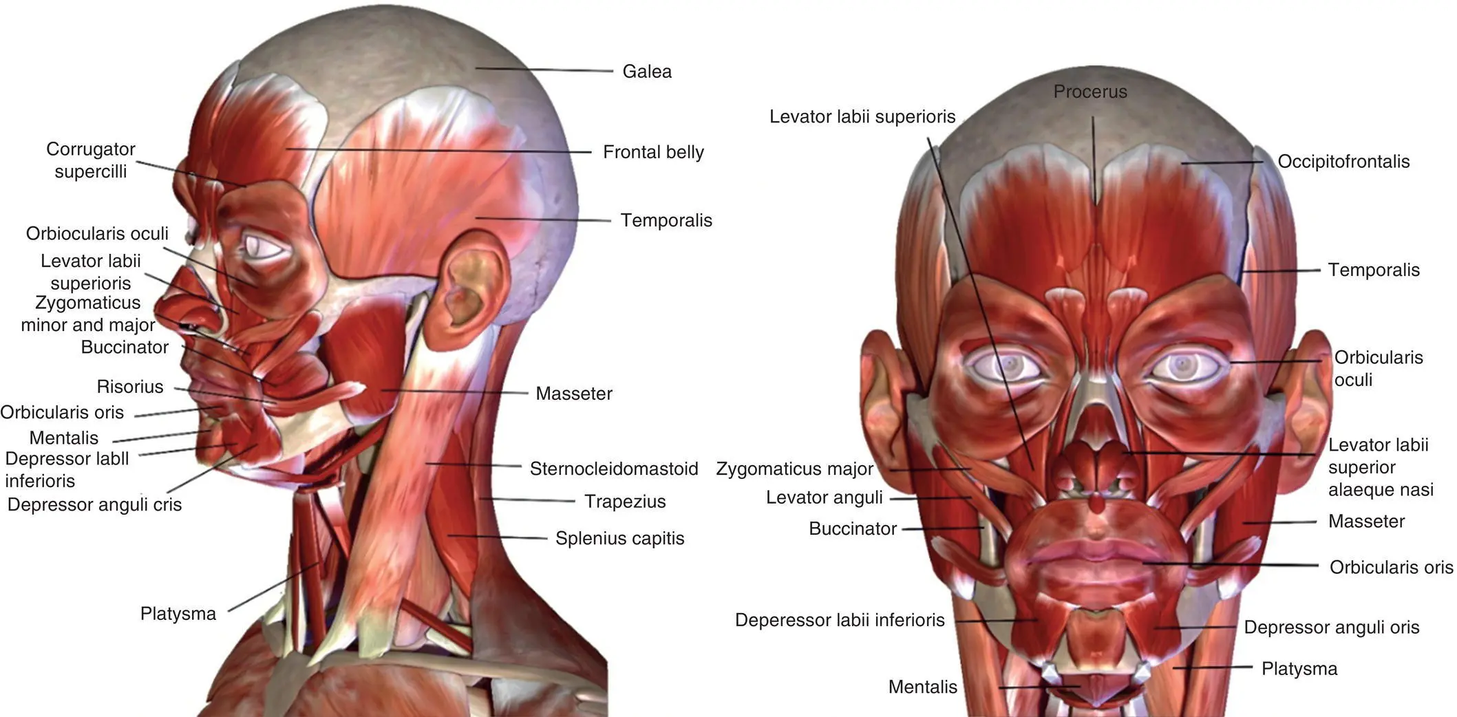
Figure 6.7 Muscles of mastication and facial expression.
Source: Life science/Shutterstock.com.
6.2 Procedures
Begin the patient assessment with some light conversation with your patient comfortably seated in the dental chair. Discuss the patient's chief complaint and motivations for seeking treatment. Observe the functionality of the muscles of facial expression during your exchange, being sure to note any perceived abnormalities as well as the patient's smile dynamics and dentofacial aesthetics.
Читать дальшеИнтервал:
Закладка:
Похожие книги на «Practical Procedures in Implant Dentistry»
Представляем Вашему вниманию похожие книги на «Practical Procedures in Implant Dentistry» списком для выбора. Мы отобрали схожую по названию и смыслу литературу в надежде предоставить читателям больше вариантов отыскать новые, интересные, ещё непрочитанные произведения.


Обсуждение, отзывы о книге «Practical Procedures in Implant Dentistry» и просто собственные мнения читателей. Оставьте ваши комментарии, напишите, что Вы думаете о произведении, его смысле или главных героях. Укажите что конкретно понравилось, а что нет, и почему Вы так считаете.