Practical Procedures in Implant Dentistry
Здесь есть возможность читать онлайн «Practical Procedures in Implant Dentistry» — ознакомительный отрывок электронной книги совершенно бесплатно, а после прочтения отрывка купить полную версию. В некоторых случаях можно слушать аудио, скачать через торрент в формате fb2 и присутствует краткое содержание. Жанр: unrecognised, на английском языке. Описание произведения, (предисловие) а так же отзывы посетителей доступны на портале библиотеки ЛибКат.
- Название:Practical Procedures in Implant Dentistry
- Автор:
- Жанр:
- Год:неизвестен
- ISBN:нет данных
- Рейтинг книги:3 / 5. Голосов: 1
-
Избранное:Добавить в избранное
- Отзывы:
-
Ваша оценка:
Practical Procedures in Implant Dentistry: краткое содержание, описание и аннотация
Предлагаем к чтению аннотацию, описание, краткое содержание или предисловие (зависит от того, что написал сам автор книги «Practical Procedures in Implant Dentistry»). Если вы не нашли необходимую информацию о книге — напишите в комментариях, мы постараемся отыскать её.
covers core topics such as:
Rationale and assessment for implant placement and restoration, including the diagnostic records and surgical considerations required for optimal planning and risk management Incision design considerations and flap management, with an essential knowledge of regional neuro-vascular structures Implant placement, encompassing the timing of the placement, bone requirements and understanding the importance of the peri-implant interface for soft tissue stability Impression techniques, loading protocols, digital workflows and the aesthetic considerations of implants Prosthetic rehabilitation of single tooth implants to fully edentulous workflows, including discussions of soft tissue support, biomechanics and occlusal verification Perfect for both general dental practitioners and specialists in implant dentistry,
is also a valuable reference to senior undergraduate and postgraduate dental students.
Practical Procedures in Implant Dentistry — читать онлайн ознакомительный отрывок
Ниже представлен текст книги, разбитый по страницам. Система сохранения места последней прочитанной страницы, позволяет с удобством читать онлайн бесплатно книгу «Practical Procedures in Implant Dentistry», без необходимости каждый раз заново искать на чём Вы остановились. Поставьте закладку, и сможете в любой момент перейти на страницу, на которой закончили чтение.
Интервал:
Закладка:
| Bone | Paired | Single | Cranial/Facial | Articulation |
|---|---|---|---|---|
| Frontal | X | Cranial | Maxilla, zygomatic, sphenoid, parietal, ethmoid, nasal, lacrimal | |
| Parietal | X | Cranial | Temporal, frontal, parietal, occipital, sphenoid | |
| Temporal | X | Cranial | Mandible, zygomatic, sphenoid, parietal, occipital | |
| Occipital | X | Cranial | Temporal, atlas (C1), parietal, sphenoid | |
| Sphenoid | X | Cranial | Maxilla, ethmoid, palatine, vomer, frontal, parietal, temporal, occipital, zygomatic | |
| Ethmoid | X | Cranial | Maxilla, palatine, vomer, nasal, lacrimal, inferior nasal concha, frontal, sphenoid | |
| Zygomatic | X | Facial | Maxilla, frontal, temporal | |
| Maxilla | X | Facial | Maxilla, zygomatic, frontal, sphenoid, ethmoid, palatine, vomer, nasal, lacrimal, inferior nasal concha | |
| Palatine | X | Facial | Maxilla, palatine, vomer, inferior nasal concha, ethmoid, sphenoid | |
| Vomer | X | Facial | Maxilla, palatine, ethmoid, sphenoid | |
| Nasal | X | Facial | Maxilla, nasal, frontal | |
| Lacrimal | X | Facial | Maxilla, frontal, ethmoid, inferior nasal concha | |
| Inferior nasal concha | X | Facial | Maxilla, palatine, lacrimal, ethmoid | |
| Mandible | X | Facial | Temporal |
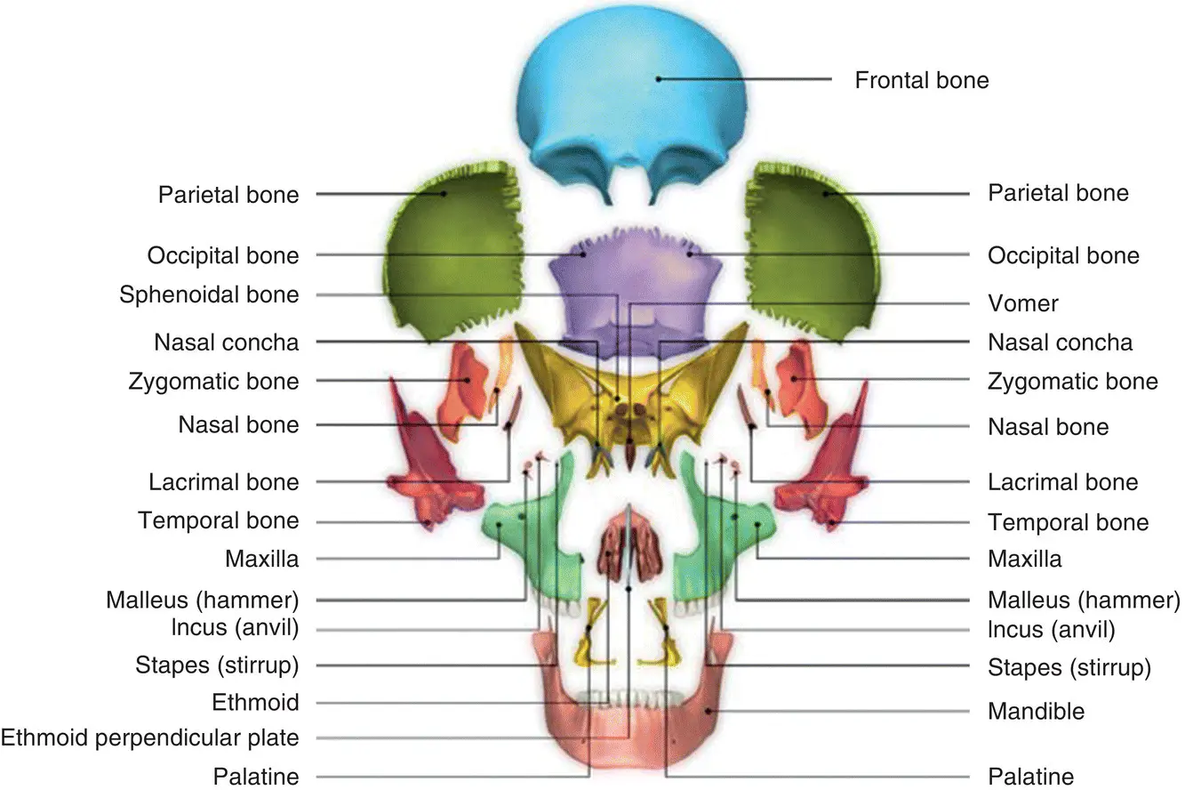
Figure 6.1 Osteology of the skull (exploded view).
Source : sciencepics/Shutterstock.com.
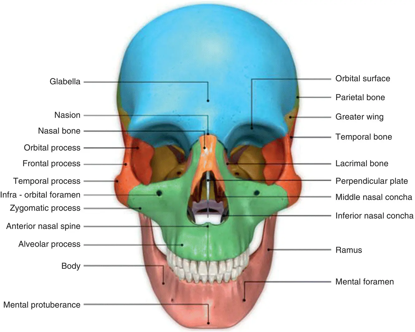
Figure 6.2Osteology of the skull (frontal view).
Source : sciencepics/Shutterstock.com.
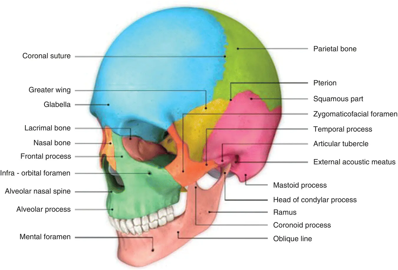
Figure 6.3 Osteology of the skull (lateral view).
Source : sciencepics/Shutterstock.com.
6.1.2 Innervation and Vascular Supply
The innervation and vascular supply of the maxillary and mandibular dentition are reliant upon the nerves and blood vessels that supply the bones in which they are housed, namely the paired bones of the maxilla and the singular mandible. The maxilla is an immobile part of the midface, with a neurovasculature separate from that of the mobile mandible, which is considered part of the lower face.
The second division of the trigeminal nerve (cranial nerve V division II, CN V2) is called the maxillary nerve, which is responsible for the sensory innervation of the maxillary dentition. It branches off the trigeminal nerve at the trigeminal ganglion and exits the skull via the foramen rotundum. It then branches into four major divisions: the posterior superior alveolar nerve (PSA), the infraorbital nerve, the zygomatic nerve, and branches to the pterygoid plexus. The infraorbital nerve further branches into the middle superior alveolar nerve (MSA) and the anterior superior alveolar nerve (ASA) [1].
The PSA, MSA, and ASA form the superior dental plexus. The PSA innervates the maxillary molars and posterior aspect of the maxillary sinus. The MSA innervates the premolars, the medial and lateral aspects of the maxillary sinus, and sometimes the mesiobuccal root of the first molar. The ASA innervates the incisors, canines, and the anterior aspect of the maxillary sinus.
The infraorbital nerve also exits the infraorbital foramen before branching off into the nasal, inferior palpebral, and superior labial nerves. These branches supply the cartilaginous ala of the nose, the dermal surface of the lower eyelid, and the upper lip, respectively.
Both maxillary and mandibular dentitions derive their blood supply from the external carotid artery via its maxillary artery branch. The maxillary arch is supplied by a plexus of three arteries: the PSA artery, the MSA artery, and the ASA artery. The PSA artery supplies the maxillary molars, premolars, and posterior maxillary sinus. The MSA artery (when present) and ASA artery both branch off of the infraorbital artery, and supply the premolar/canine region with medial and lateral aspects of the maxillary sinus, and anterior dentition with anterior aspects of the maxillary arch and sinus, respectively [2].
Venous drainage of the maxilla occurs via the PSA vein, the MSA vein, and the ASA vein, which converge to form the pterygoid venous plexus. The pterygoid venous plexus is drained via the relatively short maxillary vein to the retromandibular vein [1].
The third and largest division of the trigeminal nerve (cranial nerve V division III, CN V3) is called the mandibular nerve and is responsible for both sensory and motor innervation of the mandible after branching off the trigeminal ganglion and exiting the skull via the foramen ovale. The mandibular nerve then further divides into a meningeal branch, followed by another split into the anterior division and posterior division.
The anterior division is the smaller of the two divisions and mainly motor in function, with the exception of the buccal branch which remains sensory in nature. The other branches of the anterior division are the masseteric nerve, the anterior and posterior deep temporal nerves, the medial pterygoid nerve, and finally the lateral pterygoid nerve.
The larger posterior division conversely is mainly sensory in function, with the exception of the mylohyoid branch which remains motor in nature. The other branches of the posterior division are the auriculotemporal nerve, the lingual nerve, and the inferior alveolar nerve. The lingual nerve supplies sensory innervation to the mucous membranes of the anterior two‐thirds of the tongue and the gingiva on the lingual aspect of the mandibular teeth. The inferior alveolar nerve is the largest branch of the mandibular nerve, running between the sphenomandibular ligament and mandibular ramus before entering the mandible at the mandibular foramen. The inferior alveolar nerve emerges from the mandible in the vicinity of the second premolar via the mental foramen. It provides sensory innervation to all mandibular teeth, associated periodontal ligaments, and the gingiva from the premolars anteriorly to the midline. The inferior alveolar nerve terminates into the mental and incisive nerves at the approximate level of the second premolar. The mental nerve supplies the chin, lip, facial gingiva, and mucosa from the second premolar anteriorly to the midline. The incisive nerve continues anteriorly supplying the teeth and periodontal ligaments from approximately the first premolar to the midline, depending on the location of where the branching of the inferior alveolar nerve into the mental and incisive nerves occurs [1].
The mandibular arch is supplied by a single branch of the maxillary artery called the inferior alveolar artery. It mirrors the path of the inferior alveolar nerve through the mandible via the mandibular foramen. Likewise, it terminates into the branches of the mental and incisive arteries at approximately the level of the second premolar. The mental and incisive arteries supply the labial gingiva of the anterior dentition and the anterior dentition themselves, respectively. Venous drainage of the mandible occurs via the singular inferior alveolar vein, which drains into the pterygoid venous plexus. Figures 6.4– 6.6give a detailed overview of the innervation and vascular supply of the skull.
Читать дальшеИнтервал:
Закладка:
Похожие книги на «Practical Procedures in Implant Dentistry»
Представляем Вашему вниманию похожие книги на «Practical Procedures in Implant Dentistry» списком для выбора. Мы отобрали схожую по названию и смыслу литературу в надежде предоставить читателям больше вариантов отыскать новые, интересные, ещё непрочитанные произведения.


Обсуждение, отзывы о книге «Practical Procedures in Implant Dentistry» и просто собственные мнения читателей. Оставьте ваши комментарии, напишите, что Вы думаете о произведении, его смысле или главных героях. Укажите что конкретно понравилось, а что нет, и почему Вы так считаете.