Diatom Morphogenesis
Здесь есть возможность читать онлайн «Diatom Morphogenesis» — ознакомительный отрывок электронной книги совершенно бесплатно, а после прочтения отрывка купить полную версию. В некоторых случаях можно слушать аудио, скачать через торрент в формате fb2 и присутствует краткое содержание. Жанр: unrecognised, на английском языке. Описание произведения, (предисловие) а так же отзывы посетителей доступны на портале библиотеки ЛибКат.
- Название:Diatom Morphogenesis
- Автор:
- Жанр:
- Год:неизвестен
- ISBN:нет данных
- Рейтинг книги:5 / 5. Голосов: 1
-
Избранное:Добавить в избранное
- Отзывы:
-
Ваша оценка:
Diatom Morphogenesis: краткое содержание, описание и аннотация
Предлагаем к чтению аннотацию, описание, краткое содержание или предисловие (зависит от того, что написал сам автор книги «Diatom Morphogenesis»). Если вы не нашли необходимую информацию о книге — напишите в комментариях, мы постараемся отыскать её.
A unique book presenting the range of silica structures formed by diatoms, theories and hypotheses of how they are made, and applications to nanotechnology by use or imitation of diatom morphogenesis.
Audience
Diatom Morphogenesis — читать онлайн ознакомительный отрывок
Ниже представлен текст книги, разбитый по страницам. Система сохранения места последней прочитанной страницы, позволяет с удобством читать онлайн бесплатно книгу «Diatom Morphogenesis», без необходимости каждый раз заново искать на чём Вы остановились. Поставьте закладку, и сможете в любой момент перейти на страницу, на которой закончили чтение.
Интервал:
Закладка:
Diatoms reproduce both asexually (visible in Figure 1.6) and sexually. Most of the time, they reproduce asexually via binary fission through adding new hypovalves to the parent valves. Those new hypovalves are synthesized inside the silica deposition vesicle (SDV). Only after the new hypovalves have completely synthesized and the protoplast cleavage, as well as the exocytosis of siliceous parts, has occurred, the final splitting apart will occur, leaving two daughter diatoms in place. Because the SDV forms inside of each new cell before splitting into two, each new cell creates a new interior of the petri-dish structure. What this means is that the cell that originally contained the upper part of the petri dish (the epitheca) remains the same size, whereas the cell that originally contained the lower part of the petri-dish (the hypotheca) becomes smaller, since it has now built a smaller hypovalve to fit into it. Repeated cell division, therefore, leads to some part of the resulting population becoming smaller and smaller. Were asexual reproduction the only method by which diatoms reproduces, this could lead the population eventually to become vulnerable to dying out, but diatoms are ingenious and have gotten around this problem. At some point, sexual reproduction is initiated by a number of steps, including meiotic divisions to produce male and female gametes. These cells can find each other, fuse to form a zygote and create a structure known as an auxospore, out of which a new large cell of the diatom species will form, restoring its optimal size, which also depends on the environmental circumstances surrounding the auxospores. Some new research proposes chemical communication with pheromones between the male and female gametes [1.20].
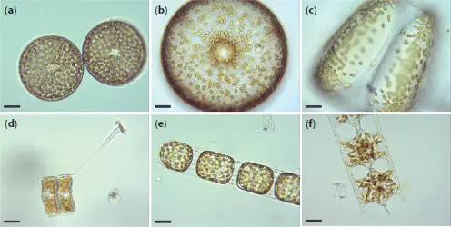
Figure 1.1Living diatoms as observed under LM, brightfield. (a) Two living cells of Actinoptychus senarius (Ehrenberg) Ehrenberg at the valve view. (b) The valve view of a single living cell of Coscinodiscus wailesii Gran and Angst. (c) The girdle view of a single living cell of Coscinodiscus granii L.F. Gough. (d) Two living cells of Achnanthes brevipes C. Agardh at the girdle view attached to each other with a prolonged stalk for the attachment to the substrate. (e) A living colony of Stephanopyxis turris (Greville) Ralfs with visible linking spines. (f) A living colony of Odontella longicruris (Greville) M.A. Hoban with discoid chloroplasts. Copyright reserved Mary Ann Tiffany, used with her permission. The identification was carried out by Mary Ann Tiffany. All the scale bars are 50 μm.
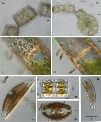
Figure 1.2Live centric (a, b) and pennate (c–h) diatoms. (a, b) Pleurosira laevis (Ehrenberg) Compère shown from girdle view, frustules with numerous girdle bands in straight filaments with discoid chloroplasts, chains connected with mucilage pads released from ocelli; in (b), visible diameter size restoration within the chain; (c, d) Epiphytic diatoms on Cladophora glomerata (Linnaeus) Kützing, in (c) focus on Cocconeis spp. With visible one flat C-shaped plastid; in (d) focus on Rhoicosphenia spp.; (e) Cymbella sp. partial valve and girdle views, visible chloroplast bridge connecting the chloroplast plates; (f) Eunotia cf. camelus Ehrenberg in girdle view with visible discoid chloroplasts; (g) Amphora ovalis (Kützing) Kützing with H shaped chloroplast; (h) Rhoicosphenia sp. girdle view with visible lobes of the plastid. Scale bars, 10 μm. These micrographs were obtained and identified by KMM.
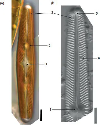
Figure 1.3Specific diatom morphology gleaned from images with whole and partial valves views of Navicula oblonga (Kützing) Kützing; (a) live linear-lanceolate cell with visible two plates like brown chloroplasts, visible linear striae, and proximal raphe ends deflected slightly toward the secondary side. (b) Valve view after cleaning, axial area is linear, widening toward the central area and about twice the width of the raphe. The central area orbicular. The raphe is lateral, becoming filiform near the proximal ends, which are simple. Central striae do not reach valve edge. These micrographs were obtained and identified by KMM.
Details shown:
1 Central area is more or less orbicular and two to three times wider than the axial area. Proximal raphe ends are simple and barely wider than the raphe. Striae are finely lineate and the individual areolae are difficult to distinguish.
2 Round, subsidiary vacuoles on each side of the nucleus visible behind the glass cell wall and chloroplasts; axial area outlines by lineate striae.
3 Terminal bent striae (terminal striae convergent at the margins and bent back toward the central area). Striae are radiate next to the axial area.
4 Voigt discontinuity identifies the secondary side of the valve morphogenesis. Ontogeny in diatoms varies with morphology; in Naviculoid diatoms, the secondary side shows the completion of silica deposition around the raphe.
5 Distal raphe positioned on the broad, rounded apices and curved toward the primary side of the valve in the opposite direction when compared to the proximal raphe ends. Scale bars, 10 μm.
Frustule morphogenesis, deposits SDVs and needs more research with new tools. However, it has been established that the silica morphogenesis of centric species will begin at the center of the valve, and it begins by creating a primary rib in pennate species [1.21]. Completion of the sternum around the raphe slit morphologically can be identified with the Voight discontinuity (Figure 1.3b). From that onset within the mother frustule, the silica will continue to form outward to complete the shape as well as inward to create more layers, with the oldest silica being on the most outside layer [1.46]. The silicic acid (or its anions) is taken from the environment, condensed, associated with proteins synthesized by the endoplasmic reticulum and packaged in a globular vesicle in the Golgi apparatus. Then finally, these vesicles (silica deposition vesicles) are transported by microtubules, likely in a genetically predetermined pattern, and delivered to the new valve interface. These are not the only groups that pull silicic acid (an inorganic compound contains silicon) out of the water and use it to make a frustule, but diatoms do it uniquely.
Diatom frustules are porous with multilayer, multiscalar porosity, a property that is unique for each species, giving frustules their beautiful ornamentation [1.17]. The major bigger pores within the valves are called “areolae” and usually arranged in rows known as “striae”, which could be either branched or not. In the most general way, diatoms can be divided into centric and pennate diatoms, which are classified based on the valve symmetry. Centric diatoms are radially symmetric and lack raphes. Pennate diatoms usually have bilateral symmetry and there can be no, one, or two raphes. Pennate diatoms can further be classified based on variations in the position of the raphe on valve. The raphe is used for motility [1.4] and attachment [1.12]. Sometimes, the frustules are also covered in spines, which can allow some species to hook together and form chains (Figure 1.1e).
Читать дальшеИнтервал:
Закладка:
Похожие книги на «Diatom Morphogenesis»
Представляем Вашему вниманию похожие книги на «Diatom Morphogenesis» списком для выбора. Мы отобрали схожую по названию и смыслу литературу в надежде предоставить читателям больше вариантов отыскать новые, интересные, ещё непрочитанные произведения.


Обсуждение, отзывы о книге «Diatom Morphogenesis» и просто собственные мнения читателей. Оставьте ваши комментарии, напишите, что Вы думаете о произведении, его смысле или главных героях. Укажите что конкретно понравилось, а что нет, и почему Вы так считаете.