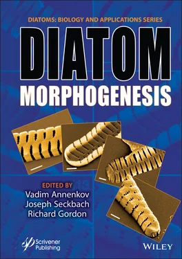
Diatom Morphogenesis
Здесь есть возможность читать онлайн «Diatom Morphogenesis» — ознакомительный отрывок электронной книги совершенно бесплатно, а после прочтения отрывка купить полную версию. В некоторых случаях можно слушать аудио, скачать через торрент в формате fb2 и присутствует краткое содержание. Жанр: unrecognised, на английском языке. Описание произведения, (предисловие) а так же отзывы посетителей доступны на портале библиотеки ЛибКат.
- Название:Diatom Morphogenesis
- Автор:
- Жанр:
- Год:неизвестен
- ISBN:нет данных
- Рейтинг книги:5 / 5. Голосов: 1
-
Избранное:Добавить в избранное
- Отзывы:
-
Ваша оценка:
- 100
- 1
- 2
- 3
- 4
- 5
Diatom Morphogenesis: краткое содержание, описание и аннотация
Предлагаем к чтению аннотацию, описание, краткое содержание или предисловие (зависит от того, что написал сам автор книги «Diatom Morphogenesis»). Если вы не нашли необходимую информацию о книге — напишите в комментариях, мы постараемся отыскать её.
A unique book presenting the range of silica structures formed by diatoms, theories and hypotheses of how they are made, and applications to nanotechnology by use or imitation of diatom morphogenesis.
Audience
Diatom Morphogenesis — читать онлайн ознакомительный отрывок
Ниже представлен текст книги, разбитый по страницам. Система сохранения места последней прочитанной страницы, позволяет с удобством читать онлайн бесплатно книгу «Diatom Morphogenesis», без необходимости каждый раз заново искать на чём Вы остановились. Поставьте закладку, и сможете в любой момент перейти на страницу, на которой закончили чтение.
Интервал:
Закладка:
3 Chapter 3Figure 3.1 Pascal’s triangle, which shows the development of the sizes of diato...Figure 3.2 An early illustration of a diatom chain with a sketch of the size se...Figure 3.3 In the left half of the figure, diatoms of the chain L 0R 1L 2R 1are...Figure 3.4 The string L 0R 1L 2R 1is in the left branch first mirrored and then...Figure 3.5 Bar chart of the size indices from the 7th generation (128 diatoms) ...Figure 3.6 Elementary step in the development of a substring A of any generatio...Figure 3.7 The Pascal triangle illustrates the development of a substring W. W ...Figure 3.8 Dragon curve of the 13-th generation. It contains 2 13- 1 = 8,192 an...Figure 3.9 Eunotia chain at the bottom of a Petri dish of a culture. Looking at...Figure 3.10 Bar chart of differences in lengths of adjacent diatoms within the ...Figure 3.11 Interpretation of the observed sequence of differences (Figure 3.10...Figure 3.12 Eunotia chain at the bottom of a Petri dish of a culture. A chain w...Figure 3.13 Probability P k( t ) that the k-th division has occurred at the time t k...
4 Chapter 4Figure 4.1-4.6 Amphitetras antediluviana, mature valves, S.E.M. Figure 4.1 Comp...Figure 4.7-4.12 Amphitetras antediluviana, forming valves, S.E.M. Figure 4.7 Ea...Figure 4.13-4.18 Amphitetras antediluviana, forming ocelli, S.E.M. Figure 4.13 ...Figure 4.19-.24 Amphitetras antediluviana, forming cribra, S.E.M. Figure 4.19 E...Figure 4.25-4.30 Amphitetras antediluviana, S.E.M. Figure 4.25 Detail of nearly...
5 Chapter 5Figure 5.1 (a and b) Concentric pattern of areolae on the valve of Aulacodiscus ...Figure 5.2 Figure 5.2 (a) An artificially generated concentric pattern where all...Figure 5.3 The repeating stages of the formation of the rows of a concentric pat...Figure 5.4 Natural (b) and artificial (a, c) unidirectional spiral patterns. (a,...Figure 5.5 Natural (b) and artificial (a, c) bidirectional spiral pattern. (a) T...
6 Chapter 6Figure 6.1 A flow chart illustrating the methodology for assigning the most like...Figure 6.2 Diagram showing the frustule structure of (a) a centric frustule and ...Figure 6.3 Diagram illustrating two types of 2D lattices, the hexagonal and rect...Figure 6.4 A segmented micrograph of the external view of a single valve of Rope...Figure 6.5 Two SEMs of the internal view of a single valve of Pleurosigma sp. (a...Figure 6.6 SEM of the external view of a single valve of Roperia tesselata (1,02...Figure 6.7 SEM of the internal view of a single valve of Asteromphalus hookeri (...Figure 6.8 SEM of an internal view of a single valve of Roperia tesselata in upr...Figure 6.9 (a) A segmented SEM of the internal view of a single valve of Asterom...Figure 6.10 A segmented SEM of the internal view of a single valve of Asterompha...Figure 6.11 A threshold of the same micrograph of the internal view of a single ...Figure 6.12 A segmented selected part of the SEM of an internal view of a single...Figure 6.13 A segmented micrograph of a zoom in the internal view of a single va...Figure 6.14 (a) A thresholded micrograph of the internal view of a single valve ...Figure 6.15 A selected circle of an SEM of the internal view of a single valve o...Figure 6.16 (a) SEM of the external view at the center of a single valve of Thal...Figure 6.17 (a) A thresholded SEM of a zoom-in of the internal view of a single ...Figure 6.18 (a) A segmented SEM of the external view of a single immature valve ...Figure 6.19 (a) A segmented SEM micrograph of a zoom-in external view of Asterol...Figure 6.20 (a) A segmented SEM micrograph of the external view of Roperia tesse...Figure 6.21 (a) A segmented SEM micrograph of a selected 2D pore group at the ex...Figure 6.22 (a) A segmented SEM micrograph of the internal view of Haslea sp. (1...Figure 6.23 A segmented SEM of a zoom-in of a Gyrosigma fasciola valve (276 × 17...Figure 6.24 (a) A segmented SEM of a zoom-in of a valve of Gyrosigma balticum, a...Figure 6.25 (a) The areolae chambers appearing from a crack in the valve of Aste...Figure 6.26 An artificial honeycomb-like structure and its correpsonding 2D FFT ...Figure 6.27 A zoom-in of the external view of a single slightly immature valve o...Figure 6.28 (a) SEM of an external view of the central area of a single valve of...Figure 6.29 (a) SEM of the external view of a single valve of Cocconeis sp. illu...
7 Chapter 7Figure 7.1 Ensemble surface features—examples: (a) smooth, but not flat; (b) fla...Figure 7.2 Actinoptychus senarius modeled valve formation sequence steps 1 throu...Figure 7.3 Arachnoidiscus ehrenbergii modeled valve formation sequence steps 1 t...Figure 7.4 Cyclotella meneghiniana modeled valve formation sequence steps 1 thro...Figure 7.5 Single-linkage cluster analysis using Hamming distance for non-zero v...Figure 7.6 Contribution percentage of ensemble surface measures Jacobian, Hessia...Figure 7.7 Jacobian, Hessian, and Laplacian from z -terms and Christoffel symbols...Figure 7.8 Contribution of the u and v parameters of the z -term of the Jacobian,...Figure 7.9 All Christoffel symbols for the three contravariant indices: top, k =...Figure 7.10 Plot of all Christoffel symbols shows change in contribution for  , ...Figure 7.11 Ensemble surface measures as indicators of ensemble surface features...Figure 7.12 A 3D morphospace of ensemble surface measures of valve formation for...Figure 7.13 A 3D morphospace of ensemble surface measures of valve formation for...
8 Chapter 8Figure 8.1 Top row: A non-buckled (a) and buckled (b) microtubule filament or bu...Figure 8.2 (a) Ray tracing valve formation on the valve face from annulus to mar...Figure 8.3 From left to right, centric diatom 3D surface models of Actinoptychus...Figure 8.4 Change in tangent lines and planes during valve formation indicate th...Figure 8.5 Plots of Actinoptychus, Arachnoidiscus, and Cyclotella buckling durin...Figure 8.6 The Jacobian determinant as the coefficient of deformation during val...Figure 8.7 Strain rate tensor deformation for Actinoptychus, Arachnoidiscus , and...Figure 8.8 Combined strain rate tensor deformation for Actinoptychus, Arachnoidi...Figure 8.9 Upper Hessenberg matrix eigenvalues for Actinoptychus, Arachnoidiscus ...Figure 8.10 Non-buckling (blue curve) and buckling (orange curve) for Actinoptyc...Figure 8.11 Upper Hessenberg matrices for all taxa based on the Jacobian (a) and...Figure 8.12 Multiscale buckling with respect to association of valve surface, mi...
9 Chapter 9Figure 9.1 Centric diatom schematic with valve profile. Axial load at the point ...Figure 9.2 Various potential valve profiles undergoing buckling.Figure 9.3 Measurement of buckling forces with respect to two fixed points along...Figure 9.4 Least-squares polynomial curve fits of six profiles. In the x-directi...Figure 9.5 Tracings of diatom profiles after those depicted in Figures 26a–e fro...Figure 9.6 Curve fit of a sixth-order polynomial in Cartesian coordinates to the...Figure 9.7 Plot of approximately constant profile length from six polynomial lea...Figure 9.8 Valve profile buckling forces at the valve mantle ordered from lowest...Figure 9.9 Plot of size reduction step during diatom vegetative reproduction usi...Figure 9.10 Plot of valve profile versus buckling force representing Figure 9.4 ...
10 Chapter 12Figure 12.1 Generalized concept of centric and pennate frustule morphotypes amon...Figure 12.2 Silica valves of different diatoms in light microscopy. Micrographs ...Figure 12.3 (a) Morphotypes, habitats and lifestyles of centric diatoms. Centric...Figure 12.4 Variation of PAR attenuation in diatom dominated environments. In th...Figure 12.5 Example of frustule structural variation in centric and pennate diat...Figure 12.6 Permanent preparation diatom slide in dark field microscopy. (a) Mos...Figure 12.7 Chord diagram of studied optical properties of diatom frustules. Lef...
Читать дальшеИнтервал:
Закладка:
Похожие книги на «Diatom Morphogenesis»
Представляем Вашему вниманию похожие книги на «Diatom Morphogenesis» списком для выбора. Мы отобрали схожую по названию и смыслу литературу в надежде предоставить читателям больше вариантов отыскать новые, интересные, ещё непрочитанные произведения.
Обсуждение, отзывы о книге «Diatom Morphogenesis» и просто собственные мнения читателей. Оставьте ваши комментарии, напишите, что Вы думаете о произведении, его смысле или главных героях. Укажите что конкретно понравилось, а что нет, и почему Вы так считаете.


