Diatom Morphogenesis
Здесь есть возможность читать онлайн «Diatom Morphogenesis» — ознакомительный отрывок электронной книги совершенно бесплатно, а после прочтения отрывка купить полную версию. В некоторых случаях можно слушать аудио, скачать через торрент в формате fb2 и присутствует краткое содержание. Жанр: unrecognised, на английском языке. Описание произведения, (предисловие) а так же отзывы посетителей доступны на портале библиотеки ЛибКат.
- Название:Diatom Morphogenesis
- Автор:
- Жанр:
- Год:неизвестен
- ISBN:нет данных
- Рейтинг книги:5 / 5. Голосов: 1
-
Избранное:Добавить в избранное
- Отзывы:
-
Ваша оценка:
Diatom Morphogenesis: краткое содержание, описание и аннотация
Предлагаем к чтению аннотацию, описание, краткое содержание или предисловие (зависит от того, что написал сам автор книги «Diatom Morphogenesis»). Если вы не нашли необходимую информацию о книге — напишите в комментариях, мы постараемся отыскать её.
A unique book presenting the range of silica structures formed by diatoms, theories and hypotheses of how they are made, and applications to nanotechnology by use or imitation of diatom morphogenesis.
Audience
Diatom Morphogenesis — читать онлайн ознакомительный отрывок
Ниже представлен текст книги, разбитый по страницам. Система сохранения места последней прочитанной страницы, позволяет с удобством читать онлайн бесплатно книгу «Diatom Morphogenesis», без необходимости каждый раз заново искать на чём Вы остановились. Поставьте закладку, и сможете в любой момент перейти на страницу, на которой закончили чтение.
Интервал:
Закладка:
The frustule’s morphological features of diatoms are required for identification. Specialized terminology has been collected in [1.5–1.7, 1.15, 1.16], and a general guide to the literature is in [1.10]. Characters continue to be discovered and new descriptive terminologies are proposed [1.23].
1.2 Tools to Explore Diatom Frustule Morphology
The beauty of diatoms was missed until the early, curious microscopists started observing ambiguous glassy microorganisms under their optical microscopes in the 18th century [1.22, 1.38]. Although the light microscope (LM) helped us to reveal the diatoms’ world, diatom frustules also helped the microscopists in developing and testing the quality and resolution of their optical microscopes [1.24, 1.25]. Since the nineteenth century, several works have been published on diatoms, its morphology, and taxonomy by remarkable workers including Kützing, Schmidt, Ehrenberg, Grunow, Hustedt, Krammer, Lange-Bertalot, and more (see references in Round et al . [1.38]). They described both living cells and clean frustules extensively using LM. The unique structure of diatom frustules under LM, with a variety of shapes and symmetries, has captured a wide interest; however, most of the diatom’s real art, at the nanoscale, was kept hidden. The limitations for observing frustule ultrastructure, especially details below 200 nm, were solved after the invention of the electron microscope [1.26]. In 1936, the transmission electron microscope (TEM) was used to capture the first micrograph of a diatom frustule [1.26, 1.27], using it as a test object for the quality and resolution of TEM. After that TEM was used to explore diatom ultrastructure. Following, the scanning electron microscope (SEM) was invented and used extensively as a more effective tool for exploring frustules morphology and ultrastructure [1.24, 1.28, 1.36, 1.38].
The details observed using the SEM and TEM reflected the beauty of diatoms when many hidden details became observable. For instance, some bright striae under an optical microscope appear as arrays of fine pores under the electron microscope (Figure 1.5a). It was, to some extent, a kind of revolution for diatom classification and taxonomy with the morphological details that became available down to 15 nm with SEM and below 10 nm with TEM (Figure 1.5b). Nowadays, the observation of diatom frustule morphology and ultrastructure using LM, SEM, and TEM became routine work for people working on ecology, environment, forensic, nanotechnological, and other applications that concern frustule ultrastructure, monitoring diatom species, and taxonomy.
Although 2D information can be collected from LM and TEM and the 3D-shape appeared under SEM, the information about the surface topology, internal ultrastructure, and siliceous element relationships within diatom frustules was missing. Therefore, more tools were evolved and involved in the exploration and understanding of the 3D complex ultrastructure of the frustule, which could be the reason for their various natural features, including unique photonic, mechanical, and hydrokinetic properties [1.9, 1.45]. The new tools include the atomic force microscope (AFM) and the focused ion beam SEM (FIBSEM) [1.32, 1.34, 1.35, 1.41].
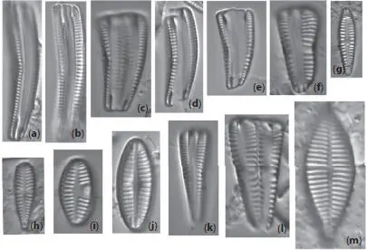
Figure 1.4Cleaned diatoms in valve (g, h–j, m–r, u, v) and girdle views (a–f, k, l, s, t, w, x). (a–e, g) Rhoicosphenia spp., frustules are clavate and strongly flexed, one valve is concave with long raphe branches and the other valve convex with shortened raphe, different depth pseudosepta visible; (f, k, l) Gomphonema spp. showing valve heterogeneity; (h) Gomphonella olivacea (Hornemann) Raben. (i) Planothidium lanceolatum (Bréb. Ex Kütz.) Lange-Bert, rapheless valve shown with asymmetrical central area containing depression; (j) Geissleria cascadensis (Sovereign) Stancheva and S. A. Spaulding, valves elliptic, with cuneate apices, coarse areolae, three pairs of annulae are present at each apex; (m) Planothidium delicatulum (Kütz.) Round and Bukht. Rapheless valve shown, lacking a central area and two middle striae spaced distantly. Cleaned diatoms in valve (g, h–j, m–r, u, v) and girdle views (a–f, k, l, s, t, w, x). (n) Gomphonema sp. valve heteropolar wider in the middle, axial area narrow, central area irregular outlined by two shortened striae and opposite to a single striae finishing with an isolated pore, striae parallel toward the headpole, radiate toward the foot pole; (o) Amphora ovalis , dorsal fascia visible and dorsal striae interrupted transapically by intercostal ribs; (p) Gomphonema micropus Reichardt lanceolate valve with headpole widely drawn out and wider than foot pole, striae radiate, central area unilaterally rectangular with shortened central stria, on the opposite side longer striae finishing with a stigmoid; (q) Navicula genovefae Fusey valve linear-lanceolate with rostrate broadly rounded apices, punctate striae radiate and curved, becoming nearly parallel at the apices, less dense around the well-defined central area; (r) Cocconeis placentula Ehrenb. Valves elliptic, striae radiate and interrupted by a hyaline ring positioned close to the valve margin, siliceous bridges (imbriae extending from valvocopula) visible; (s) Amphora pediculus (Kütz.) Grunow focus from dorsal site of two frustules; (t, u) Caloneis sp. on girdle view striae continue on valve mantle, on the linear valve view with rounded apices, axial area is narrow, broadening to a transverse fascia; (v) Navicula cryptocephala Kütz. Valve lanceolate with protracted apices and visible large, circular central area; (w) Mastogloia pseudosmithii Sylvia S. Lee, E. E. Gaiser, Van de Vijver, Edlund, and S. A. Spaulding, evenly sized partecta (chambers on the valvocopula) on both valves; (x) Navicula cf. tripunctata (O.F. Müll.) Bory. Scale bar, 10 μm. These micrographs were obtained and identified by KMM.
In 1992, the first observation of diatoms using an AFM has been done [1.33]. In general, AFM is used as an advanced tool to explore diatom ultrastructure providing information in the Z-direction, with the ability to understand the surface topology of the frustule parts with a nanoresolution. For instance, AFM observations of Coscinodiscus sp. clean valves revealed a distinct dome topology for the cribellum, which was not observed before [1.34]. At the beginning of the current century, AFM was used in several works for understanding the nanoscale ultrastructure and topology of frustule surfaces in a 3D manner. Today, AFM is also used to explore the organic envelope, micromechanical properties, and to understand the biomineralization processes of diatom frustules [1.35]. Luis et al . [1.35] can be considered a good review for starting AFM studies on diatom frustules.
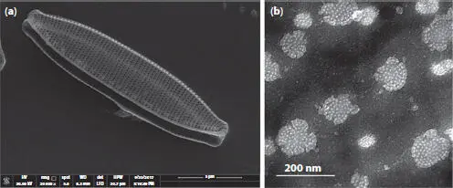
Figure 1.5(a) SEM of a single cleaned partially open frustule, two overlapping valves, of Nitzschia palea (Kützing) W. Smith, and scale bar is 5 μm. The rows of pores (striae) that observed here cannot be observed under LM for this species. (b) TEM of a close-up in Navicula sp. valve showing the hymenate pore occlusions that will not be observed under SEM; scale bar is 200 nm. These micrographs were obtained and identified by MG.
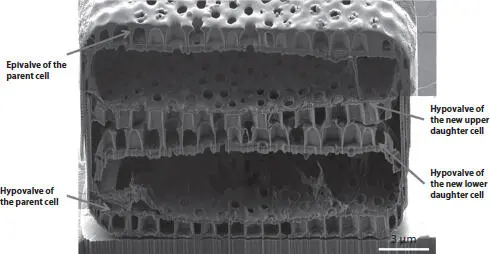
Figure 1.6A cross-section at the center of Coscinodiscus sp. cell collected and treated while binary fission process was in progress, fabricated and captured by FIB-SEM. Reproduced from Xing et al . [1.42] under a Creative Commons Attribution 4.0 International license.
Читать дальшеИнтервал:
Закладка:
Похожие книги на «Diatom Morphogenesis»
Представляем Вашему вниманию похожие книги на «Diatom Morphogenesis» списком для выбора. Мы отобрали схожую по названию и смыслу литературу в надежде предоставить читателям больше вариантов отыскать новые, интересные, ещё непрочитанные произведения.


Обсуждение, отзывы о книге «Diatom Morphogenesis» и просто собственные мнения читателей. Оставьте ваши комментарии, напишите, что Вы думаете о произведении, его смысле или главных героях. Укажите что конкретно понравилось, а что нет, и почему Вы так считаете.