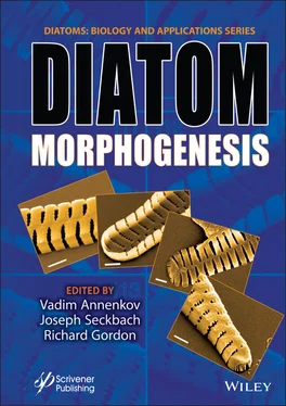
Diatom Morphogenesis
Здесь есть возможность читать онлайн «Diatom Morphogenesis» — ознакомительный отрывок электронной книги совершенно бесплатно, а после прочтения отрывка купить полную версию. В некоторых случаях можно слушать аудио, скачать через торрент в формате fb2 и присутствует краткое содержание. Жанр: unrecognised, на английском языке. Описание произведения, (предисловие) а так же отзывы посетителей доступны на портале библиотеки ЛибКат.
- Название:Diatom Morphogenesis
- Автор:
- Жанр:
- Год:неизвестен
- ISBN:нет данных
- Рейтинг книги:5 / 5. Голосов: 1
-
Избранное:Добавить в избранное
- Отзывы:
-
Ваша оценка:
- 100
- 1
- 2
- 3
- 4
- 5
Diatom Morphogenesis: краткое содержание, описание и аннотация
Предлагаем к чтению аннотацию, описание, краткое содержание или предисловие (зависит от того, что написал сам автор книги «Diatom Morphogenesis»). Если вы не нашли необходимую информацию о книге — напишите в комментариях, мы постараемся отыскать её.
A unique book presenting the range of silica structures formed by diatoms, theories and hypotheses of how they are made, and applications to nanotechnology by use or imitation of diatom morphogenesis.
Audience
Diatom Morphogenesis — читать онлайн ознакомительный отрывок
Ниже представлен текст книги, разбитый по страницам. Система сохранения места последней прочитанной страницы, позволяет с удобством читать онлайн бесплатно книгу «Diatom Morphogenesis», без необходимости каждый раз заново искать на чём Вы остановились. Поставьте закладку, и сможете в любой момент перейти на страницу, на которой закончили чтение.
Интервал:
Закладка:
3. Preparation of permanent slides should be carried out for LM observations using a suitable high refractive index mounting medium such as Naphrax and Hyrax [1.44]. Figure 1.4 considers a good example of observing cleaned frustules fixed in a permanent slide under LM.
4. Using LM to observe the 2D morphology of the cleaned valve and girdle bands to assign the most similar genus and, if possible, the species. The overall morphology, striae, valve symmetry, ribs, and the presence of raphe will be clear under LM; however, the pores, the spines, and special structures within the valve and girdle band might be not observable due to their smaller size. Though, in other few cases, the areolae might be observable even before the frustule cleaning procedure, especially for large taxa (Figure 1.1b). Valve outline, valve ends, shapes of sturdy, and hyaline silica (not penetrated with striae or other openings) should be recorded. Classical diameter in centric diatoms from valve view or length and frustule depth form girdle view allows verifications of descriptions from literature. Moreover, the frustule silicification rate can be observed under LM reflects information about the environmental condition that surrounds the living cells.
5. Using SEM, after suitable sample preparation and coating with a conductive material [1.38], to observe the ultrastructure of the valve and girdle bands and define the species accordingly. Under SEM, the valve will have two different viewpoints, the internal and external view. Some important criteria should be recorded using SEM including:
• the areolae structure from the internal and external valve view,
• striae (the periodic rows of areolae) shape,
• striae count in 10 microns or fibulae (in Nitzschioid diatoms [1.11], folds in Surirelloid diatoms [1.18]),
• sternum size and shape (in Fragilaroid and Naviculoid pennate diatoms),
• nodule zone or annulus shape and its number (if more than one present),
• raphe structure and shape of raphe at the end and in the middle of a valve,
• the pore occlusions (if observable),
• the presence of spine or other projections like fultoportulae and labiate processes,
• the shape and porosity of the girdle bands should also be recorded.
* For calculating the averages and ranges of the measured morphological data at least 10 specimens should be measured. Acid cleaned material allows observation of details but often causes change or collapse of the frustule’s three-dimensional ultrastructure, thus the selection of the most suitable cleaning procedure might be a crucial step to observe the diatom ultrastructure properly. It worthy to be noted that the observation of the frustule parts using SEM might be sufficient for genus and species identification without the need of LM; however, some important descriptions for identification are based on the LM appearance of the valve.
6. Observing the ultrastructure using TEM [1.38], if necessary and missing details did not appear under SEM, such as the hymenate pore occlusions of raphid pennate (see, for example, Figure 1.5b).
7. Study the nanotopography of the internal and external surface of the valve using AFM [1.34]. Some features within the valve could be observed through SEM, or even optical microscope if large enough, such as the frustule surface topology, which could be flat or rather undulated, wrinkled, or containing groves; however, other nanotopographical features cannot be revealed without a tool like AFM such as the dome shape of the cribellum in [1.34].
8. To understand the relationships of frustule elements and the inner ultrastructure of a given valve, or girdle bands, FIB-SEM should be considered (see example in Figure 1.6). For more advanced understanding, one of the methods mentioned for 3D reconstruction should be used [1.41] such as the 3D reconstruction of the 2D image series resulting from FIB-SEM. Through these techniques, a clear understanding of, for example, the multilayer nature of the valve could be obtained.
Afterward, all the ultrastructure details from inside and outside become well-known.
1.4 Conclusion
Diatom morphology could be an interesting topic for students; however, many challenges in the field usually keep them away from getting a deeper understanding. This is a major opportunity, since only 15,000–20,000 species have been identified [1.46], out of very large number of extant and fossil species. Recently, the developed tools, especially AFM and FIB-SEM, can make the complex 3D ultrastructure of diatoms more accessible; however, very few attempts have been recorded in literature. For both the beginners and professionals, many unrevealed secrets of diatom morphology and morphogenesis are still kept hidden, which needs more progress in the field. The study of diatom morphology could also inspire material scientists and engineers especially as the functions beyond morphology are revealed. A comprehensive tutorial suitable for different specialists could be helpful to put the beginners on the right track. Here, we only provided a potential introduction for such tutorial.
Acknowledgements
We would like to express our gratitude to the editors for their understanding and help; we are grateful for the meaningful suggestions of reviewers. KMM’s work on this project was supported by funding from EPD, State of Georgia, USA. MG is very grateful to Dr. Mary Ann Tiffany for giving him the micrographs in Figure 1.1.
References
[1.1] Bahls, L., Boynton, B., Johnston, B., Atlas of diatoms (Bacillariophyta) from diverse habitats in remote regions of western Canada. Phytokeys , 105, 1–186, 2018.
[1.2] Bishop, I.W., Esposito, R.M., Tyree, M., Spaulding, S.A., A diatom voucher flora from selected southeast rivers (USA). Phytotaxa , 332, 2, 101–140, 2017.
[1.3] Brotosudarmo, T.H.P., Wibowo, A.A., Heriyanto, Adhiwibawa, M.A.S., Single cells diatom Chaetoceros muelleri investigated by homebuilt confocal fluorescence spectro-microscopy, in: Third International Seminar on Photonics, Optics, and Its Applications , A. Nasution and A.M. Hatta (Eds.), p. 11044, 2019.
[1.4] Cohn, S.A., Manoylov, K.M., Gordon, R. (Eds.), Diatom Gliding Motility[DIGM, Volume in the series: Diatoms: Biology & Applications, series editors: Richard Gordon & Joseph Seckbach, In preparation] , Wiley-Scrivener, Beverly, MA, USA, 2020.
[1.5] Cox, E.J., Identification of Freshwater Diatoms from Live Material , Chapman & Hall, London, 1996.
[1.6] Diatom Flora of Britain and Ireland, Glossary, Wales, UK, 2020, https://naturalhistory.museumwales.ac.uk/diatoms
[1.7] Diatoms of North America, Glossary, USA, 2020, https://diatoms.org/glossary.
[1.8] Falkowski, P.G., Barber, R.T., Smetacek, V., Biogeochemical controls and feedbacks on ocean primary production. Science , 281, 5374, 200–206, 1998.
[1.9] Ghobara, M.M., Mazumder, N., Vinayak, V., Reissig, L., Gebeshuber, I.C., Tiffany, M.A., Gordon, R., On light and diatoms: A photonics and photobiology review[Chapter 7], in: Diatoms: Fundamentals & Applications[DIFA, Volume 1 in the series: Diatoms: Biology & Applications, series editors: Richard Gordon & Joseph Seckbach] , J. Seckbach and R. Gordon (Eds.), pp. 129–190, Wiley-Scrivener, Beverly, MA, USA, 2019.
[1.10] Gordon, R., Kling, H.J., Sterrenburg, F.A.S., A guide to the diatom literature for diatom nanotechnologists. J. Nanosci. Nanotechnol ., 5, 1, 175–178, 2005.
[1.11] Kaczmarska, I., Bates, S.S., Ehrman, J.M., Léger, C., Fine structure of the gamete, auxospore and initial cell in the pennate diatom Pseudo-nitzschia multiseries (Bacillariophyta). Nova Hedwig ., 71, 3–4, 337–357, 2000.
Читать дальшеИнтервал:
Закладка:
Похожие книги на «Diatom Morphogenesis»
Представляем Вашему вниманию похожие книги на «Diatom Morphogenesis» списком для выбора. Мы отобрали схожую по названию и смыслу литературу в надежде предоставить читателям больше вариантов отыскать новые, интересные, ещё непрочитанные произведения.
Обсуждение, отзывы о книге «Diatom Morphogenesis» и просто собственные мнения читателей. Оставьте ваши комментарии, напишите, что Вы думаете о произведении, его смысле или главных героях. Укажите что конкретно понравилось, а что нет, и почему Вы так считаете.


