Diatom Morphogenesis
Здесь есть возможность читать онлайн «Diatom Morphogenesis» — ознакомительный отрывок электронной книги совершенно бесплатно, а после прочтения отрывка купить полную версию. В некоторых случаях можно слушать аудио, скачать через торрент в формате fb2 и присутствует краткое содержание. Жанр: unrecognised, на английском языке. Описание произведения, (предисловие) а так же отзывы посетителей доступны на портале библиотеки ЛибКат.
- Название:Diatom Morphogenesis
- Автор:
- Жанр:
- Год:неизвестен
- ISBN:нет данных
- Рейтинг книги:5 / 5. Голосов: 1
-
Избранное:Добавить в избранное
- Отзывы:
-
Ваша оценка:
Diatom Morphogenesis: краткое содержание, описание и аннотация
Предлагаем к чтению аннотацию, описание, краткое содержание или предисловие (зависит от того, что написал сам автор книги «Diatom Morphogenesis»). Если вы не нашли необходимую информацию о книге — напишите в комментариях, мы постараемся отыскать её.
A unique book presenting the range of silica structures formed by diatoms, theories and hypotheses of how they are made, and applications to nanotechnology by use or imitation of diatom morphogenesis.
Audience
Diatom Morphogenesis — читать онлайн ознакомительный отрывок
Ниже представлен текст книги, разбитый по страницам. Система сохранения места последней прочитанной страницы, позволяет с удобством читать онлайн бесплатно книгу «Diatom Morphogenesis», без необходимости каждый раз заново искать на чём Вы остановились. Поставьте закладку, и сможете в любой момент перейти на страницу, на которой закончили чтение.
Интервал:
Закладка:
Library of Congress Cataloging-in-Publication Data
ISBN 978-1-119-487951
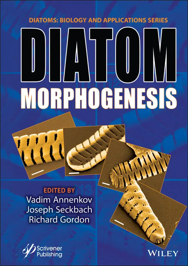
Cover image: Colored scanning electron micrographs (SEMs) of a morphogenetic sequence of the diatom Fragillaria capucina var. mesolepta by Dr. Mary Ann Tiffany, Biology Department, San Diego State University, USA Sample taken from Lake Murray (a freshwater San Diego Reservoir) on 3/18/2000.
Cover design by Russell Richardson
Set in size of 11pt and Minion Pro by Manila Typesetting Company, Makati, Philippines
Printed in the USA
10 9 8 7 6 5 4 3 2 1
Preface
Diatoms comprise a large, unicellular eukaryotic algal group that thrives mainly in aqueous environments: in fresh water, and in ponds, lakes and oceans. They may be attached to benthic substrates, in moist habitats or in floating debris and on macrophytes, and as phytoplankton; they form a substantial basis of aquatic food webs. They are ubiquitous, being distributed among various ecological locations. Among this group are some extremophiles with varying features, such as living in high temperatures, surviving desiccation, or in ice and at extreme ranges of pH. Some 20%-30% of the oxygen we breath is produced by diatom photosynthesis.
Vegetative cells of diatoms are diploid (2N), and meiosis can take place, producing male and female gametes fusing to zygotes which grow to auxospores.
One of their specific features is that their chemical composition includes siliceous (glassy) cell walls (frustules). Their exoskeleton is made of two halves called “valves” that fit inside one another, secured by silica “girdle bands”.
Diatoms’ fine structure is very impressive as revealed by transmission electron microscope, scanning microscope, and atomic force micrographs. The appearance of their cells is strikingly unique, and their shells are beautiful attractive shapes, with 60,000 to 200,000 species.
Why Valve Morphogenesis is Important?
Because there is so much detail in their silica wall shapes, spanning 8 orders of magnitude, diatoms are model organisms for single-cell morphogenesis. The problem of single cell morphogenesis has a long history, as yet unsolved, and perhaps diatoms rather than desmids and ciliates will now lead the way, especially given their 200 million years fossil record. This may further be because diatoms serve as a source of biofuel, food supplements and lipids and serve as significant material for nanotechnology. Thus, they are of very wide interest.
This volume focuses on the morphogenesis of diatoms, namely, the formation of their shape and the initial developmental steps.
The chapters were contributed by experts on morphological diatoms. The authors stem from the USA, Russia, Denmark, Germany, Greece, Israel, and Portugal.
Topics Addressed in This Volume
Topics include computer simulation of morphogenesis, silicic acid to silica frustules, inhibition in valve morphogenesis, pores within frustules, mesopores of pennate diatoms, frustule photonics and light harvesting, clonal chains, silica cell wall, geometric models of centric diatoms, morphology, surface features, buckling of valve morphogenesis, on mantle profiles, genetic-biochemical approaches, modeling silicon pools, valve morphogenesis, diatom teratology in taxonomy, phenotypic plasticity, geometric and morphometric analysis, silica morphogenesis in sister algae, and the uncanny symmetry of some diatoms.
This volume is the third book in the series Diatoms: Biology and Applications. The first book, Diatoms: Fundamentals and Applications appeared in 2019, and was edited by Joseph Seckbach and Richard Gordon. The second book, Diatom Gliding Motility , was published in September 2021 and is edited by Stanley A. Cohn, Kalina M. Manoylov and Richard Gordon.
We would like to thank the authors, the reviewers, the guest editor (Vadim V. Annenkov), and our publisher Martin Scrivener of Massachusetts, USA.
Joseph SeckbachHebrew University Jerusalem, Israel September 2021
1
Introduction for a Tutorial on Diatom Morphology
Kalina Manoylov1* and Mohamed Ghobara2
1Dept. of Biological & Environmental Sciences, Georgia College and State University, Milledgeville, GA, United States
2Department of Physics, Freie Universitat Berlin, Berlin, Germany
Abstract
Diatoms are an exceptionally successful group of unicellular microalgae with a large contribution of global primary production in aquatic environments and contributing a significant amount of oxygen to both hydro- and atmospheres. They are fascinating throughout their life and even after death, thanks to their unique cell walls made from ornamented silica. The diatoms include centric species, which may have radial or polar symmetry, and pennates, which include araphid, monoraphid, and biraphid species. Several applications have utilized diatomite, i.e., the fossil form of diatom frustules. To date, many diatoms’ secrets have been understood; however, there are still more hidden. Thus, there is a need for more research on diatom basic biology and applications. Seeking this goal, more people should be encouraged to work on diatoms. Often novice researchers are overwhelmed by the terminology associated with the diverse morphology, the discrepancy between expected features for published descriptions, and the actual observation of those complex 3D organisms, which can be a barrier for more progress. Here, we provide a brief introduction to the beginners with a guide to approach the complex diatom morphology focusing on the tools that can be used for its study.
Keywords:Diatom morphology, tutorial, LM and SEM, frustule morphology
1.1 Diatoms in Brief
Diatoms are unicellular, eukaryotic, microscopic algae (range from 1.5 μm to 5 mm in length, or diameter [1.9]), which maintain large population numbers and contribute considerably to the carbon and oxygen cycle on a global scale [1.8]. This ecologically successful group of algae is present in all aquatic habitats e.g. [1.1, 1.2] and even extends to humid terrestrial places. In aquatic habitats, diatoms are present in the photic zone, i.e., the region of water that light strongly penetrates, as well as in the benthic zone, i.e., the lowest level of water adjacent to the bottom with dim light conditions, depending on water column height and water’s turbidity. Diatoms can exist as planktonic (i.e., suspended in the water column), benthic (i.e., living near the bottom), epiphytic (i.e., adhered to aquatic plants [1.19], Figures 1.2c–d), or epizoic (i.e., adhered to a wide range of marine organisms such as crustaceans, mollusks, and vertebrates [1.19, 1.38]), or epilithic (i.e., attached completely or partially to submerged rocks). The adhesion ability of some diatoms is related to their mucilage secretion from specialized areas within their rigid cell walls (such as examples shown in Figures 1.1d and 1.2c–d). Some diatoms can form colonies in different arrangements such as chains and ribbons (examples shown in Figures 1.1 and 1.2).
Diatoms are a unique group of microalgae for several reasons, but one of the most notable and unique differences is the glass cell walls they possess [1.45]. This cell wall is called the “frustule” and is composed of amorphous hydrated silica that gives it unique properties. In general, the frustule is composed of two pieces that fit together like a petri-dish, meaning that the lower part of the frustule, called the hypotheca, sits inside of the upper part of the frustule, called the epitheca. The frustule volume extends by adding strips of silica called girdle bands (cingulum) to the mantle, i.e., the curved edge of the valve. It should be noted that there are plenty of frustule morphologies that vary between taxa.
Читать дальшеИнтервал:
Закладка:
Похожие книги на «Diatom Morphogenesis»
Представляем Вашему вниманию похожие книги на «Diatom Morphogenesis» списком для выбора. Мы отобрали схожую по названию и смыслу литературу в надежде предоставить читателям больше вариантов отыскать новые, интересные, ещё непрочитанные произведения.


Обсуждение, отзывы о книге «Diatom Morphogenesis» и просто собственные мнения читателей. Оставьте ваши комментарии, напишите, что Вы думаете о произведении, его смысле или главных героях. Укажите что конкретно понравилось, а что нет, и почему Вы так считаете.