Atlas of Endoscopic Ultrasonography
Здесь есть возможность читать онлайн «Atlas of Endoscopic Ultrasonography» — ознакомительный отрывок электронной книги совершенно бесплатно, а после прочтения отрывка купить полную версию. В некоторых случаях можно слушать аудио, скачать через торрент в формате fb2 и присутствует краткое содержание. Жанр: unrecognised, на английском языке. Описание произведения, (предисловие) а так же отзывы посетителей доступны на портале библиотеки ЛибКат.
- Название:Atlas of Endoscopic Ultrasonography
- Автор:
- Жанр:
- Год:неизвестен
- ISBN:нет данных
- Рейтинг книги:4 / 5. Голосов: 1
-
Избранное:Добавить в избранное
- Отзывы:
-
Ваша оценка:
Atlas of Endoscopic Ultrasonography: краткое содержание, описание и аннотация
Предлагаем к чтению аннотацию, описание, краткое содержание или предисловие (зависит от того, что написал сам автор книги «Atlas of Endoscopic Ultrasonography»). Если вы не нашли необходимую информацию о книге — напишите в комментариях, мы постараемся отыскать её.
Atlas of Endoscopic Ultrasonography Atlas of Endoscopic Ultrasonography, Second Edition
Atlas of Endoscopic Ultrasonography, Second Edition
Atlas of Endoscopic Ultrasonography — читать онлайн ознакомительный отрывок
Ниже представлен текст книги, разбитый по страницам. Система сохранения места последней прочитанной страницы, позволяет с удобством читать онлайн бесплатно книгу «Atlas of Endoscopic Ultrasonography», без необходимости каждый раз заново искать на чём Вы остановились. Поставьте закладку, и сможете в любой момент перейти на страницу, на которой закончили чтение.
Интервал:
Закладка:
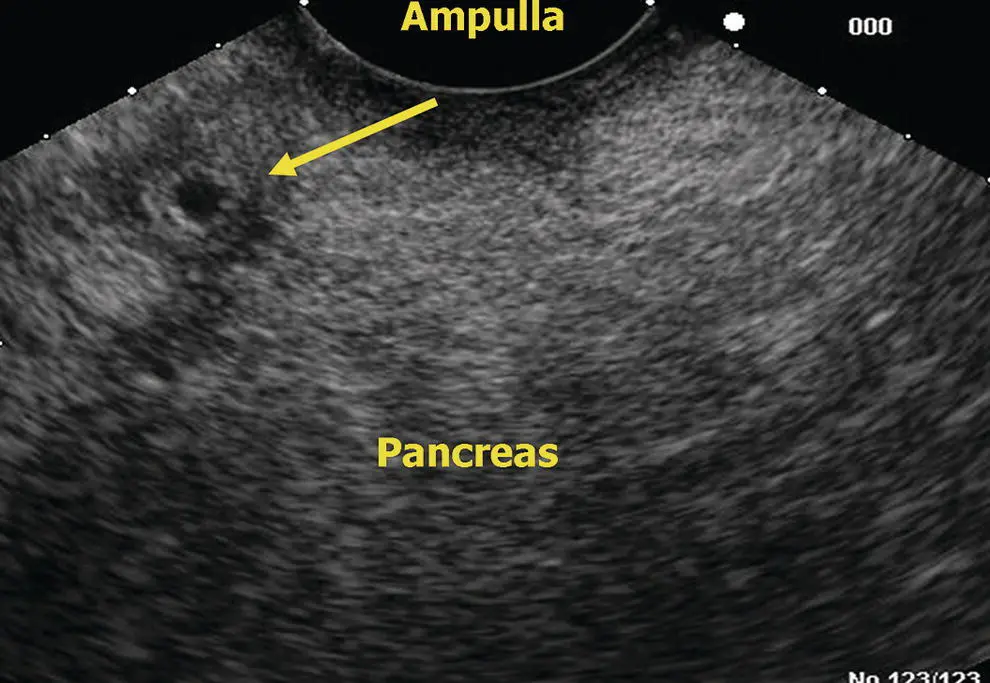
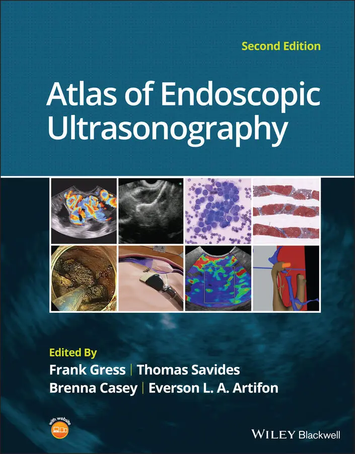
Figure 6.13 Linear EUS: ampulla.
Figure 6.14 Linear EUS: head of pancreas. CBD, common bile duct; PD, pancreatic duct.
Endosonographic appearance of the normal pancreatic parenchyma
There is considerable variability in the endosonographic appearance of the pancreatic parenchyma. Classically it has a homogeneous, fine, “salt and pepper” appearance with echogenicity similar to the spleen. The ventral anlage is more echolucent because of its different embryologic origin and its lesser content of echogenic fat. In the elderly, the pancreas can get more nodular with courser echogenicity. In obese patients, the pancreas becomes infiltrated with fat and can almost disappear into the retroperitoneal fat. Fortunately, any pathologic pancreatic lesions, such as dilated ducts, cysts, or neoplasms, will be easily visible in the bright background of retroperitoneal fat. Thin patients typically offer particularly detailed imaging of the pancreas.
Chapter video clips
Video 6.1Linear array EUS head of pancreas.
Video 6.2Linear array EUS of the pancreas neck to tail.
Video 6.3Radial array EUS head of pancreas.
Video 6.4Radial array EUS of the pancreatic neck to tail.
7 Liver, Spleen, and Kidneys: Radial and Linear
Nalini M. Guda1 and Marc F. Catalano2
1University of Wisconsin, School of Medicine and Public Health, Pancreatobiliary Services, St. Luke’s Medical Center, Milwaukee, WI, USA
2Medical College of Wisconsin, Pancreatobiliary Services, St. Luke’s Medical Center, Milwaukee, WI, USA
Introduction
This chapter describes the endosonographic features of the major organs of the abdomen: the liver, spleen, kidneys, and adrenal glands. Ultrasound features of the pancreas and bile duct are described elsewhere.
The liver, spleen, kidneys, and adrenal glands (left side) are visualized from the stomach ( Videos 7.1and 7.2).
Liver
Radial endosonography
As the radial probe is advanced through the esophagus into the gastric cardia, the liver is the predominant organ visualized. When positioning the abdominal aorta at the 6 o’clock position, the left lobe of the liver is seen anteriorly and medially to the right ( Figure 7.1). The aorta, with a dark hypoechoic band which is the diaphragmatic crux, is seen immediately adjacent to the probe. In this position, near the hiatus, the hepatic veins are seen as anechoic structures, entering the inferior vena cava (IVC). In this position, possibly with left tip deflection, the spleen can be seen on the right of the screen. As the aorta is traced distally, maintaining its 6 o’clock position, the liver may still be seen anteriorly. Vascular structures can be differentiated from ductal structures by a thicker (echogenic) wall and the presence of flow.
When advancing the echoprobe towards the antrum, the gallbladder is often visualized as an oval‐shaped anechoic structure. In this position, the porta hepatis can be seen with subtle tip deflection upwards.
Linear endosonography
With the probe at the level of the diaphragmatic hiatus, the longitudinal aorta and celiac artery origin are the most recognizable reference points ( Figure 7.2), demonstrated as tubular longitudinal structures. Here, rotation of the probe counterclockwise will bring into view the liver parenchyma and its vascular structures ( Figure 7.3).
Advancing the probe at the level of the pylorus and duodenal bulb, clockwise rotation and superior tip deflection brings into view the porta hepatis along with several vascular structures. Use of Doppler can differentiate arterial from venous structures as well as biliary structures.
The entire liver is not visualized by endoscopic ultrasound (EUS). Despite this limitation, it is useful to carefully examine the liver since metastatic processes can be easily identified and biopsied and could lead to a change in clinical staging and management of a suspected tumor.
Spleen
The spleen appears as a homogeneous structure seen between the tail of the pancreas, left kidney, and gastric wall. With a radial scope it is imaged from the gastric cardia. It is similar to liver in echogenicity except that it is devoid of any ducts and vessels ( Figure 7.4). It can be easier to follow the splenic vein after visualizing the pancreas from the gastroesophageal (GE) junction. The splenic artery, splenic vein, renal vein, and the left adrenal are usually visualized as well while attempting to scan the spleen. The splenic vein can be easily traced along the inferior aspect of the body and tail of the pancreas; however, the splenic artery is tortuous and it is difficult to follow its course to the celiac trunk. With a linear scope one has to scan inferior to the left kidney and laterally to visualize the spleen ( Figure 7.5).
Kidney
Both the right and left kidneys can be visualized by EUS. The left kidney is readily identified from the fundus of the stomach. It has a complex echogenicity with hypoechoic parenchyma and hypertonic areas within representing the calyceal system.
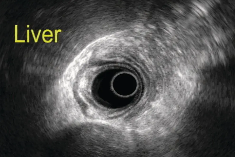
Figure 7.1 Image of the liver scanned by radial ultrasound.
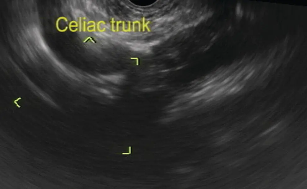
Figure 7.2 Celiac artery imaging by linear ultrasound.
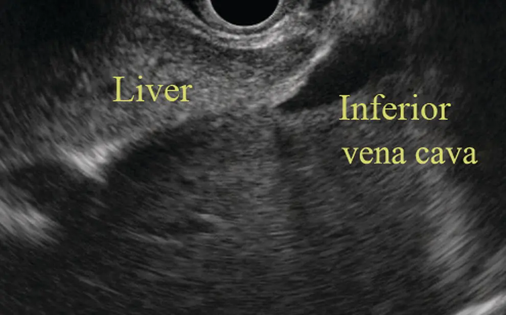
Figure 7.3 Image of liver by linear ultrasound.
Radial endosonography
With the echoendoscope in the gastric cardia the left kidney is readily visualized. One can see the calyceal system and the renal vessels ( Figure 7.6). To visualize the right kidney the echoscope is placed in the D‐3 position. Visualization of the IVC and aorta provide easily identifiable landmarks. Upon slow withdrawal, the image brings into view the right kidney immediately right of the probe along with the right renal vein and artery. As the kidney is traced proximally, lateral deflection of the tip may bring into view the right adrenal gland.
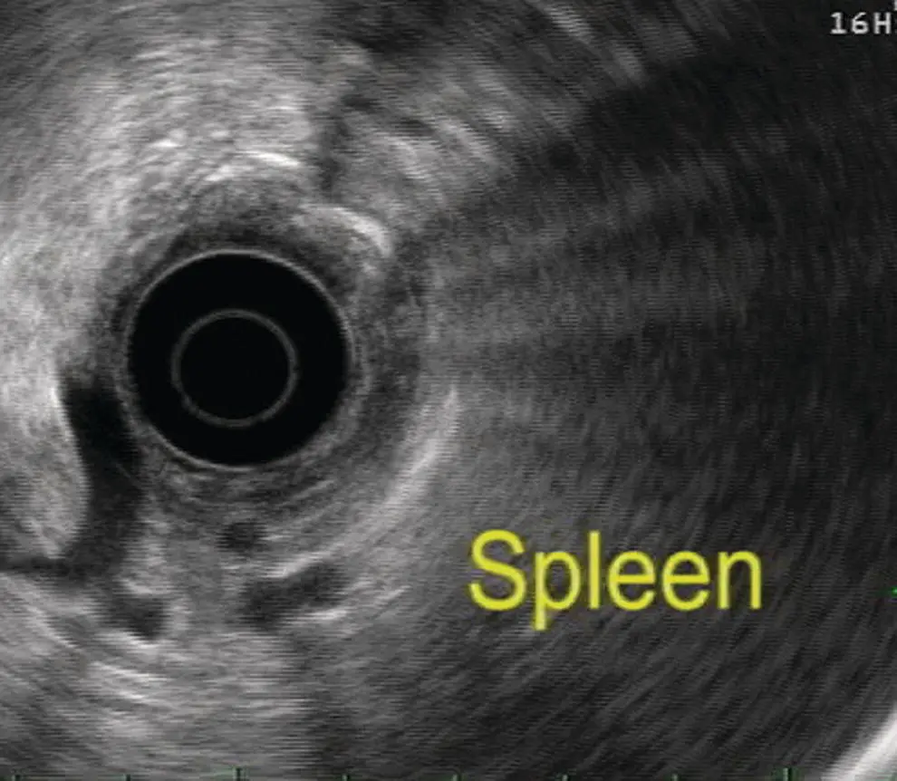
Figure 7.4 Image of spleen by radial ultrasound.
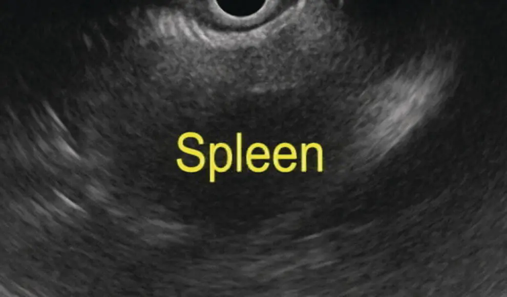
Figure 7.5 Image of spleen by linear ultrasound.
Linear endosonography
With a linear scope one can withdraw the scope in the fundus posteriorly towards the pancreas until the tail is visualized and then push the scope inferiorly to see the left kidney ( Figure 7.7). To view the right kidney, the probe is placed just below the level of the papilla and counterclockwise rotation brings into view the right kidney and its vascular structures. Slow withdrawal allows for visualization of the right adrenal glands.
Читать дальшеИнтервал:
Закладка:
Похожие книги на «Atlas of Endoscopic Ultrasonography»
Представляем Вашему вниманию похожие книги на «Atlas of Endoscopic Ultrasonography» списком для выбора. Мы отобрали схожую по названию и смыслу литературу в надежде предоставить читателям больше вариантов отыскать новые, интересные, ещё непрочитанные произведения.


Обсуждение, отзывы о книге «Atlas of Endoscopic Ultrasonography» и просто собственные мнения читателей. Оставьте ваши комментарии, напишите, что Вы думаете о произведении, его смысле или главных героях. Укажите что конкретно понравилось, а что нет, и почему Вы так считаете.