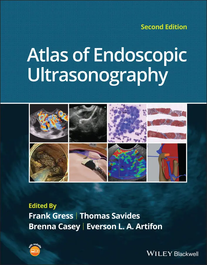
Atlas of Endoscopic Ultrasonography
Здесь есть возможность читать онлайн «Atlas of Endoscopic Ultrasonography» — ознакомительный отрывок электронной книги совершенно бесплатно, а после прочтения отрывка купить полную версию. В некоторых случаях можно слушать аудио, скачать через торрент в формате fb2 и присутствует краткое содержание. Жанр: unrecognised, на английском языке. Описание произведения, (предисловие) а так же отзывы посетителей доступны на портале библиотеки ЛибКат.
- Название:Atlas of Endoscopic Ultrasonography
- Автор:
- Жанр:
- Год:неизвестен
- ISBN:нет данных
- Рейтинг книги:4 / 5. Голосов: 1
-
Избранное:Добавить в избранное
- Отзывы:
-
Ваша оценка:
Atlas of Endoscopic Ultrasonography: краткое содержание, описание и аннотация
Предлагаем к чтению аннотацию, описание, краткое содержание или предисловие (зависит от того, что написал сам автор книги «Atlas of Endoscopic Ultrasonography»). Если вы не нашли необходимую информацию о книге — напишите в комментариях, мы постараемся отыскать её.
Atlas of Endoscopic Ultrasonography Atlas of Endoscopic Ultrasonography, Second Edition
Atlas of Endoscopic Ultrasonography, Second Edition
Atlas of Endoscopic Ultrasonography — читать онлайн ознакомительный отрывок
Ниже представлен текст книги, разбитый по страницам. Система сохранения места последней прочитанной страницы, позволяет с удобством читать онлайн бесплатно книгу «Atlas of Endoscopic Ultrasonography», без необходимости каждый раз заново искать на чём Вы остановились. Поставьте закладку, и сможете в любой момент перейти на страницу, на которой закончили чтение.
Интервал:
Закладка:
Table of Contents
1 Cover
2 Title Page
3 Copyright Page
4 Contributors
5 Preface
6 About the Companion Website
7 1 Normal EUS Anatomy 1 Normal Human Anatomy Introduction Normal EUS anatomy from the esophagus Normal EUS anatomy from the stomach Normal EUS anatomy from the duodenum Normal EUS anatomy from the rectum Vascular videos 2 Esophagus Layers of the esophageal wall Normal radial extraesophageal anatomy (Video 2.1) Normal linear thoracic anatomy 3 Normal Mediastinal Anatomy by EUS and EBUS Introduction Anatomical definitions Equipment Endoscopic ultrasound technique Endobronchial ultrasound Complications and safety Conclusions 4 Stomach 5 Bile Duct Normal bile duct anatomy Normal anatomy of the bile duct and gallbladder with radial echoendoscope Normal anatomy of the bile duct and gallbladder with linear echoendoscope 6 EUS of the Normal Pancreas Radial examination of the pancreas Linear examination of the pancreas Endosonographic appearance of the normal pancreatic parenchyma 7 Liver, Spleen, and Kidneys Introduction Liver Spleen Kidney Adrenal glands 8 Anatomy of the Anorectum Introduction Examination technique Normal anatomy
8 2 Upper and Lower GI EUS 9 Esophageal Cancer Introduction Updated American Joint Committee on Cancer staging guidelines for esophageal cancer 2017 and implications for endosonographers Role of EUS in staging of esophageal cancer Limitations Impact of EUS staging on management Technique References 10 EUS for Achalasia Introduction Clinical presentation and diagnosis Role of EUS in achalasia 11 Malignant Mediastinal Lesions 12 Benign Mediastinal Lesions 13 Gastric Cancer 14 Gastric and Esophageal Subepithelial Masses Introduction Lipoma Carcinoid tumors Granular cell tumor Duplication cyst Pancreatic rest Varices Gastrointestinal stromal cell tumors and leiomyomas Glomus tumor Gastritis cystica profunda Extrinsic compression lesions 15 Anorectal Neoplasia Colorectal cancer staging by EUS Endoscopic ultrasound for local recurrence of colorectal carcinoma Submucosal tumors of the colorectal wall 16 Anal Sphincter Disease Introduction Fecal incontinence Perianal fistula 17 Endometriosis Introduction Definition and location Epidemiology and risk factors Clinical picture Diagnosis Classification Imaging methods 18 Vascular Anomalies and Abnormalities Introduction Aortic arch anomalies Vascular calcification and plaques Aneurysms and pseudoaneurysms Venous thrombosis Dieulafoy lesions Neoplasms Miscellaneous aberrancies
9 3 Pancreatico‐biliary 19 Duodenal and Ampullary Neoplasia 20 Biliary Tract Pathology 21 Gallbladder Pathology Introduction Gallbladder stones Gallbladder polyps Gallbladder carcinoma 22 Pancreatic Adenocarcinoma Introduction Tumor identification and diagnosis via fine needle aspiration or fine needle biopsy Evaluation of vascular invasion Evaluation of peripancreatic lymphadenopathy Limitations and complications of EUS in patients with pancreatic cancer Conclusion References 23 Pancreatic Malignancy (Non‐adenocarcinoma) Introduction Endocrine pancreatic tumors (Figures 23.1 and 23.2) Primary pancreatic lymphoma (Figure 23.3) Solid pseudopapillary tumors (Figure 23.4) Acinar cell carcinoma (Figure 23.5) Secondary metastatic tumors (Figure 23.6) Summary 24 Autoimmune Pancreatitis Introduction Endoscopic ultrasound imaging (Video 24.1) Image‐enhancing techniques during EUS EUS‐FNA and EUS‐FNB Histologic features Summary 25 Pancreatic Cystic Lesions Introduction Pseudocyst Serous lesions Mucinous lesions Other cystic neoplasms 26 Intraductal Papillary Mucinous Neoplasms Introduction Clinical features Role of imaging Cross‐sectional imaging Endoscopic ultrasound evaluation Management of small IPMN (≤3 cm) 27 Chronic Pancreatitis Introduction Clinical overview of chronic pancreatitis Endoscopic ultrasound imaging of the normal pancreas EUS imaging in chronic pancreatitis: historical perspectives EUS imaging in chronic pancreatitis: the Rosemont Criteria Endoscopic ultrasound imaging in chronic pancreatitis: the future 28 Liver Pathology Introduction Cirrhosis Fatty liver disease Hepatic cysts Neoplasms Dilated intrahepatic ducts
10 4 How to Section 29 How to Interpret EUS‐FNA Cytology Introduction Technical quality of EUS biopsy material Quality of the interpretation Integration of pathologic and clinical information 30 How to do Mediastinal FNA 31 How to do Pancreatic Mass FNA Introduction The technique 32 How to do Pancreatic Cyst FNA Introduction Technique (Video 32.1) Summary 33 How to do Pancreatic Pseudocyst Drainage Introduction Patient selection Requisite instruments and accessories Assessment of the pseudocyst by EUS prior to drainage Technique for placement of plastic endoprosthesis Technique for lumen‐apposing metal stent placement Post‐procedure follow‐up 34 How to do EUS‐guided Pancreatic Cyst Chemoablation Background Pretreatment evaluation Patient selection Technical aspects of the procedure Postoperative care and follow‐up Conclusions References 35 How to do Celiac Plexus Block Introduction Technique (Video 35.1) Complications 36 How to Place Fiducials for Radiation Therapy Introduction Equipment Techniques Periprocedural care 37 How to Inject Chemotherapeutic Agents 38 How to do EUS‐guided Pelvic Abscess Drainage Introduction Patient preparation Devices and accessories Procedural technique Clinical outcomes Technical limitations Conclusions 39 How to do Doppler Probe EUS for Bleeding Background and equipment Practical application of DopUS probe Conclusions 40 How to do Endoscopic Ultrasound‐guided Portal Pressure Gradient Measurement Introduction Endoscopic ultrasound‐guided PPGM technique References 41 How to do Endoscopic Ultrasound‐guided Liver Biopsy Indications and contraindications EUS‐LB technique Postprocedure recovery after EUS‐LB Adverse effects Conclusions References 42 How to do EUS‐guided Treatment of Gastric Varices Introduction Technique (Video 42.1) Complicatons 43 How to do EUS‐guided Arterial Embolization Background Technique Postprocedural management and complications References 44 How to do EUS‐guided Radiofrequency Ablation of Pancreatic Neuroendocrine Tumors Introduction Methods for EUS‐guided RFA Indications Results for EUS‐guided RFA studies Conclusion References 45 How to do EUS Pancreatic Duct Access and Drainage Introduction Indications Contraindications Before the procedure Techniques for drainage Outcomes Algorithm Controversies and future directions 46 How to do EUS Gallbladder Drainage Introduction Background concept Choice of stents: metal stents versus plastic stents EUS‐GBD with LAMS Postprocedural management Long‐term management Conclusion References 47 How to do an EUS‐guided Gastrojejunostomy Introduction Technique (Video 47.1) Complications References 48 How to do EUS Elastography Introduction Technique Indications Complications Further reading 49 How to do Contrast‐enhanced EUS Introduction Principle of CH‐EUS Critical points for performing CH‐EUS Advantages of CH‐EUS compared to other contrast‐enhanced modalities Advantages of CH‐EUS compared to conventional EUS Evaluation of CH‐EUS image for lesions in different organs 50 How to do EUS‐guided Ablation of Pancreatic Neurendocrine Tumors Introduction and indications Techniques Conclusion and future perspectives 51 How to do EUS‐guided Needle Confocal Laser Endomicroscopy of Pancreatic Cysts Confocal laser endomicroscopy technique 52 How to use ex vivo Models in Teaching Therapeutic Endoscopic Ultrasound Introduction Ex vivo models Conclusion 53 How to do Endoscopic Necrosectomy Introduction Preprocedure assessment EUS evaluation of walled‐off pancreatic necrosis Access creation: cystgastrostomy or cystenterostomy Necrosectomy tools and technique Complication management Conclusion References 54 How to Perform Pancreatic Mass Fine Needle Biopsy Introduction Technique (Video 54.1) Summary 55 How to Perform Endoscopic Ultrasound‐directed Transgastric Endoscopic Retrograde Cholangiopancreatography (EDGE) Introduction Technique Post‐EDGE fistula management Summary
Читать дальшеИнтервал:
Закладка:
Похожие книги на «Atlas of Endoscopic Ultrasonography»
Представляем Вашему вниманию похожие книги на «Atlas of Endoscopic Ultrasonography» списком для выбора. Мы отобрали схожую по названию и смыслу литературу в надежде предоставить читателям больше вариантов отыскать новые, интересные, ещё непрочитанные произведения.


Обсуждение, отзывы о книге «Atlas of Endoscopic Ultrasonography» и просто собственные мнения читателей. Оставьте ваши комментарии, напишите, что Вы думаете о произведении, его смысле или главных героях. Укажите что конкретно понравилось, а что нет, и почему Вы так считаете.