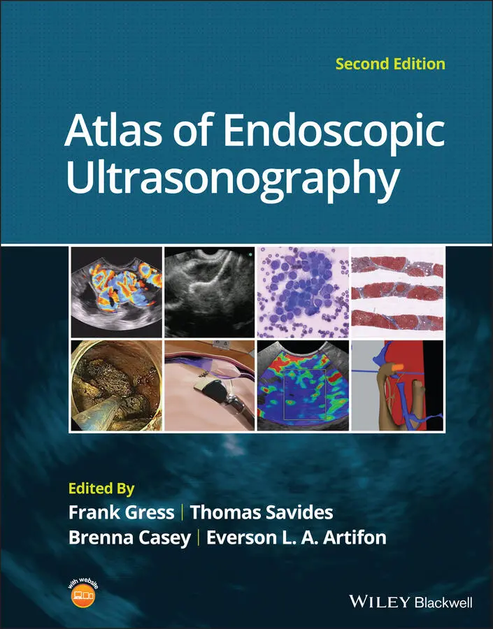
Atlas of Endoscopic Ultrasonography
Здесь есть возможность читать онлайн «Atlas of Endoscopic Ultrasonography» — ознакомительный отрывок электронной книги совершенно бесплатно, а после прочтения отрывка купить полную версию. В некоторых случаях можно слушать аудио, скачать через торрент в формате fb2 и присутствует краткое содержание. Жанр: unrecognised, на английском языке. Описание произведения, (предисловие) а так же отзывы посетителей доступны на портале библиотеки ЛибКат.
- Название:Atlas of Endoscopic Ultrasonography
- Автор:
- Жанр:
- Год:неизвестен
- ISBN:нет данных
- Рейтинг книги:4 / 5. Голосов: 1
-
Избранное:Добавить в избранное
- Отзывы:
-
Ваша оценка:
Atlas of Endoscopic Ultrasonography: краткое содержание, описание и аннотация
Предлагаем к чтению аннотацию, описание, краткое содержание или предисловие (зависит от того, что написал сам автор книги «Atlas of Endoscopic Ultrasonography»). Если вы не нашли необходимую информацию о книге — напишите в комментариях, мы постараемся отыскать её.
Atlas of Endoscopic Ultrasonography Atlas of Endoscopic Ultrasonography, Second Edition
Atlas of Endoscopic Ultrasonography, Second Edition
Atlas of Endoscopic Ultrasonography — читать онлайн ознакомительный отрывок
Ниже представлен текст книги, разбитый по страницам. Система сохранения места последней прочитанной страницы, позволяет с удобством читать онлайн бесплатно книгу «Atlas of Endoscopic Ultrasonography», без необходимости каждый раз заново искать на чём Вы остановились. Поставьте закладку, и сможете в любой момент перейти на страницу, на которой закончили чтение.
Интервал:
Закладка:
25 Chapter 25Figure 25.1 Thick‐walled cystic lesion undergoing fine needle aspiration (FN...Figure 25.2 Serous cystadenoma. Multicystic lesion in the body of the pancre...Figure 25.3 (a) Mucinous cystic neoplasm with multiple septations. The lesio...Figure 25.4 (a) Thinly septated intraductal papillary mucinous neoplasm (IPM...Figure 25.5 (a) Pancreatic cystic endocrine tumor. Note the presence of the ...
26 Chapter 26Figure 26.1 Reformatted computed tomography scan demonstrating a side‐branch...Figure 26.2 Magnetic resonance cholangiopancreatography demonstrating a side...Figure 26.3 Linear endoscopic ultrasound image demonstrating a dilated main ...Figure 26.4 Linear endoscopic ultrasound image revealing a small cystic lesi...Figure 26.5 Linear endoscopic ultrasound image of a malignant‐appearing 2.9‐...Figure 26.6 Fine needle aspiration of a malignant cystic mass.Figure 26.7 International Association of Pancreatology (IAP) algorithm for m...
27 Chapter 27Figure 27.1 Endoscopic ultrasound (linear) showing features of chronic pancr...Figure 27.2 Endoscopic ultrasound showing a hyperechoic duct wall (crossmark...
28 Chapter 28Figure 28.1 (a) Cirrhosis. The cirrhotic liver is characterized by diffuse, ...Figure 28.2 Fatty liver is described as diffuse or as foci of hyperechoic or...Figure 28.3 Simple hepatic cyst can be seen here as an anechoic fluid‐filled...Figure 28.4 (a, b) Metastatic liver disease. Lesions found within the hepati...Figure 28.5 Dilated intrahepatic ducts (arrows) are visualized here and are ...
29 Chapter 29Figure 29.1 (a) Core biopsies demonstrate intact tissue architecture and str...Figure 29.2 Diagnostic features of autoimmune pancreatitis rely on the demon...Figure 29.3 Aspirate smears demonstrate fine cellular detail such as squamou...Figure 29.4 Cell blocks, prepared from fine needle aspirate material and pla...Figure 29.5 Clotted blood within the needle obscures lesional tissue (a). Op...Figure 29.6 Air‐dried slides stained with a modified Romanowsky stain demons...Figure 29.7 Distinctive or commonly encountered in endoscopic ultrasound‐gui...
30 Chapter 30Figure 30.1 The International Association for the Study of Lung Cancer (IASL...
31 Chapter 31Figure 31.1 (a) Hypoechoic, irregular, 3‐cm adenocarcinoma in the head of pa...Figure 31.2 (a) Small adenocarcinoma in the head of pancreas adjacent to the...Figure 31.3 (a) Large 5‐cm adenocarcinoma in the tail of pancreas. (b) Mass ...Figure 31.4 (a) A 2‐cm mass in the head of pancreas with acoustic interferen...Figure 31.5 (a) Small 1‐cm hypoechoic neuroendocrine tumor in the head of pa...Figure 31.6 (a) Cystic neoplasm with malignant degeneration in the head of p...Figure 31.7 Hepatic metastases visualized during EUS examination for a pancr...
32 Chapter 32Figure 32.1 EUS of left lobe of liver.Figure 32.2 EUS of aorta, celiac artery (CA), and superior mesenteric artery...Figure 32.3 Transgastric EUS examination of pancreas (genu, body, tail).Figure 32.4 Transduodenal EUS examination of pancreas (head and uncinate pro...Figure 32.5 (a) EUS of anechoic septated lobular cyst in tail of pancreas. (...Figure 32.6 Position lesion at 6 o’clock for FNA/FNB.Figure 32.7 EUS with Doppler showing blood vessels (purple) in path of needl...Figure 32.8 EUS needle handle.Figure 32.9 FNA needle within pancreatic cyst (yellow arrow indicates tip of...Figure 32.10 Vacuum syringe.Figure 32.11 Serous fluid aspirated from large pancreatic cyst.Figure 32.12 String sign consistent with mucinous cyst.
33 Chapter 33Figure 33.1 A large unilocular pseudocyst with clear contents as visualized ...Figure 33.2 Two pseudocysts are seen at EUS: the promixal lesion on FNA anal...Figure 33.3 Presence of intervening vasculature between the EUS transducer a...Figure 33.4 Endoscopic ultrasound guidance enables access to the pseudocyst ...Figure 33.5 Pseudocyst accessed with a 19‐gauge FNA needle.Figure 33.6 (a) Acute angulation encountered when the echoendoscope is locat...Figure 33.7 A 0.035‐inch guidewire is coiled within the pseudocyst under flu...Figure 33.8 The transmural tract is dilated using a 5‐Fr endoscopic retrogra...Figure 33.9 The transmural tract is subsequently dilated using an 8 mm over‐...Figure 33.10 Multiple 7‐Fr transmural stents are deployed (fluoroscopy view)...Figure 33.11 (a) Endoscopic view of the proximal flange of the LAMS in the s...
34 Chapter 34Figure 34.1 The alcohol free EUS‐guided cyst ablation process. The FNA needl...Figure 34.2 A high‐pressure gun, syringe, and short connector tubing assembl...Figure 34.3 (a) Magnetic resonance image showing a 76‐year‐old female with a...Figure 34.4 (a) MRI ( top ) and magnetic resonance cholangiopancreatography (M...
35 Chapter 35Figure 35.1 Hypoechoic pancreatic mass as seen by EUS.Figure 35.2 Endoscopic ultrasound demonstration of lobularity, one of the ma...Figure 35.3 Celiac axis (CA) as seen by EUS. AO, aorta.Figure 35.4 Celiac axis confirmed by the aorta, celiac artery (CA) and super...Figure 35.5 Insertion of the EUS‐guided fine needle aspiration needle into t...
36 Chapter 36Figure 36.1 Gold coil fiducial marker is (a) flexible and (b) has a coiled d...Figure 36.2 Gold coil fiducial preloaded on needle‐carrier delivery device....Figure 36.3 Endoscopic ultrasound image (linear array) of fiducial within an...Figure 36.4 Abdominal radiograph (anteroposterior view) demonstrating three ...
37 Chapter 37Figure 37.1 Initial assessment of tumor size by endoscopic ultrasound (EUS) ...Figure 37.2 Endoscopic ultrasound (EUS)‐guided fine needle injection (FNI) o...Figure 37.3 Tumor assessment during weekly injections of chemotherapeutic ag...Figure 37.4 Gross specimen of the resected pancreas in Figure 37.3 revealing...Figure 37.5 Histology from the specimen in Figure 37.4 demonstrating a compl...
38 Chapter 38Figure 38.1 Computed tomography (CT) scan of the pelvis revealing an 8 × 7 c...Figure 38.2 Passage of a 19‐gauge fine needle aspiration (FNA) needle into t...Figure 38.3 A 0.035‐inch guidewire is then coiled within the abscess cavity:...Figure 38.4 Fluoroscopy view revealing dilation of the tract using an endosc...Figure 38.5 Fluoroscopic view revealing the presence of a stent within the p...
39 Chapter 39Figure 39.1 VTI endoscopic Doppler system. (a) A 20‐MHz pulsed‐wave Doppler ...Figure 39.2 Endo‐Dop, 16‐MHz pulsed‐wave, multigated Doppler ultrasound (Dop...Figure 39.3 Illustration of a bleeding peptic ulcer with a non‐bleeding visi...Figure 39.4 Doppler ultrasound (DopUS) in acute peptic ulcer bleeding. (a) E...Figure 39.5 Doppler ultrasound‐guided hemostasis in acute peptic ulcer bleed...Figure 39.6 Atypical peptic ulcer bleeding: recurrent major bleeding from a ...Figure 39.7 Doppler‐negative non‐bleeding visible vessel. Endoscopic image o...Figure 39.8 Atypical appearance of a bleeding gastric varix. Mild active ooz...Figure 39.9 Doppler ultrasound examination of a gastrointestinal stromal tum...Figure 39.10 Dieulafoy lesion in gastric cardia. (a) Active bleeding. (b) Do...Figure 39.11 Dieulafoy lesion in rectum, Doppler with strong arterial signal...
40 Chapter 40Figure 40.1 Compact manometer for EUS‐guided portal pressure measurement....Figure 40.2 EUS‐guided portal pressure measurement apparatus showing noncomp...Figure 40.3 EUS image (a) and schematic (b) of needle puncture of the middle...Figure 40.4 EUS Doppler flow image of the middle hepatic vein demonstrating ...Figure 40.5 EUS image (a) and schematic (b) of needle puncture of the left p...Figure 40.6 EUS Doppler flow image of the left portal vein demonstrating typ...
41 Chapter 41Figure 41.1 Visualization of the left hepatic lobe from the proximal stomach...Figure 41.2 View of liver (on the left) and spleen (on the right) from the p...Figure 41.3 Right lobe biopsies are obtained with the echoendoscope tip plac...Figure 41.4 Close‐up of the tips of the core needles used for EUS‐LB: ( left )...Figure 41.5 Comparison of a 19G FNB needle to 19G FNA needle in terms of (a...Figure 41.6 Excellent core of cirrhotic liver obtained with the 19G FNB nee...Figure 41.7 Priming the needle with heparin for wet suction technique.Figure 41.8 (a) Use of tissue sieve (CoreCatcher; ProAct Ltd, Center Hall, P...Figure 41.9 Long liver core is placed in formalin jar without excessive hand...
Читать дальшеИнтервал:
Закладка:
Похожие книги на «Atlas of Endoscopic Ultrasonography»
Представляем Вашему вниманию похожие книги на «Atlas of Endoscopic Ultrasonography» списком для выбора. Мы отобрали схожую по названию и смыслу литературу в надежде предоставить читателям больше вариантов отыскать новые, интересные, ещё непрочитанные произведения.


Обсуждение, отзывы о книге «Atlas of Endoscopic Ultrasonography» и просто собственные мнения читателей. Оставьте ваши комментарии, напишите, что Вы думаете о произведении, его смысле или главных героях. Укажите что конкретно понравилось, а что нет, и почему Вы так считаете.