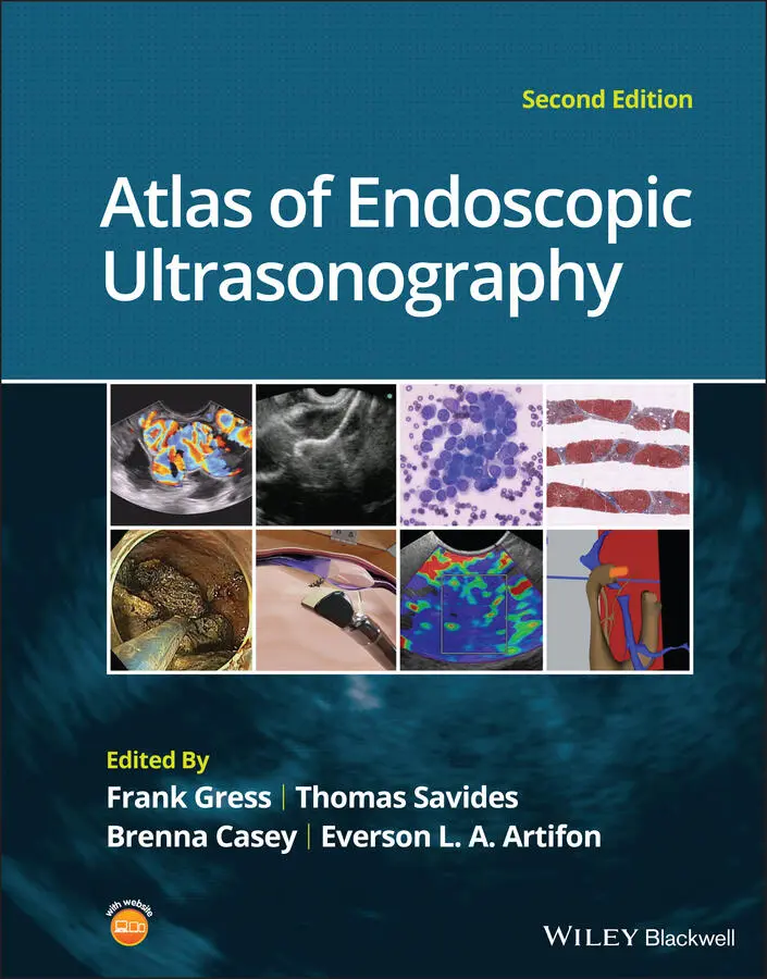
Atlas of Endoscopic Ultrasonography
Здесь есть возможность читать онлайн «Atlas of Endoscopic Ultrasonography» — ознакомительный отрывок электронной книги совершенно бесплатно, а после прочтения отрывка купить полную версию. В некоторых случаях можно слушать аудио, скачать через торрент в формате fb2 и присутствует краткое содержание. Жанр: unrecognised, на английском языке. Описание произведения, (предисловие) а так же отзывы посетителей доступны на портале библиотеки ЛибКат.
- Название:Atlas of Endoscopic Ultrasonography
- Автор:
- Жанр:
- Год:неизвестен
- ISBN:нет данных
- Рейтинг книги:4 / 5. Голосов: 1
-
Избранное:Добавить в избранное
- Отзывы:
-
Ваша оценка:
Atlas of Endoscopic Ultrasonography: краткое содержание, описание и аннотация
Предлагаем к чтению аннотацию, описание, краткое содержание или предисловие (зависит от того, что написал сам автор книги «Atlas of Endoscopic Ultrasonography»). Если вы не нашли необходимую информацию о книге — напишите в комментариях, мы постараемся отыскать её.
Atlas of Endoscopic Ultrasonography Atlas of Endoscopic Ultrasonography, Second Edition
Atlas of Endoscopic Ultrasonography, Second Edition
Atlas of Endoscopic Ultrasonography — читать онлайн ознакомительный отрывок
Ниже представлен текст книги, разбитый по страницам. Система сохранения места последней прочитанной страницы, позволяет с удобством читать онлайн бесплатно книгу «Atlas of Endoscopic Ultrasonography», без необходимости каждый раз заново искать на чём Вы остановились. Поставьте закладку, и сможете в любой момент перейти на страницу, на которой закончили чтение.
Интервал:
Закладка:
12 Chapter 12Figure 12.1 Benign subcarinal lymph nodes. Note the oval and triangular‐shap...Figure 12.2 Sarcoid lymph nodes. Note the matted together, draping nodes wit...Figure 12.3 Cryptococcal lymph node. These were incidental lymph nodes found...Figure 12.4 Duplication cyst. Note the anechoic appearance with acoustic enh...Figure 12.5 Enlarged lymph node due to Blastocystis hominis infection.
13 Chapter 13Figure 13.1 T1 gastric cancer. This is a 2.2 × 0.5 cm thick tumor confined t...Figure 13.2 T2 gastric cancer. This large 4.2 × 3.6 cm exophytic tumor invad...Figure 13.3 T3 gastric cancer. This 2.5 × 1.6 cm tumor invades through all l...Figure 13.4 T4 gastric cancer. This invasive poorly differentiated cancer in...Figure 13.5 Diffuse gastric cancer. The stomach is thickened to 0.8 cm (norm...Figure 13.6 Pseudo‐linitis plastica. A patient with breast cancer has metast...Figure 13.7 Gastric lymphoma causing a linitis plastica appearance. This pat...Figure 13.8 Gastric lymphoma. The five‐layer wall pattern is obliterated and...Figure 13.9 Perigastric lymphadenopathy in a patient with primary gastric ly...Figure 13.10 Ascites. A large pocket of hypoechoic material is seen next to ...Figure 13.11 Endoscopic ultrasound‐guided fine needle aspiration (EUS‐FNA) o...
14 Chapter 14Figure 14.1 Lipoma. (a) Endoscopic view of lipoma with smooth overlying muco...Figure 14.2 Carcinoid tumor. (a) Endoscopic view of a carcinoid tumor. (b) E...Figure 14.3 Granular cell tumor. (a) Endoscopic view of esophageal granular ...Figure 14.4 Esophageal duplication cysts. (a) Endoscopic view of several eso...Figure 14.5 Pancreatic rest. (a) Endoscopic view of a gastric antral pancrea...Figure 14.6 Esophageal varices. (a) Endoscopic view of grade 3 esophageal va...Figure 14.7 Gastric gastrointestinal stromal tumor (GIST). (a) Endoscopic vi...Figure 14.8 Leiomyoma. (a) Endoscopic view of an esophageal leiomyoma. (b) E...Figure 14.9 Glomus tumor. (a) Endoscopic view of a gastric antral glomus tum...Figure 14.10 Gastritis cystica profunda (GCP). (a) Endoscopic view showing G...Figure 14.11 Extrinsic compression. (a) Endoscopic and EUS view of extrinsic...
15 Chapter 15Figure 15.1 (a) A T1 rectal adenocarcinoma (by radial EUS) arising in a vill...Figure 15.2 (a) A rectal adenocarcinoma that appears to be T2 (penetration i...Figure 15.3 Radial EUS of a T3 lesion showing the primary rectal tumor penet...Figure 15.4 Radial EUS of the patient in Figure 15.3 showing a 7‐mm round, p...Figure 15.5 EUS‐guided fine needle aspiration of a perirectal lymph node. Th...Figure 15.6 Recurrent rectal cancer. During surveillance colonoscopy in a pa...Figure 15.7 (a) A subepithelial bulge in the rectum from a large intramural ...Figure 15.8 Rectal carcinoid. (a) A rectal bulge was noted during routine co...
16 Chapter 16Figure 16.1 Schematic diagram of the anal canal. EAS, external anal sphincte...Figure 16.2 Normal anal sphincter complex. The internal anal sphincter (IAS)...Figure 16.3 Normal anal sphincter complex. The internal anal sphincter (IAS)...Figure 16.4 Forty percent defect in both the internal hypoechoic anal sphinc...Figure 16.5 Forty percent defect in external anal sphincter (EAS), likely du...Figure 16.6 Thirty percent defect in both the internal anal sphincter (IAS) ...Figure 16.7 Transperineal three‐dimensional ultrasound images showing the an...Figure 16.8 Transverse (cross‐sectional) 1‐mm multislice imaging of the norm...Figure 16.9 Perianal fistula. Note that the anechoic tubular structure is ou...Figure 16.10 Perianal fistula with small perianal abscess measuring 11 × 7 m...Figure 16.11 Perianal abscess and fistula.Figure 16.12 Post‐hydrogen peroxide injection image of the perianal fistula ...Figure 16.13 Perianal fistula. A hyperechoic path is visible, starting on th...Figure 16.14 The path of a complex perianal fistula is identified by inserti...Figure 16.15 Perirectal abscess. A 36‐year‐old woman with history of Crohn’s...Figure 16.16 Anastomotic abscess. A patient with history of sigmoidectomy fo...
17 Chapter 17Figure 17.1 (a, b) Classification of intestinal endometriosis according to R...Figure 17.2 Endometriosis sites with alteration of the colonic wall of the r...Figure 17.3 (a, b) Endoscopic view of the area of sigmoid substenosis. (c) P...Figure 17.4 Radial scanning with two foci in different locations: (a, b) rec...Figure 17.5 (a) Bulging of the upper rectum wall. (b–d) Radial exploration o...Figure 17.6 (a, b) Radial exploration of sites of hypoechoic, homogeneous, s...Figure 17.7 (a, b) Radial exploration of a heterogeneous hypoechoic focus wi...Figure 17.8 (a, b) Endoscopic view of the upper rectum showing bulging of th...Figure 17.9 (a, b) Extrinsic sigmoid compression of a left ovary endometriom...Figure 17.10 (a) Colonoscopy showing a subepithelial lesion as sigmoid bulgi...Figure 17.11 (a) Bulging of the rectosigmoid junction. (b, c) Sectoral explo...Figure 17.12 Endometrial gland and endometrial stroma and superficial coloni...Figure 17.13 Radial scanning EUS of two foci of endometriosis located in two...Figure 17.14 Glandular cells aspirated from endometrial lining.
18 Chapter 18Figure 18.1 A radial array endoscopic ultrasound image of a calcific aortic ...Figure 18.2 A linear array image of the aortopulmonary window with an isoech...Figure 18.3 A linear array image showing the aortic root with clot due to an...Figure 18.4 Radial array Doppler image of a clot in the splenic vein.Figure 18.5 A linear array image of a metastasis (T) from breast cancer to t...Figure 18.6 A linear array image of a rhabdomyosarcoma infiltrating into the...
19 Chapter 19Figure 19.1 The linear array echoendoscope is in the duodenum directly adjac...Figure 19.2 A hypoechoic mass is seen here arising from the ampulla. The mal...Figure 19.3 An ampullary lesion is noted endoscopically. On EUS, water is in...
20 Chapter 20Figure 20.1 In this example of a small bile duct malignancy, a focal hypoech...Figure 20.2 The linear array echoendoscope is positioned in the second porti...Figure 20.3 This 78‐year‐old woman has unresectable pancreatic cancer. Endos...Figure 20.4 This 46‐year‐old woman presented with elevated liver function te...
21 Chapter 21Figure 21.1 Gallbladder stone (arrow) with posterior shadowing.Figure 21.2 Gallbladder stone (arrow) with posterior shadowing.Figure 21.3 Gallbladder sludge (arrow).Figure 21.4 Gallbladder microlithiasis (arrows).Figure 21.5 Gallbladder polyps (arrows). Notice the lack of shadowing.Figure 21.6 Gallbladder polyps (arrows). Notice the lack of shadowing.Figure 21.7 Gallbladder carcinoma (arrow).Figure 21.8 Thickened gallbladder wall (arrow).
22 Chapter 22Figure 22.1 (a) Representative EUS image of a large pancreatic adenocarcinom...Figure 22.2 EUS‐guided FNA of a pancreatic adenocarcinoma with a 22‐gauge ne...Figure 22.3 EUS image of a pancreatic adenocarcinoma in the body of the panc...Figure 22.4 A 2‐cm pancreatic head mass seen to abut and compress the common...Figure 22.5 Large pancreatic cancer in the head of the pancreas seen to enca...Figure 22.6 A 15‐mm peripancreatic lymph node. Node is hypoechoic, round, we...Figure 22.7 EUS‐guided FNA of a large, 4‐cm peripancreatic node in a patient...
23 Chapter 23Figure 23.1 Images obtained from a patient with a gastrinoma following helic...Figure 23.2 Following a negative computed tomography (CT) to search for a pr...Figure 23.3 Linear endoscopic ultrasound imaging of a patient with a pancrea...Figure 23.4 Endoscopic ultrasound revealed a large well‐circumscribed hypoec...Figure 23.5 A pancreatic head acinar cell tumor is identified by endoscopic ...Figure 23.6 The varied appearance of metastatic tumors to the pancreas is de...
24 Chapter 24Figure 24.1 Classic endoscopic ultrasound appearance of autoimmune pancreati...Figure 24.2 Almost classic endoscopic ultrasound appearance of autoimmune pa...Figure 24.3 Almost classic endoscopic ultrasound appearance of autoimmune pa...Figure 24.4 Endoscopic ultrasound finding of a mass‐like lesion in a patient...Figure 24.5 Endoscopic ultrasound features of nonspecific chronic pancreatit...Figure 24.6 Histologic examination of endoscopic ultrasound‐guided Tru‐cut b...Figure 24.7 IgG 4immunostaining of the Tru‐cut biopsy specimen reveals the f...
Читать дальшеИнтервал:
Закладка:
Похожие книги на «Atlas of Endoscopic Ultrasonography»
Представляем Вашему вниманию похожие книги на «Atlas of Endoscopic Ultrasonography» списком для выбора. Мы отобрали схожую по названию и смыслу литературу в надежде предоставить читателям больше вариантов отыскать новые, интересные, ещё непрочитанные произведения.


Обсуждение, отзывы о книге «Atlas of Endoscopic Ultrasonography» и просто собственные мнения читателей. Оставьте ваши комментарии, напишите, что Вы думаете о произведении, его смысле или главных героях. Укажите что конкретно понравилось, а что нет, и почему Вы так считаете.