Bone and Soft Tissue Augmentation in Implantology
Здесь есть возможность читать онлайн «Bone and Soft Tissue Augmentation in Implantology» — ознакомительный отрывок электронной книги совершенно бесплатно, а после прочтения отрывка купить полную версию. В некоторых случаях можно слушать аудио, скачать через торрент в формате fb2 и присутствует краткое содержание. Жанр: unrecognised, на английском языке. Описание произведения, (предисловие) а так же отзывы посетителей доступны на портале библиотеки ЛибКат.
- Название:Bone and Soft Tissue Augmentation in Implantology
- Автор:
- Жанр:
- Год:неизвестен
- ISBN:нет данных
- Рейтинг книги:4 / 5. Голосов: 1
-
Избранное:Добавить в избранное
- Отзывы:
-
Ваша оценка:
Bone and Soft Tissue Augmentation in Implantology: краткое содержание, описание и аннотация
Предлагаем к чтению аннотацию, описание, краткое содержание или предисловие (зависит от того, что написал сам автор книги «Bone and Soft Tissue Augmentation in Implantology»). Если вы не нашли необходимую информацию о книге — напишите в комментариях, мы постараемся отыскать её.
R. Gruber, Th. Hanser, Ph. Keeve, Ch. Khoury, J. Neugebauer, J. E. Zöller
Bone and Soft Tissue Augmentation in Implantology addresses useful methods of bone grafting procedures in implant treatment based on current biologic principles and constitutes a unique reference in this field. The book describes, in over 760 pages and 2837 mostly color illustrations, the different possibilities available to augment the bone volume in width and height. The information presented includes not only the underlying scientific concepts of the different augmentation techniques with autogenous bone, but also the associated soft tissue management, from safe approaches to different possibilities for soft tissue augmentation and papilla reconstruction techniques.
The book provides surgeons with a basic understanding of the biologic response to bone grafting procedures. Experienced implantologists will benefit from the in-depth background information, details of high-level surgical techniques, and scientific results, which will enable them to optimize their surgical procedures. Each chapter offers a wealth of information on the specific topic covered, with much attention given to the scientific concepts behind each one. Extensive case reports with step-by-step documentation allow readers to gain an impression of what is possible today in the 3D reconstruction procedures of the alveolar crest. Important criteria for success are presented as well as possible complications and their treatment.
Bone and Soft Tissue Augmentation in Implantology is a must-read for every implantologist, oral and maxillofacial surgeon, and any dentist interested in surgery.
Bone and Soft Tissue Augmentation in Implantology — читать онлайн ознакомительный отрывок
Ниже представлен текст книги, разбитый по страницам. Система сохранения места последней прочитанной страницы, позволяет с удобством читать онлайн бесплатно книгу «Bone and Soft Tissue Augmentation in Implantology», без необходимости каждый раз заново искать на чём Вы остановились. Поставьте закладку, и сможете в любой момент перейти на страницу, на которой закончили чтение.
Интервал:
Закладка:
 In the rare case of vertical bony defects where the remaining bone still has a wide platform of > 8 mm, independent of the region, sandwich grafting or distraction osteogenesis can be considered as an alternative to 3D bone reconstruction. For this purpose, a residual bone height of at least 6 mm is necessary so that the distractor can be sufficiently anchored in the local bone and a segment can be mobilized. Distraction osteogenesis is also suitable for the entire mandible under the same conditions. The patient acceptance for such a treatment is limited due to the 2- to 3-month disturbance caused by distraction devices.
In cases of vertical bony defects in the posterior mandible, the elongation of the teeth in the adjacent jaw and the available space for prosthetic reconstruction should be checked on the basis of the clinical situation and articulated models ( Fig 2-22ato f). If a sufficient maxillomandibular distance is available for an absolute reconstruction of the alveolar crest for the future prosthetic construction, a choice exists between a 3D bone augmentation, a sandwich grafting or a distraction osteogenesis to restore the necessary bone volume. If the maxillomandibular distance is restricted and does not allow for any correction, e.g. reducing the volume of the antagonist teeth, then the method of nerve lateralization in connection with the implant placement could be considered.
In cases of vertical bony defects in the posterior maxilla, the maxillomandibular distance should be checked, as in the mandible. If there is sufficient maxillomandibular distance for a vertical bone augmentation, then a 3D augmentation with autogenous bone blocks with or without sinus floor elevation is recommended ( Fig 2-23ato d). This is important so that the later crowns obtain a normal dimension, which also has not only esthetic but also hygienic significance in this region. Implants inserted in cases of important vertical bone loss in the posterior maxilla without vertical bone augmentation are very difficult to restore and to clean ( Fig 2-23eand f). After a short time, this will lead to peri-implantitis. If the maxillomandibular distance is limited, implant placement is performed in conjunction with or after a sinus floor elevation.
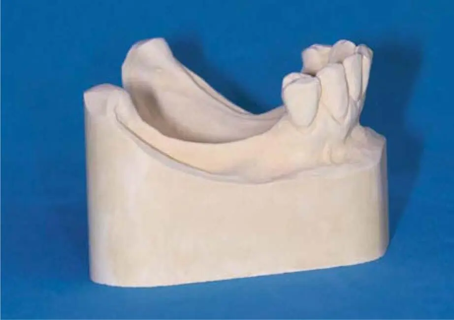
Fig 2-22aBone atrophy in the bilateral free-end situation.
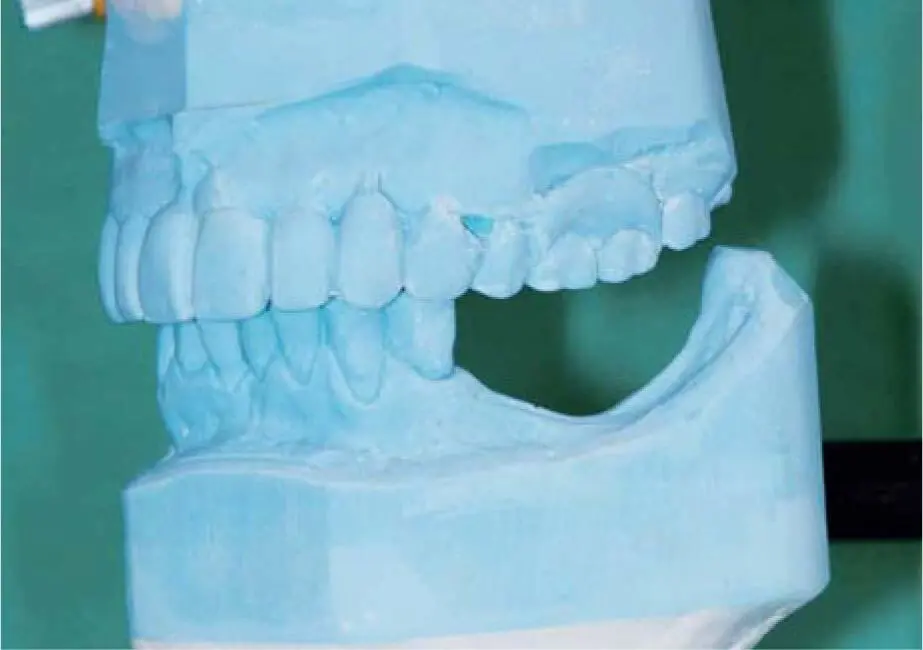
Fig 2-22bThe scope of the vertical defect is clearly visible in the articulator.
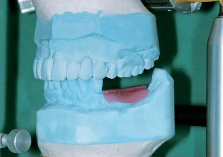
Fig 2-22cSimulation of the necessary grafting volume in wax.

Fig 2-22dWax-up of correct tooth length in the left mandible.
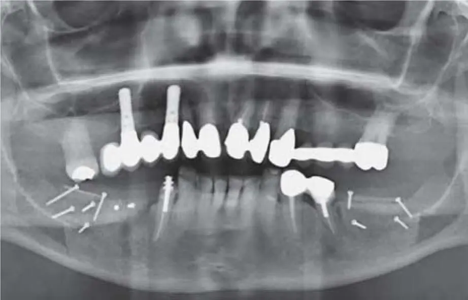
Fig 2-22ePanoramic radiograph documenting a bilateral vertical bone augmentation in the right and left posterior mandible. The right bone graft is very close to the antagonist elongated second molar.
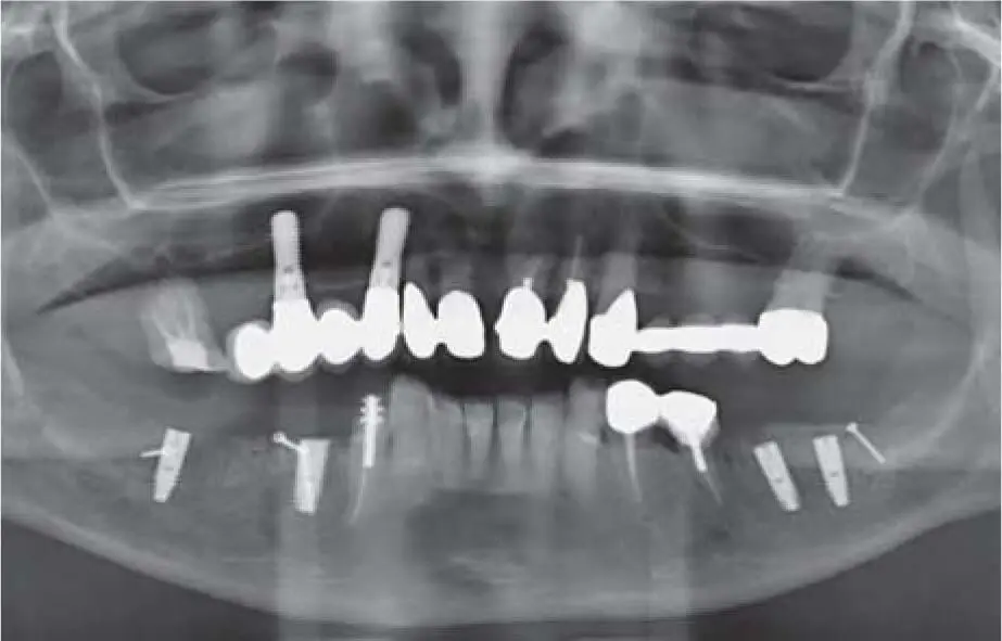
Fig 2-22fPanoramic radiograph documenting the implant insertion after reducing the graft volume. An endodontic treatment was also performed on the antagonist elongated tooth after reducing its volume.
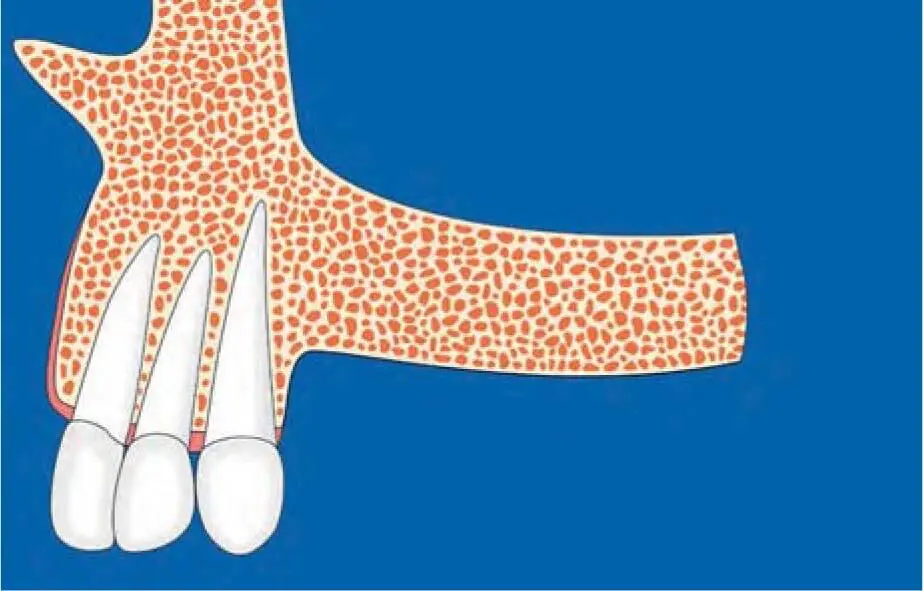
Fig 2-23aSchematic illustration of a class C vertical bone defect according to Chiapasco et al. 18
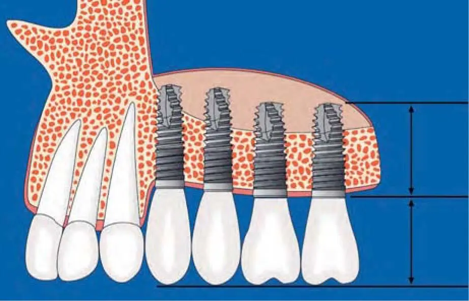
Fig 2-23bSimultaneous implantation with sinus floor elevation in class C, with the consequence of an unfavorable crown–implant ratio.
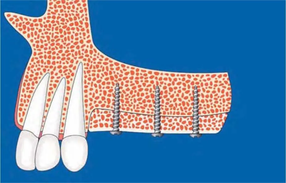
Fig 2-23cSchematic illustration of vertical grafting in a class E defect to create a physiologic course for the alveolar ridge.
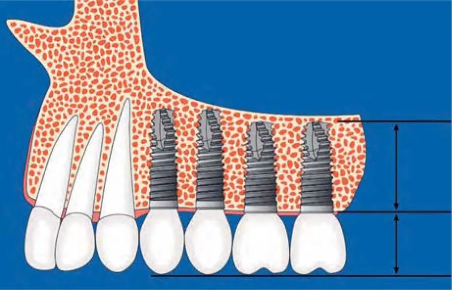
Fig 2-23dSchematic illustration of implants placed after vertical reconstruction with a balanced crown–implant ratio.
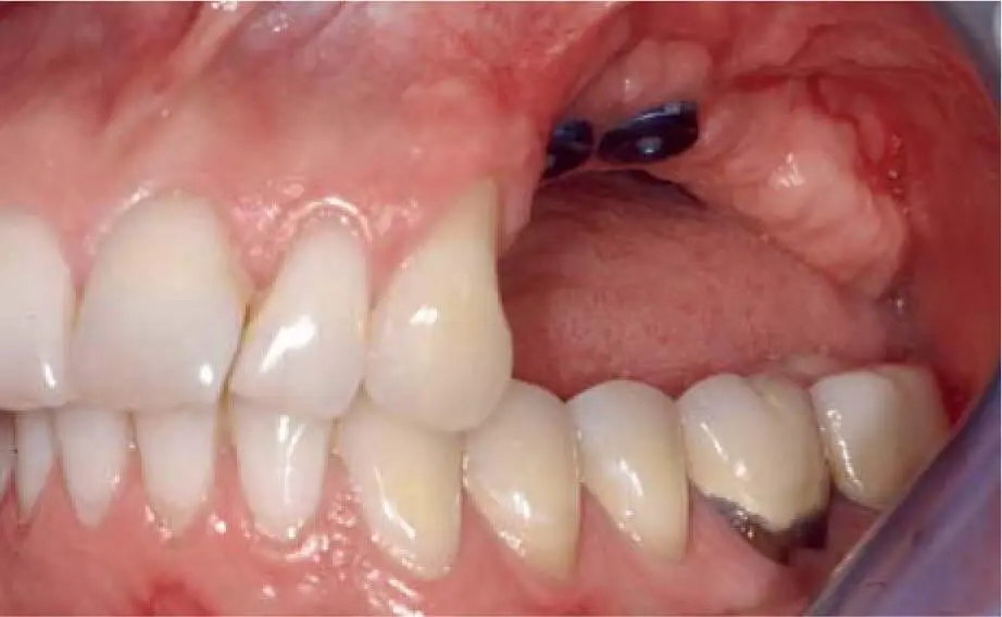
Fig 2-23eIncorrect implant insertion in the posterior left maxilla.
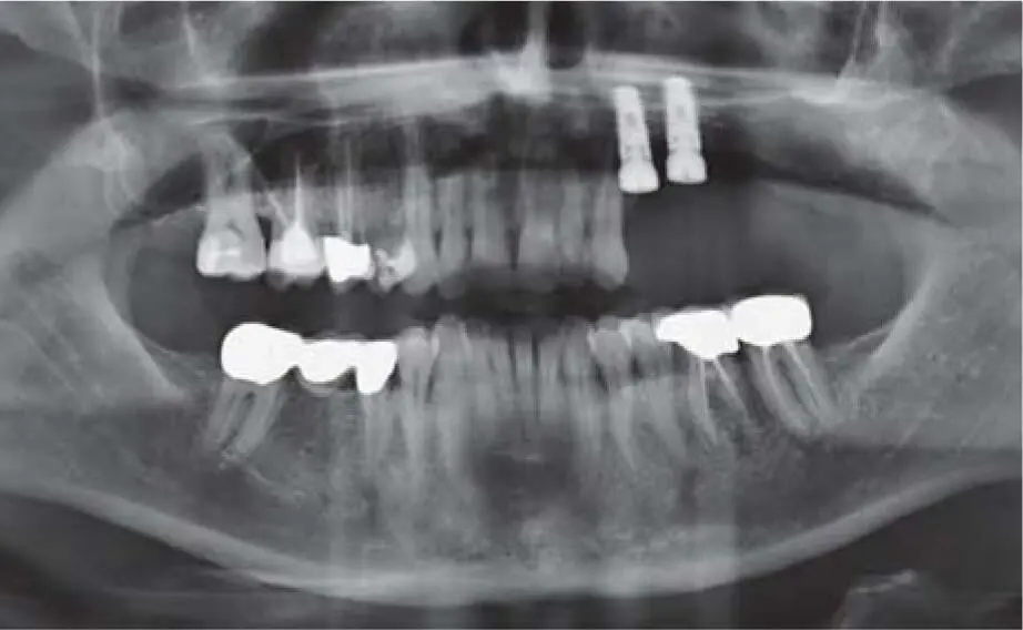
Fig 2-23fPanoramic radiograph documenting the extreme apical position of the implants.
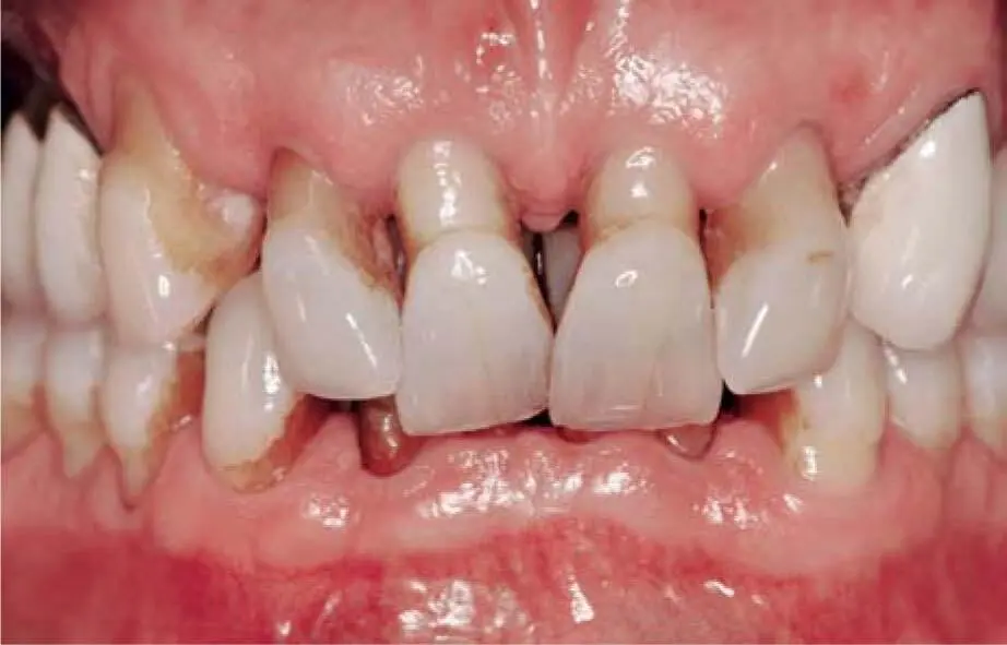
Fig 2-24aClinical situation before treatment: closed bite and severe periodontal disease.
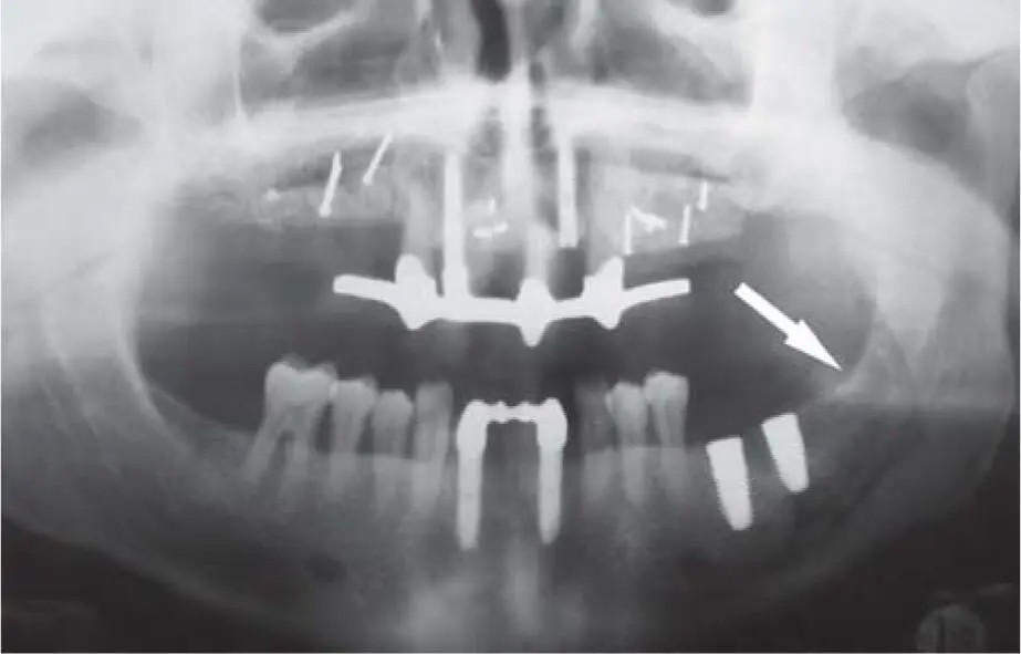
Fig 2-24bAfter periodontal treatment involving the extraction of the hopeless teeth and a fixed temporary restoration, vertical bone grafting in different areas of the maxilla with bone blocks from the mandibular left retromolar area. The external oblique was so pronounced that only one intraoral donor site was sufficient for the entire maxillary reconstruction.
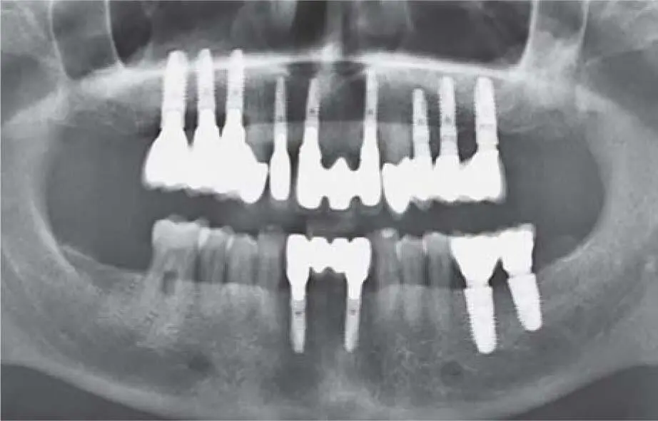
Fig 2-24cControl radiograph 7 years postoperatively with a stable peri-implant bone level.
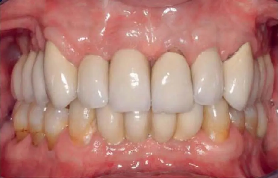
Fig 2-24dClinical situation 7 years postoperatively.
Читать дальшеИнтервал:
Закладка:
Похожие книги на «Bone and Soft Tissue Augmentation in Implantology»
Представляем Вашему вниманию похожие книги на «Bone and Soft Tissue Augmentation in Implantology» списком для выбора. Мы отобрали схожую по названию и смыслу литературу в надежде предоставить читателям больше вариантов отыскать новые, интересные, ещё непрочитанные произведения.


Обсуждение, отзывы о книге «Bone and Soft Tissue Augmentation in Implantology» и просто собственные мнения читателей. Оставьте ваши комментарии, напишите, что Вы думаете о произведении, его смысле или главных героях. Укажите что конкретно понравилось, а что нет, и почему Вы так считаете.