Bone and Soft Tissue Augmentation in Implantology
Здесь есть возможность читать онлайн «Bone and Soft Tissue Augmentation in Implantology» — ознакомительный отрывок электронной книги совершенно бесплатно, а после прочтения отрывка купить полную версию. В некоторых случаях можно слушать аудио, скачать через торрент в формате fb2 и присутствует краткое содержание. Жанр: unrecognised, на английском языке. Описание произведения, (предисловие) а так же отзывы посетителей доступны на портале библиотеки ЛибКат.
- Название:Bone and Soft Tissue Augmentation in Implantology
- Автор:
- Жанр:
- Год:неизвестен
- ISBN:нет данных
- Рейтинг книги:4 / 5. Голосов: 1
-
Избранное:Добавить в избранное
- Отзывы:
-
Ваша оценка:
Bone and Soft Tissue Augmentation in Implantology: краткое содержание, описание и аннотация
Предлагаем к чтению аннотацию, описание, краткое содержание или предисловие (зависит от того, что написал сам автор книги «Bone and Soft Tissue Augmentation in Implantology»). Если вы не нашли необходимую информацию о книге — напишите в комментариях, мы постараемся отыскать её.
R. Gruber, Th. Hanser, Ph. Keeve, Ch. Khoury, J. Neugebauer, J. E. Zöller
Bone and Soft Tissue Augmentation in Implantology addresses useful methods of bone grafting procedures in implant treatment based on current biologic principles and constitutes a unique reference in this field. The book describes, in over 760 pages and 2837 mostly color illustrations, the different possibilities available to augment the bone volume in width and height. The information presented includes not only the underlying scientific concepts of the different augmentation techniques with autogenous bone, but also the associated soft tissue management, from safe approaches to different possibilities for soft tissue augmentation and papilla reconstruction techniques.
The book provides surgeons with a basic understanding of the biologic response to bone grafting procedures. Experienced implantologists will benefit from the in-depth background information, details of high-level surgical techniques, and scientific results, which will enable them to optimize their surgical procedures. Each chapter offers a wealth of information on the specific topic covered, with much attention given to the scientific concepts behind each one. Extensive case reports with step-by-step documentation allow readers to gain an impression of what is possible today in the 3D reconstruction procedures of the alveolar crest. Important criteria for success are presented as well as possible complications and their treatment.
Bone and Soft Tissue Augmentation in Implantology is a must-read for every implantologist, oral and maxillofacial surgeon, and any dentist interested in surgery.
Bone and Soft Tissue Augmentation in Implantology — читать онлайн ознакомительный отрывок
Ниже представлен текст книги, разбитый по страницам. Система сохранения места последней прочитанной страницы, позволяет с удобством читать онлайн бесплатно книгу «Bone and Soft Tissue Augmentation in Implantology», без необходимости каждый раз заново искать на чём Вы остановились. Поставьте закладку, и сможете в любой момент перейти на страницу, на которой закончили чтение.
Интервал:
Закладка:
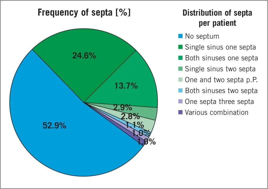
Fig 2-19dDistribution of frequency of septa in the maxillary sinus per patient.
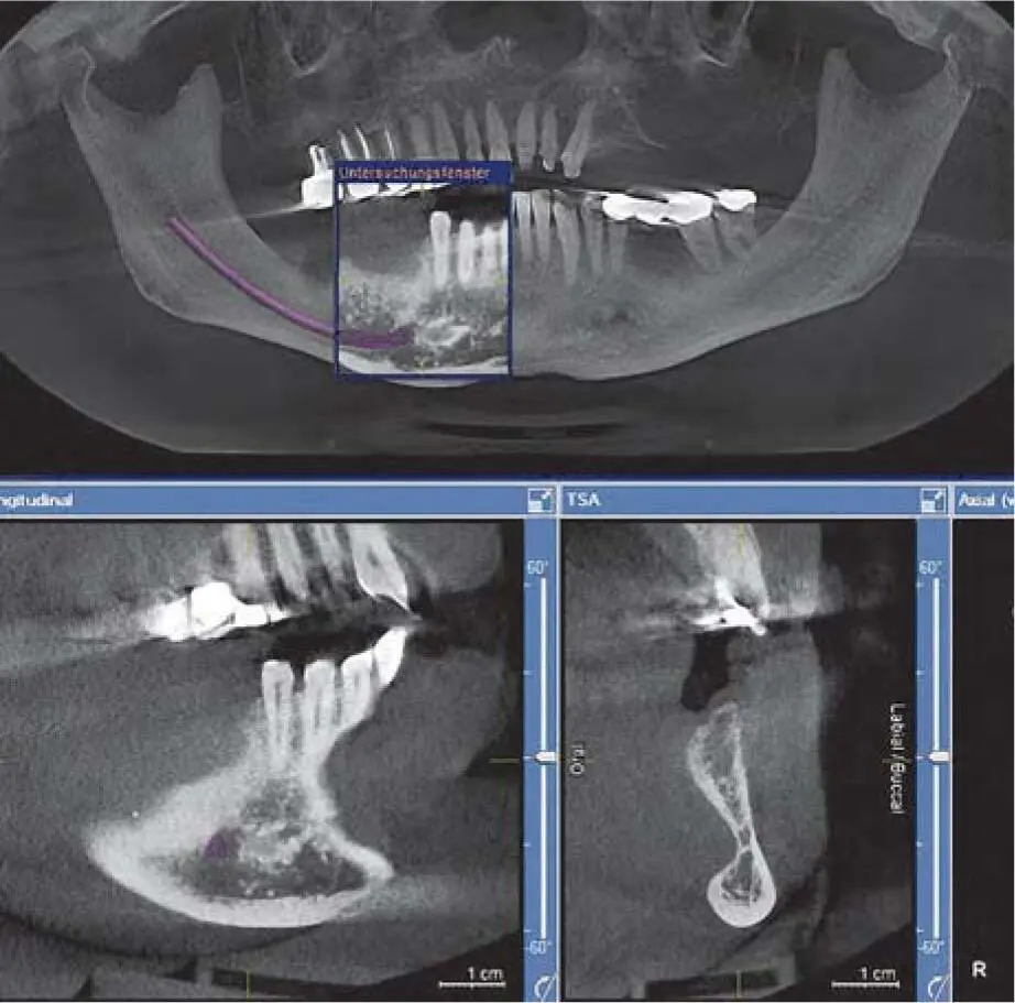
Fig 2-19eCBCT detection of an important undercut in the mandibular premolar area.
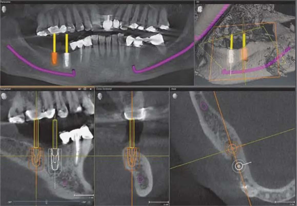
Fig 2-19fCBCT of area of undercut in the mandibular molar area for the purposes of 3D planning.
2.4.4.2 Indications for 3D diagnostics
When using any radiologic technique, the benefit and risk of ionizing exposure should be calculated. 1,2Therefore, depending on the type of device, whether spiral CT or CBCT, with or without image intensifier technology, the indication can be limited or passed on. 21,72Before a CBCT scan is taken, a clinical examination and evaluation of the available radiograph images is necessary to ensure that the patient will have a diagnostic or therapeutic benefit greater than the potential hazard from radiation exposure. It should also be considered that the risk of radiation hazard in children is three times greater than in adults, although most patients requiring bone graft surgery are over 50 years of age, at which age the relative risk decreases to 30% to 50% compared with a 30-year-old patient. 23
It has been shown that in more than 40% of patients, a mostly asymptomatic change in the maxillary sinus mucosa (e.g. swelling, mucocele) is present. The prevalence of at least one septum is 46.8% per patient 67( Fig 2-19d). Three-dimensional diagnostics can be used to explore areas of undercut on the lingual side of the mandible that are not detectable on a panoramic radiograph ( Fig 2-19e). This avoids lingual perforations during implant bed preparation ( Fig 2-19f).
The evaluation of the retromolar triangle has shown that the thickness of the buccal cortical structure is approximately 3 to 4 mm. The location of the mandibular canal in moderately atrophied jaws is usually > 10 mm under the alveolar crest, so that sufficient bone is normally available for the harvesting procedure. However, it was also found that in 10.2% of cases, the nerve was very superficial to the buccal site, with very close contact with the vestibular cortical bone plate, which requires a very careful approach for bone harvesting. 63
Simulation of the prosthetic setup
Depending on the number of remaining teeth and the software used to accomplish implant planning, the prosthetic setup for implant placement can be performed using a radiologic template, or digitally by matching computer-assisted design/computer-assisted manufacturing (CAD/CAM) data. 68With this digital technique, it is possible to find the ideal position and direction of the implants in a simple way in relation to the ideal position of the implants and to fix them in a surgical guide ( Fig 2-20ato m). If necessary, the bone can be reconstructed in different directions according to the surgical guide.
Construction parameters for a 3D radiographic template
If the implant insertion is planned simultaneously with the grafting procedure and with the software utilizing a reference plate, it is recommended to convert the prosthetic setup into a radiologic template. To obtain information about the planned crown position, it is necessary to implement radiopaque structures in the template. This can be done by gutta-percha pins, which represent the ideal axes of the implant abutments. With this approach, no specific software is necessary for the implant planning. After the analysis of the position and the axis of the future implant, the dental technician will replace the corrected position of the guttapercha pin through a hole for the pilot bur.
More orientation is given if the crowns are converted into barium sulfate resin. For the best possible information, the crowns should rest on the soft tissue with the natural anatomical profile. This allows the determination of the thickness of the soft tissue during the scan. For the exact determination of the position, it is important that the crowns are separated at the mucosal level and partly reduced to the diameter of the putative root ( Fig 2-21). If a mucosa-supported surgical guide has to be used, a different doping with barium sulphate is recommended for the template base and the setup teeth in order to visualize the soft tissue, so that the soft tissue contours and the prosthetic orientation are reproduced in the image. Depending on the software, a double scan of the patient with the prosthetic setup and the setup alone is sometimes recommended. 27Both scans are matched to give the best visualization of the prosthetic setup for the orientation of the implants and the amount of grafting.
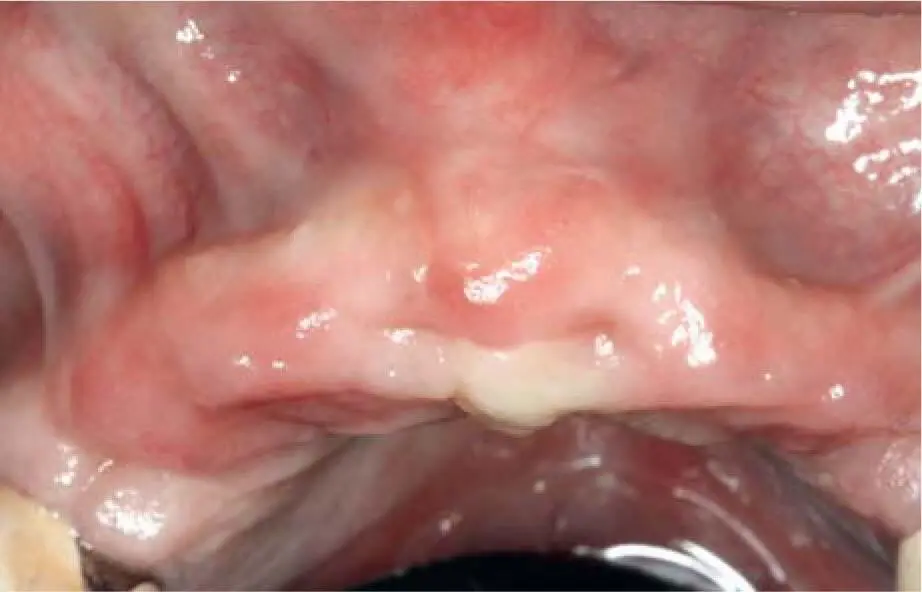
Fig 2-20aSignificant atrophy of the anterior maxilla after several infections.
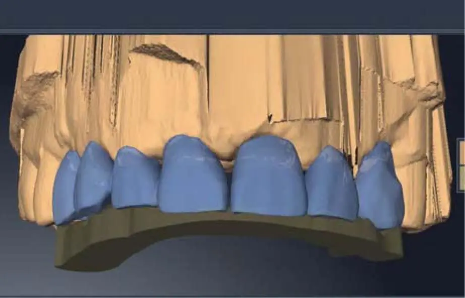
Fig 2-20bImporting the digital wax-up into the implant planning software (Sicat Implant 2.0; Sicat, Bonn, Germany).
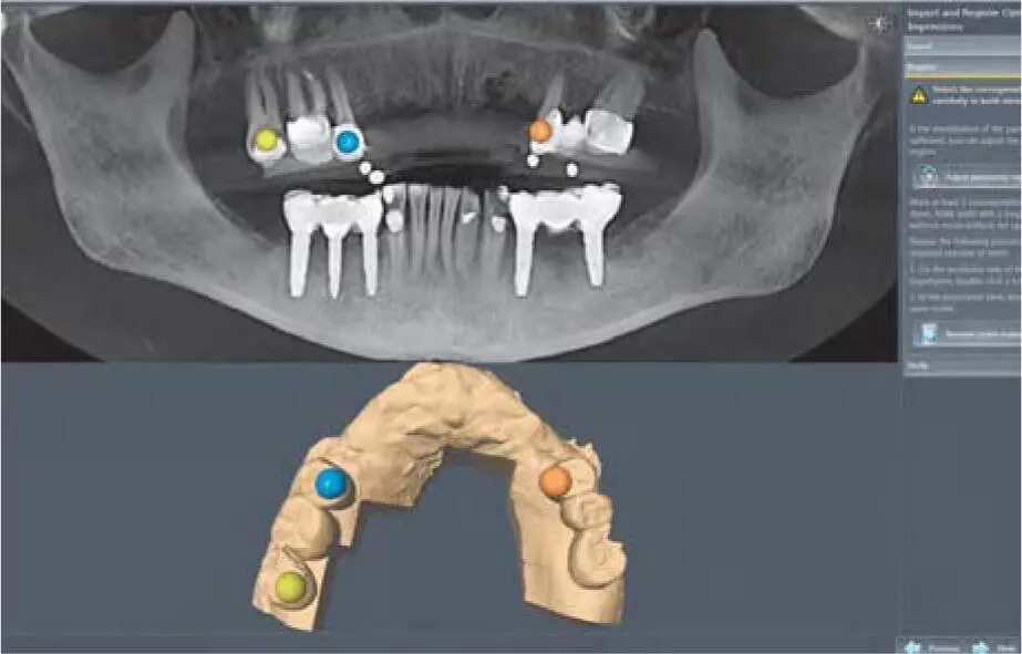
Fig 2-20cSelection of marker point on residual teeth of the digital model and corresponding structure within the CBCT to match the data sets.
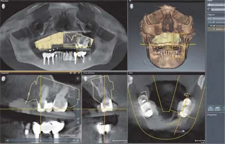
Fig 2-20dControl of the contours of the optical model over the crowns of the corresponding teeth in the matched data set.
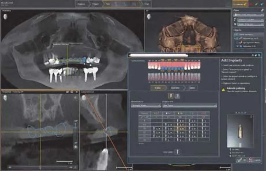
Fig 2-20eSelection of implant size, abutments, and sleeves for the detailed plan for simultaneous implant placement and lateral grafting.
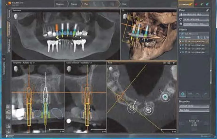
Fig 2-20fDigital planning with volumetric samples of implant bodies and drill sleeves in the position of the planned implants in the anterior maxilla.
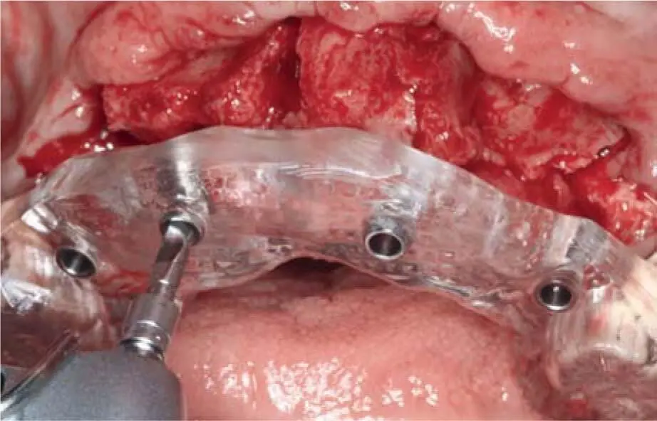
Fig 2-20gGuided pilot drilling with the 3D surgical guide (Optiguide; Sicat).
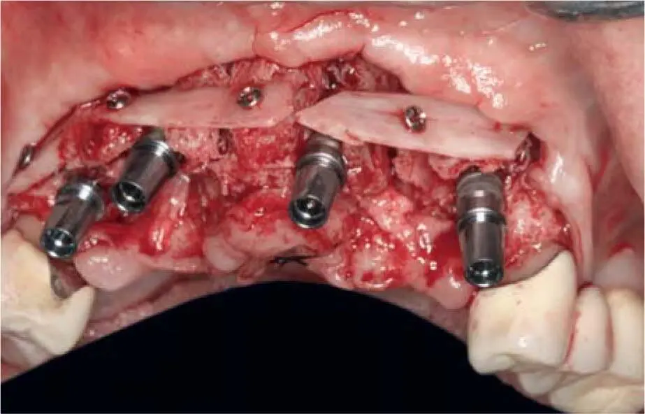
Fig 2-20hImplant insertion (XiVE; Dentsply Sirona) with simultaneous lateral block grafting.
Читать дальшеИнтервал:
Закладка:
Похожие книги на «Bone and Soft Tissue Augmentation in Implantology»
Представляем Вашему вниманию похожие книги на «Bone and Soft Tissue Augmentation in Implantology» списком для выбора. Мы отобрали схожую по названию и смыслу литературу в надежде предоставить читателям больше вариантов отыскать новые, интересные, ещё непрочитанные произведения.


Обсуждение, отзывы о книге «Bone and Soft Tissue Augmentation in Implantology» и просто собственные мнения читателей. Оставьте ваши комментарии, напишите, что Вы думаете о произведении, его смысле или главных героях. Укажите что конкретно понравилось, а что нет, и почему Вы так считаете.