Bone and Soft Tissue Augmentation in Implantology
Здесь есть возможность читать онлайн «Bone and Soft Tissue Augmentation in Implantology» — ознакомительный отрывок электронной книги совершенно бесплатно, а после прочтения отрывка купить полную версию. В некоторых случаях можно слушать аудио, скачать через торрент в формате fb2 и присутствует краткое содержание. Жанр: unrecognised, на английском языке. Описание произведения, (предисловие) а так же отзывы посетителей доступны на портале библиотеки ЛибКат.
- Название:Bone and Soft Tissue Augmentation in Implantology
- Автор:
- Жанр:
- Год:неизвестен
- ISBN:нет данных
- Рейтинг книги:4 / 5. Голосов: 1
-
Избранное:Добавить в избранное
- Отзывы:
-
Ваша оценка:
Bone and Soft Tissue Augmentation in Implantology: краткое содержание, описание и аннотация
Предлагаем к чтению аннотацию, описание, краткое содержание или предисловие (зависит от того, что написал сам автор книги «Bone and Soft Tissue Augmentation in Implantology»). Если вы не нашли необходимую информацию о книге — напишите в комментариях, мы постараемся отыскать её.
R. Gruber, Th. Hanser, Ph. Keeve, Ch. Khoury, J. Neugebauer, J. E. Zöller
Bone and Soft Tissue Augmentation in Implantology addresses useful methods of bone grafting procedures in implant treatment based on current biologic principles and constitutes a unique reference in this field. The book describes, in over 760 pages and 2837 mostly color illustrations, the different possibilities available to augment the bone volume in width and height. The information presented includes not only the underlying scientific concepts of the different augmentation techniques with autogenous bone, but also the associated soft tissue management, from safe approaches to different possibilities for soft tissue augmentation and papilla reconstruction techniques.
The book provides surgeons with a basic understanding of the biologic response to bone grafting procedures. Experienced implantologists will benefit from the in-depth background information, details of high-level surgical techniques, and scientific results, which will enable them to optimize their surgical procedures. Each chapter offers a wealth of information on the specific topic covered, with much attention given to the scientific concepts behind each one. Extensive case reports with step-by-step documentation allow readers to gain an impression of what is possible today in the 3D reconstruction procedures of the alveolar crest. Important criteria for success are presented as well as possible complications and their treatment.
Bone and Soft Tissue Augmentation in Implantology is a must-read for every implantologist, oral and maxillofacial surgeon, and any dentist interested in surgery.
Bone and Soft Tissue Augmentation in Implantology — читать онлайн ознакомительный отрывок
Ниже представлен текст книги, разбитый по страницам. Система сохранения места последней прочитанной страницы, позволяет с удобством читать онлайн бесплатно книгу «Bone and Soft Tissue Augmentation in Implantology», без необходимости каждый раз заново искать на чём Вы остановились. Поставьте закладку, и сможете в любой момент перейти на страницу, на которой закончили чтение.
Интервал:
Закладка:
2.5.1.3 Multiple horizontal and vertical bony defects
With the use of the SBB technique, it is possible to harvest sufficient bone, allowing for the grafting of multiple horizontal and vertical bony defects. Since with this technique every harvested bone block is split into two or three blocks, one can multiply the number of blocks available for the grafting procedure. In some situations, with a well-pronounced external oblique line, and when following the SBB technique, block harvesting from one retromolar area can be sufficient to graft large areas ( Fig 2-24ato d). If there is a need for more bone blocks, the harvesting procedure can be performed from both the right and left mandibular retromolar areas ( Fig 2-25ato e). For a complete oral rehabilitation with multiple bone grafting in the maxilla and mandible, bone grafts can also be harvested from the chin and molar areas in addition to the mandibular retromolar areas ( Fig 2-26ato h).
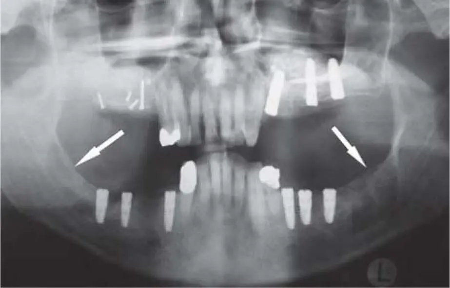
Fig 2-25aTwo donor sites in the retromolar area of the right and left mandible for two augmentation procedures in the right and left maxilla.
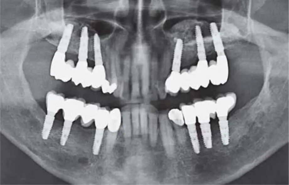
Fig 2-25bRadiographic control 14 years postoperatively.
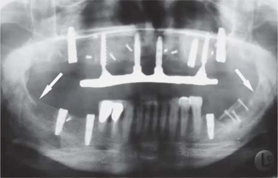
Fig 2-25cTwo donor sites in the retromolar area of the right and left mandible for multiple augmentation areas in the maxilla and mandible.
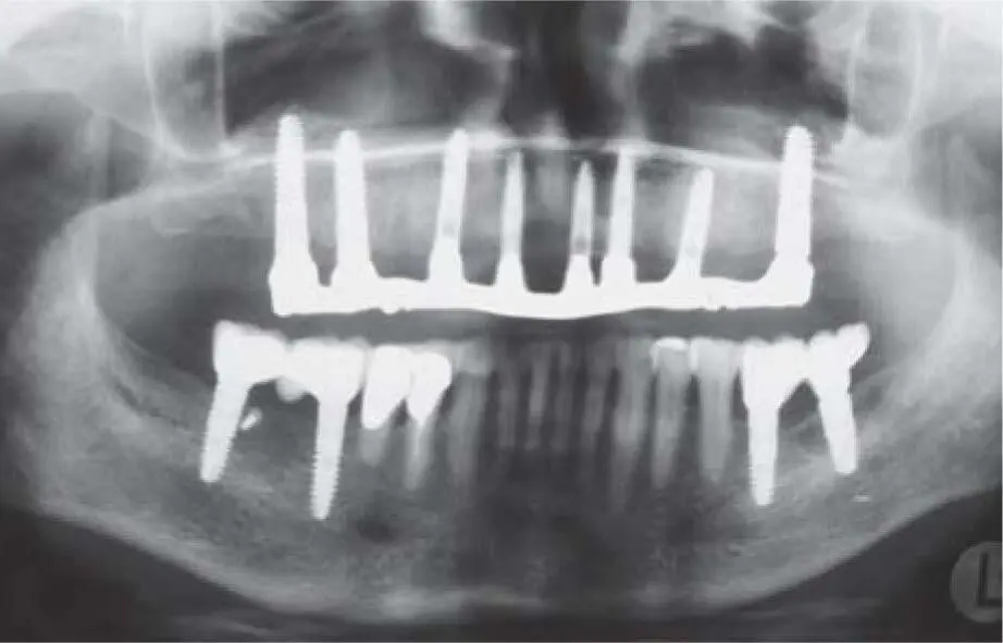
Fig 2-25dRecall radiograph 2 years postoperatively with implants that show good osseointegration.
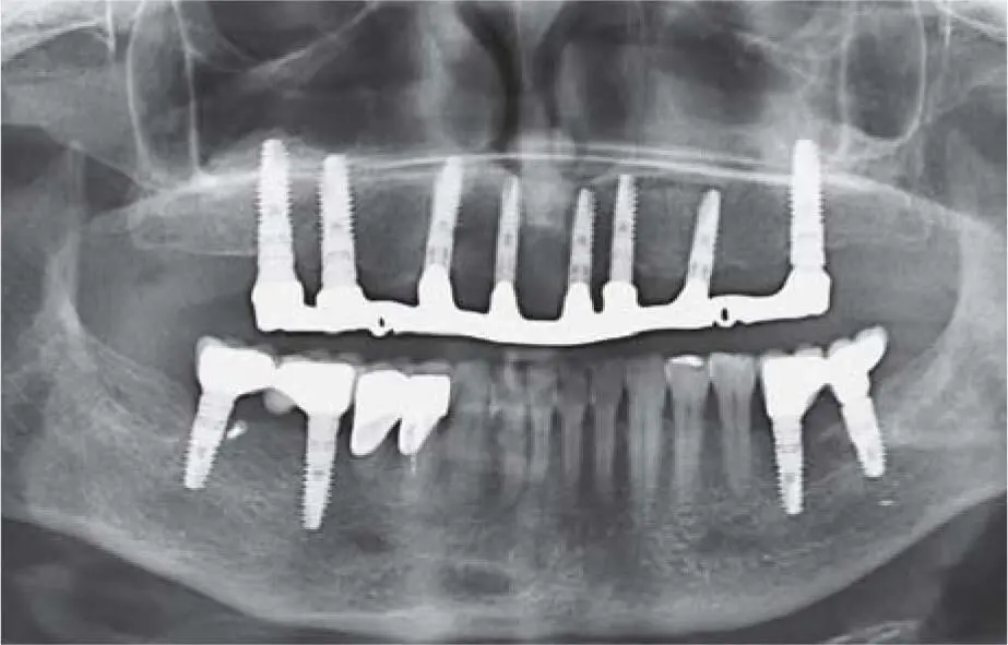
Fig 2-25eControl radiograph 12 years postoperatively.
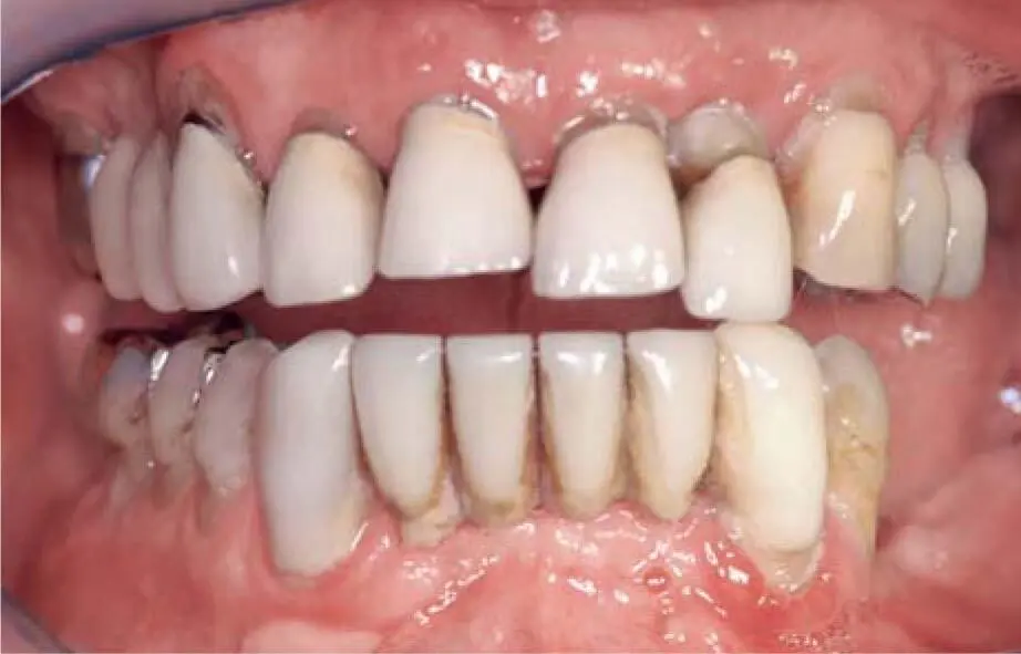
Fig 2-26aSevere periodontal disease with many mobile teeth.
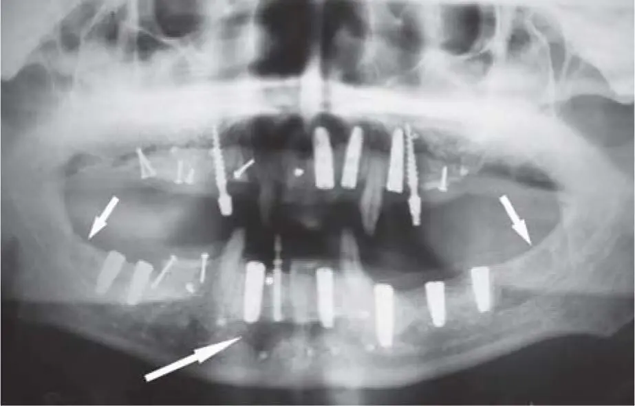
Fig 2-26bMultiple bone grafting in various areas of the maxilla and mandible after periodontal rehabilitation with extraction of the hopeless teeth and a fixed temporary restoration using provisional implants. The bone grafts were harvested from the retromolar area on the right and left mandible as well as from the chin area.
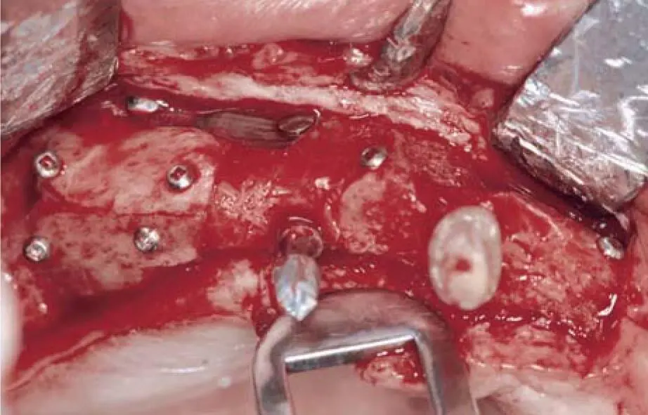
Fig 2-26cClinical situation of the vertical grafted bone in the right maxilla (3D reconstruction) 4 months postoperatively.
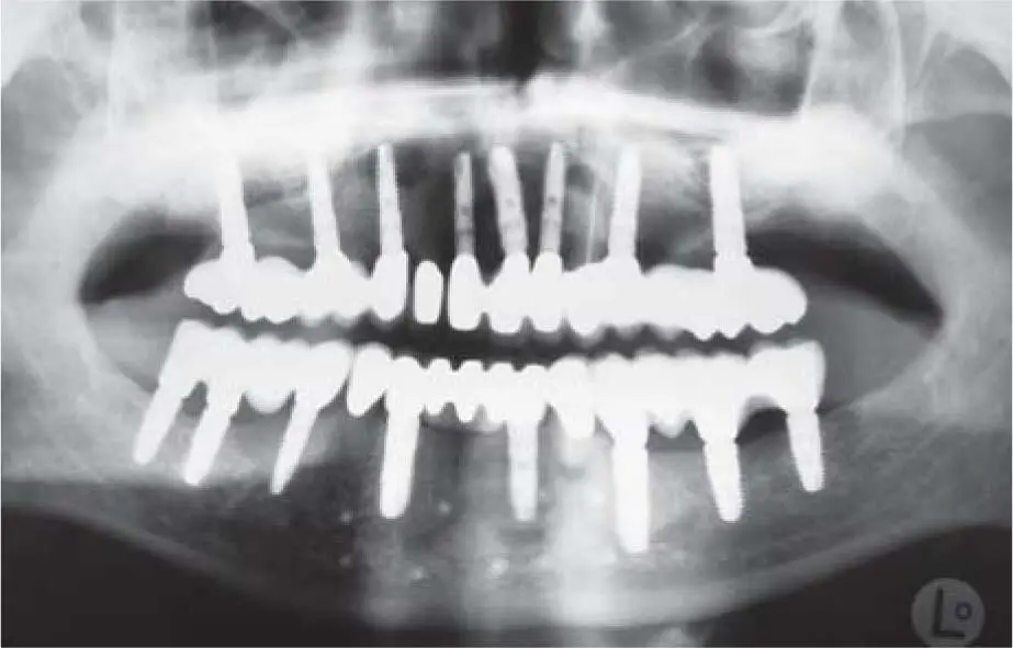
Fig 2-26dControl radiograph 7 years postoperatively.
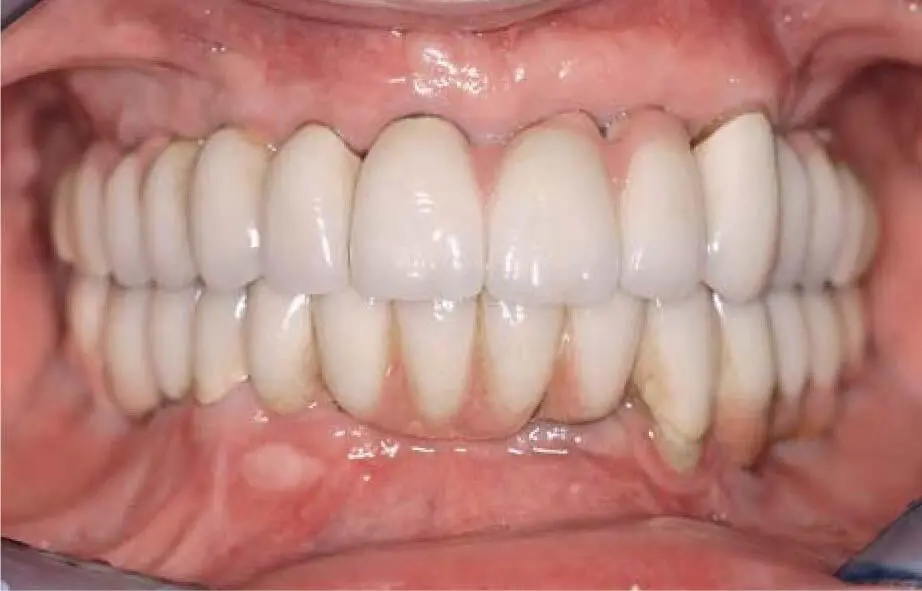
Fig 2-26eClinical situation 7 years postoperatively.
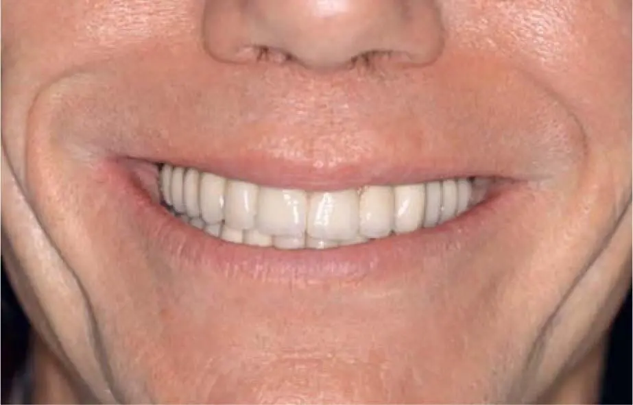
Fig 2-26fThe long upper lip covers the pink ceramic and the cleaning canals.

Fig 2-26gClinical appearance 17 years postoperatively with poor oral hygiene: patients must stay in a regular recall program to help them to maintain good oral hygiene.
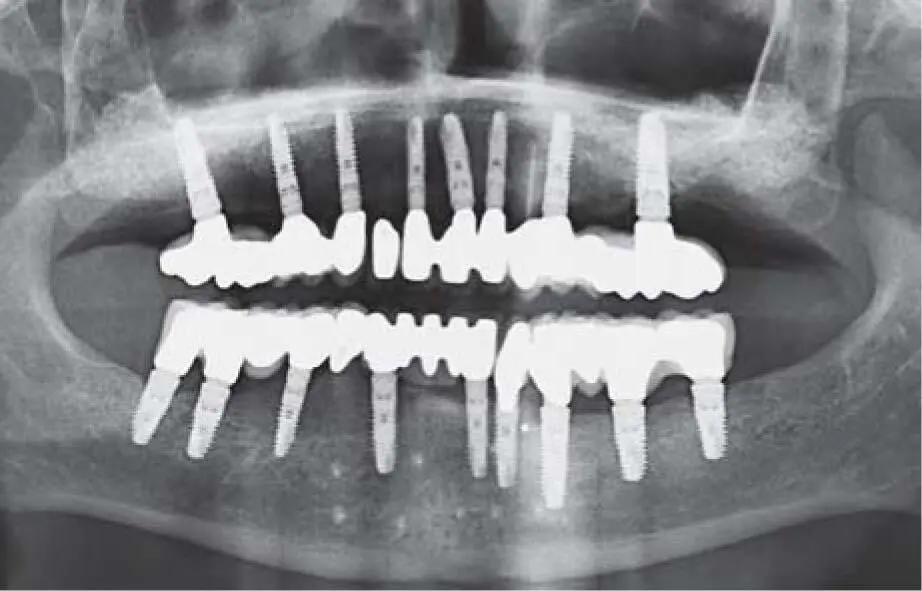
Fig 2-26hControl radiograph 17 years postoperatively.
Only in exceptional cases, such as extensive vertical bone defects with severely scarred soft tissue and a history of uncontrollable smoking, is it advisable to use a cortico-spongy iliac crest graft due to its high regeneration potential. In addition, if there is insufficient bone in the mandible for harvesting in the case of extensive defects, a sufficient donor area is always available in the iliac crest to enable the reconstruction of difficult defect geometries ( Fig 2-27ato f).
The selection of the appropriate method is thus determined by the various parameters and depends on the individual patient findings.
2.6 Conclusion
Targeted implant therapy requires a precise survey of the treatment-relevant, individual patient findings, especially when severe bone loss is present. Due to the exact knowledge of the anamnesis and the anatomical structures, patients can be provided with a sufficient alveolar crest for the intended implant placement to achieve an appropriate prosthetic restoration. Different grafting procedures are available, their use depending on the soft tissue situation, the defect configuration, and the desires of the patient. In the atrophied edentulous maxilla, the surgical part of hard and soft tissue augmentation can be less invasive in case of a removable definitive restoration compared with a fixed restoration. In this situation, the goal of the bone grafting procedure is to provide enough bone volume for the insertion of implants. In case of a planned fixed restoration, there are additional goals apart from the aim of inserting the implant, which include having a significant volume of bone and soft tissue to guarantee an esthetic restoration with definitive crowns of a normal length and adequate soft tissue volume, margins, and papillae.
It is important to pay special attention in cases of reconstructions in the esthetic area. In some situations, there is a need for preoperative orthodontic treatment to create sufficient space for a symmetric restoration without injury to the neighboring teeth, something which is not always easy. In such a situation, where the orthodontic treatment was not able to fulfill the expected result, alternatives need to be discussed with the patient ( Fig 2-28ato k).
Precise preoperative diagnostics allow for the exact positioning of implants even under difficult anatomical conditions. Especially in the situation with looseness of the vertical dimension, e.g. in the case of hypodontia, the planning as well as the surgical guide must be the result of teamwork between the surgeon, prosthodontist, and dental technician ( Fig 2-29ato y). In routine cases, the use of tooth-borne orientation guides on the basis of a 2D radiologic diagnosis is sufficient.
Читать дальшеИнтервал:
Закладка:
Похожие книги на «Bone and Soft Tissue Augmentation in Implantology»
Представляем Вашему вниманию похожие книги на «Bone and Soft Tissue Augmentation in Implantology» списком для выбора. Мы отобрали схожую по названию и смыслу литературу в надежде предоставить читателям больше вариантов отыскать новые, интересные, ещё непрочитанные произведения.


Обсуждение, отзывы о книге «Bone and Soft Tissue Augmentation in Implantology» и просто собственные мнения читателей. Оставьте ваши комментарии, напишите, что Вы думаете о произведении, его смысле или главных героях. Укажите что конкретно понравилось, а что нет, и почему Вы так считаете.