Bone and Soft Tissue Augmentation in Implantology
Здесь есть возможность читать онлайн «Bone and Soft Tissue Augmentation in Implantology» — ознакомительный отрывок электронной книги совершенно бесплатно, а после прочтения отрывка купить полную версию. В некоторых случаях можно слушать аудио, скачать через торрент в формате fb2 и присутствует краткое содержание. Жанр: unrecognised, на английском языке. Описание произведения, (предисловие) а так же отзывы посетителей доступны на портале библиотеки ЛибКат.
- Название:Bone and Soft Tissue Augmentation in Implantology
- Автор:
- Жанр:
- Год:неизвестен
- ISBN:нет данных
- Рейтинг книги:4 / 5. Голосов: 1
-
Избранное:Добавить в избранное
- Отзывы:
-
Ваша оценка:
Bone and Soft Tissue Augmentation in Implantology: краткое содержание, описание и аннотация
Предлагаем к чтению аннотацию, описание, краткое содержание или предисловие (зависит от того, что написал сам автор книги «Bone and Soft Tissue Augmentation in Implantology»). Если вы не нашли необходимую информацию о книге — напишите в комментариях, мы постараемся отыскать её.
R. Gruber, Th. Hanser, Ph. Keeve, Ch. Khoury, J. Neugebauer, J. E. Zöller
Bone and Soft Tissue Augmentation in Implantology addresses useful methods of bone grafting procedures in implant treatment based on current biologic principles and constitutes a unique reference in this field. The book describes, in over 760 pages and 2837 mostly color illustrations, the different possibilities available to augment the bone volume in width and height. The information presented includes not only the underlying scientific concepts of the different augmentation techniques with autogenous bone, but also the associated soft tissue management, from safe approaches to different possibilities for soft tissue augmentation and papilla reconstruction techniques.
The book provides surgeons with a basic understanding of the biologic response to bone grafting procedures. Experienced implantologists will benefit from the in-depth background information, details of high-level surgical techniques, and scientific results, which will enable them to optimize their surgical procedures. Each chapter offers a wealth of information on the specific topic covered, with much attention given to the scientific concepts behind each one. Extensive case reports with step-by-step documentation allow readers to gain an impression of what is possible today in the 3D reconstruction procedures of the alveolar crest. Important criteria for success are presented as well as possible complications and their treatment.
Bone and Soft Tissue Augmentation in Implantology is a must-read for every implantologist, oral and maxillofacial surgeon, and any dentist interested in surgery.
Bone and Soft Tissue Augmentation in Implantology — читать онлайн ознакомительный отрывок
Ниже представлен текст книги, разбитый по страницам. Система сохранения места последней прочитанной страницы, позволяет с удобством читать онлайн бесплатно книгу «Bone and Soft Tissue Augmentation in Implantology», без необходимости каждый раз заново искать на чём Вы остановились. Поставьте закладку, и сможете в любой момент перейти на страницу, на которой закончили чтение.
Интервал:
Закладка:
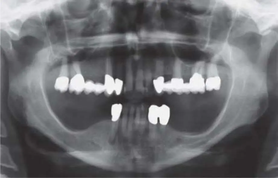
Fig 2-27aPronounced atrophy in the mandibular bilateral free-end situation after long-term restoration with removable dentures.
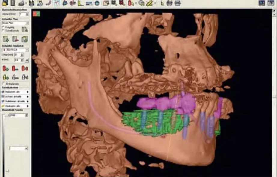
Fig 2-27bThree-dimensional illustration of the necessary autologous graft for implant insertion into the mandible. It was planned to harvest bone grafts from the iliac crest due to the poor bone volume of the mandibular external oblique line.
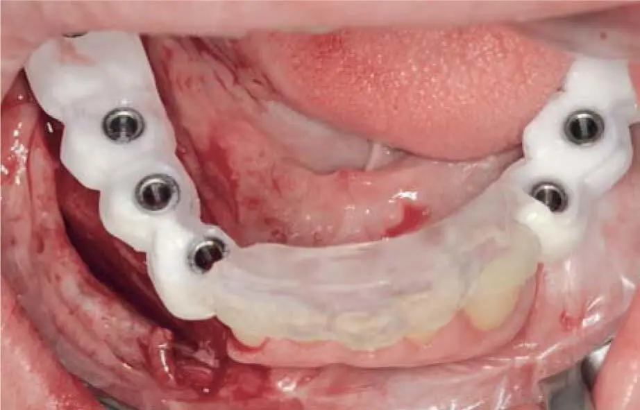
Fig 2-27cA surgical guide based on CBCT data for optimum utilization of the grafted bone 3 months after vertical augmentation using transplants from the iliac crest.
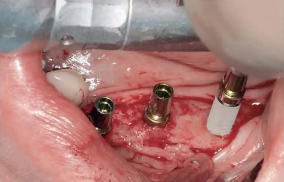
Fig 2-27dInsertion of three implants in the left mandible in a fully regenerated iliac crest graft. Another four implants were inserted in the grafted right mandible.
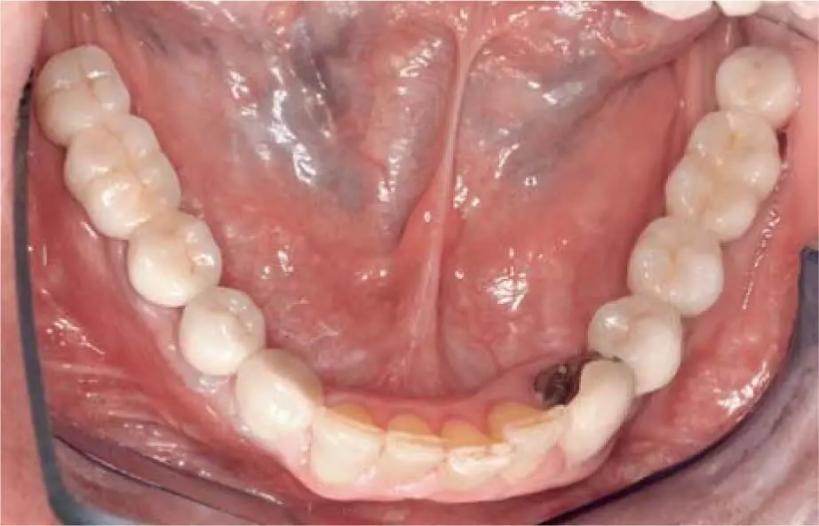
Fig 2-27eDefinitive prosthetic restoration.
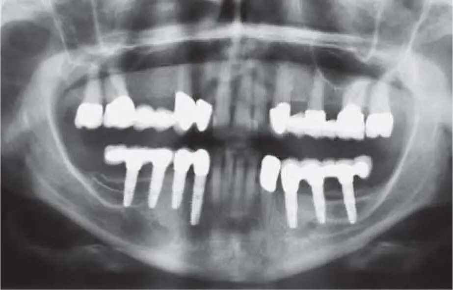
Fig 2-27fControl radiograph 2 years postoperatively.
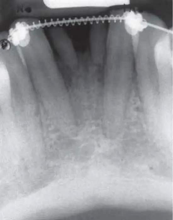
Fig 2-28aPersistent primary teeth in the absence of the right and left mandibular central incisors. Despite many years of orthodontic treatment, it was not possible to obtain sufficient space for two implants.
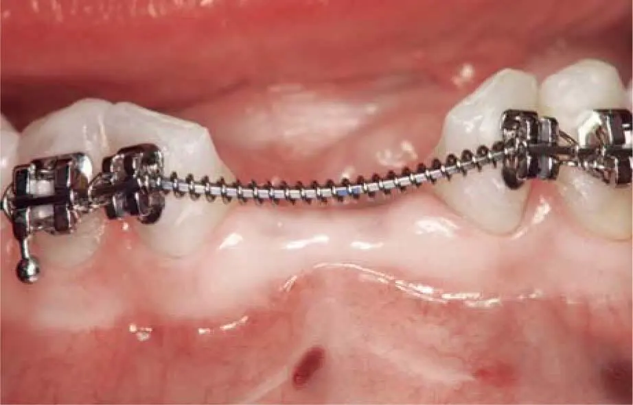
Fig 2-28bClinical situation 4 weeks after extraction of the primary teeth.
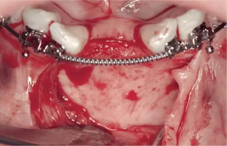
Fig 2-28cSevere atrophy of the alveolar ridge.
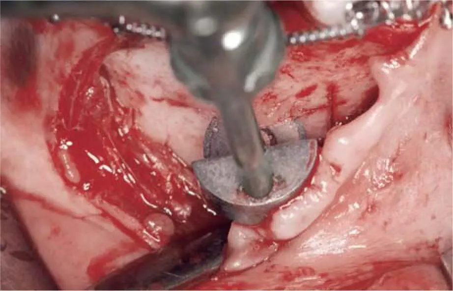
Fig 2-28dA bone block is harvested from the apical region of the chin with a micro saw.
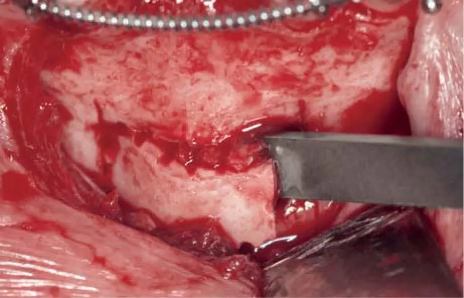
Fig 2-28eRemoval of the bone block with a thin chisel.
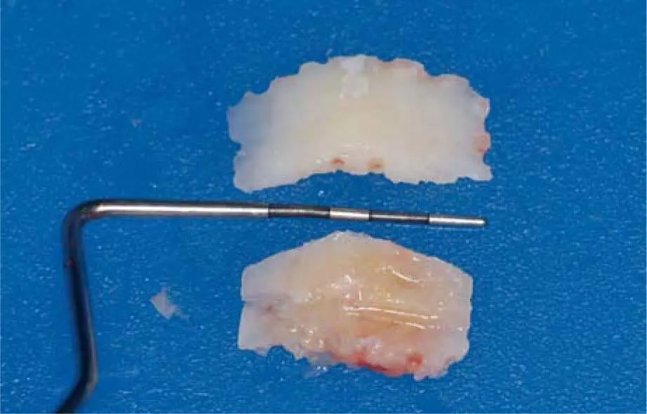
Fig 2-28fLongitudinal split of the bone block into two thin blocks.
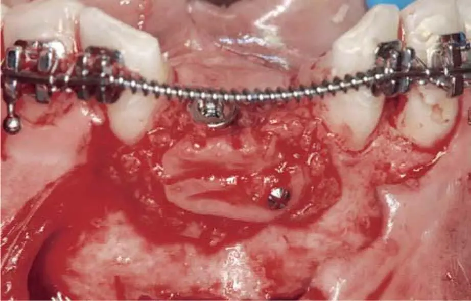
Fig 2-28gInsertion of one implant after bone spreading and grafting of one of the blocks on the vestibular side to support the mobile and thin vestibular bone wall.
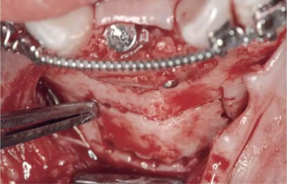
Fig 2-28hReplantation of the second bone block back in its original donor site.
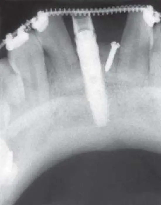
Fig 2-28iControl radiograph 4 months postoperatively.
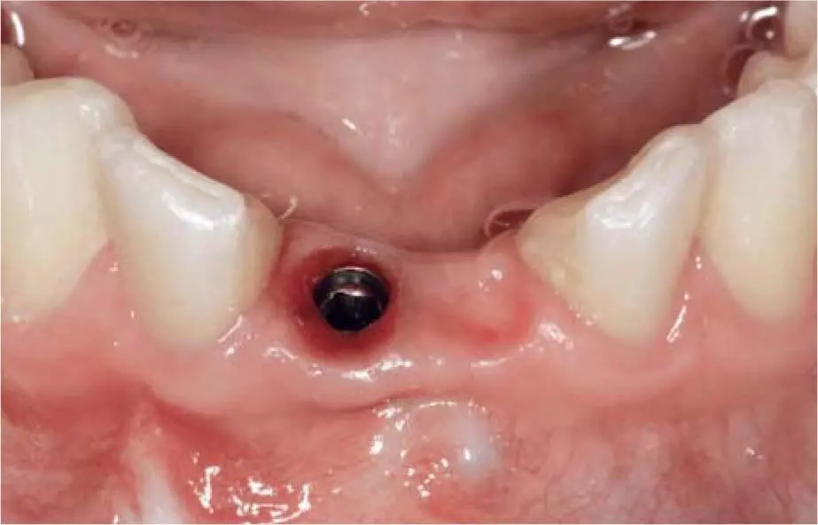
Fig 2-28jClinical situation 6 months postoperatively after conditioning the soft tissue with the temporary restoration.
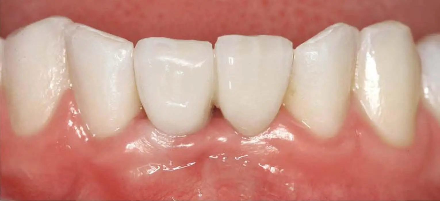
Fig 2-28kClinical situation after definitive prosthetic restoration with a pontic for the left central incisor supported by the implant of the right central incisor.
The use of 3D imaging, especially with prepared radiopaque, prosthetically oriented structures or the superimposition of digitally generated prosthetic proposals, allows for the detailed planning from the anatomical and prosthetic points of view. 66
Finally, a greater planning effort is rewarded by fewer prosthetic and laboratory/technical complications or problems for a precise implant placement in order to achieve an optimal prosthetic restoration after an extensive grafting procedure. Despite the intensive preoperative diagnostics, it is necessary to pay close attention to the recommended protocols in order to avoid complications and failures. 96
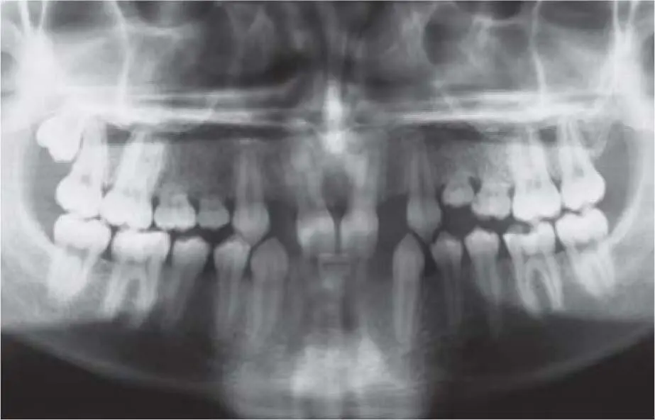
Fig 2-29aMultiple hypodontia in the maxilla and mandible of a 19-year-old female patient.
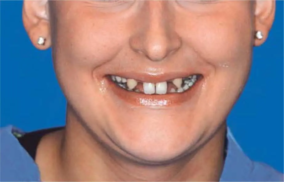
Fig 2-29bClinical situation with a relatively high smile line and a slightly reduced vertical dimension.
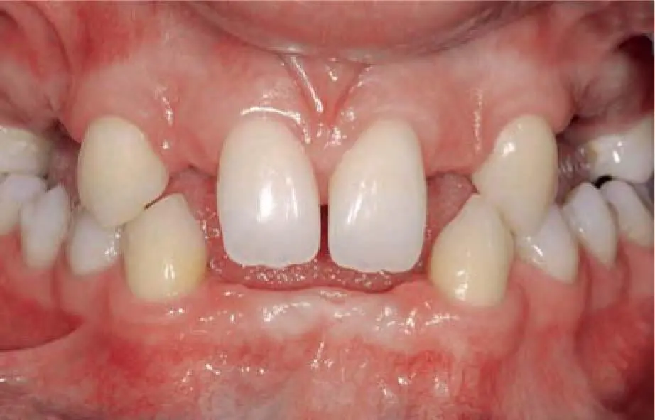
Fig 2-29cIntraoral situation documenting a looseness of the vertical dimension.
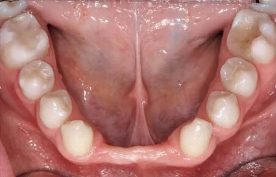
Fig 2-29dSevere alveolar ridge atrophy in the anterior mandible.
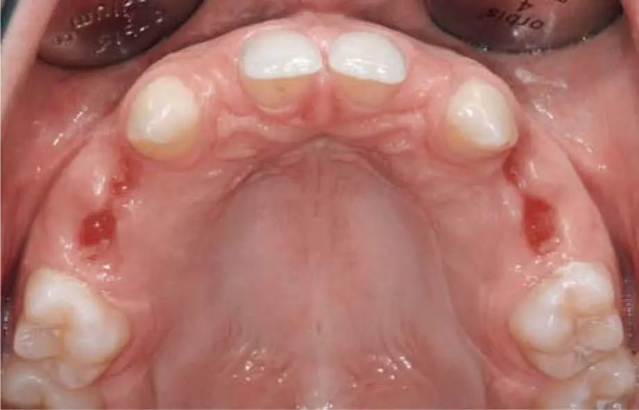
Fig 2-29eClinical situation 3 weeks after extraction of the primary teeth in the maxilla.
Читать дальшеИнтервал:
Закладка:
Похожие книги на «Bone and Soft Tissue Augmentation in Implantology»
Представляем Вашему вниманию похожие книги на «Bone and Soft Tissue Augmentation in Implantology» списком для выбора. Мы отобрали схожую по названию и смыслу литературу в надежде предоставить читателям больше вариантов отыскать новые, интересные, ещё непрочитанные произведения.


Обсуждение, отзывы о книге «Bone and Soft Tissue Augmentation in Implantology» и просто собственные мнения читателей. Оставьте ваши комментарии, напишите, что Вы думаете о произведении, его смысле или главных героях. Укажите что конкретно понравилось, а что нет, и почему Вы так считаете.