Bone and Soft Tissue Augmentation in Implantology
Здесь есть возможность читать онлайн «Bone and Soft Tissue Augmentation in Implantology» — ознакомительный отрывок электронной книги совершенно бесплатно, а после прочтения отрывка купить полную версию. В некоторых случаях можно слушать аудио, скачать через торрент в формате fb2 и присутствует краткое содержание. Жанр: unrecognised, на английском языке. Описание произведения, (предисловие) а так же отзывы посетителей доступны на портале библиотеки ЛибКат.
- Название:Bone and Soft Tissue Augmentation in Implantology
- Автор:
- Жанр:
- Год:неизвестен
- ISBN:нет данных
- Рейтинг книги:4 / 5. Голосов: 1
-
Избранное:Добавить в избранное
- Отзывы:
-
Ваша оценка:
Bone and Soft Tissue Augmentation in Implantology: краткое содержание, описание и аннотация
Предлагаем к чтению аннотацию, описание, краткое содержание или предисловие (зависит от того, что написал сам автор книги «Bone and Soft Tissue Augmentation in Implantology»). Если вы не нашли необходимую информацию о книге — напишите в комментариях, мы постараемся отыскать её.
R. Gruber, Th. Hanser, Ph. Keeve, Ch. Khoury, J. Neugebauer, J. E. Zöller
Bone and Soft Tissue Augmentation in Implantology addresses useful methods of bone grafting procedures in implant treatment based on current biologic principles and constitutes a unique reference in this field. The book describes, in over 760 pages and 2837 mostly color illustrations, the different possibilities available to augment the bone volume in width and height. The information presented includes not only the underlying scientific concepts of the different augmentation techniques with autogenous bone, but also the associated soft tissue management, from safe approaches to different possibilities for soft tissue augmentation and papilla reconstruction techniques.
The book provides surgeons with a basic understanding of the biologic response to bone grafting procedures. Experienced implantologists will benefit from the in-depth background information, details of high-level surgical techniques, and scientific results, which will enable them to optimize their surgical procedures. Each chapter offers a wealth of information on the specific topic covered, with much attention given to the scientific concepts behind each one. Extensive case reports with step-by-step documentation allow readers to gain an impression of what is possible today in the 3D reconstruction procedures of the alveolar crest. Important criteria for success are presented as well as possible complications and their treatment.
Bone and Soft Tissue Augmentation in Implantology is a must-read for every implantologist, oral and maxillofacial surgeon, and any dentist interested in surgery.
Bone and Soft Tissue Augmentation in Implantology — читать онлайн ознакомительный отрывок
Ниже представлен текст книги, разбитый по страницам. Система сохранения места последней прочитанной страницы, позволяет с удобством читать онлайн бесплатно книгу «Bone and Soft Tissue Augmentation in Implantology», без необходимости каждый раз заново искать на чём Вы остановились. Поставьте закладку, и сможете в любой момент перейти на страницу, на которой закончили чтение.
Интервал:
Закладка:
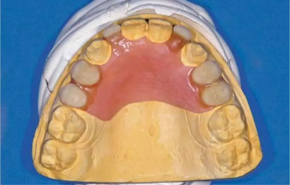
Fig 2-29fDiagnostic wax-up for the maxilla.
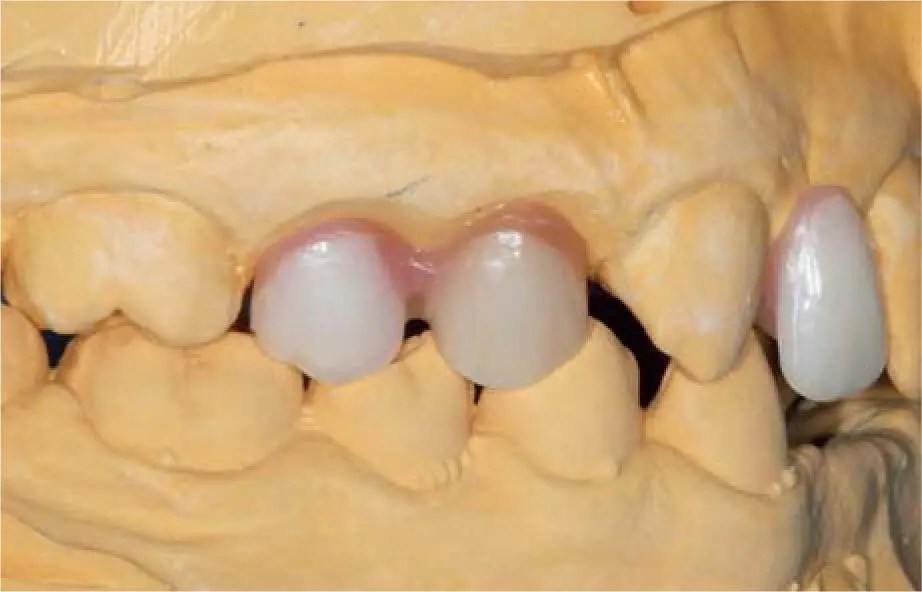
Fig 2-29gFollowing the wax-up, a vertical bone augmentation is not necessary in the right maxilla.
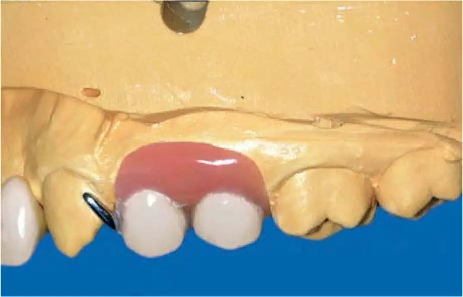
Fig 2-29hWax-up of the left maxilla shows the extent of the required vertical bone augmentation.
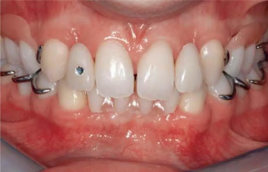
Fig 2-29iSlight increase of the vertical bite dimension with temporary restorations and composite reconstructions on the remaining teeth.
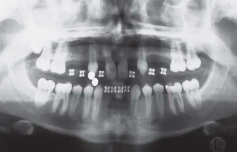
Fig 2-29jPanoramic radiograph with surgical templates: the vertical bone atrophy is clearly visible in the premolar area of the left maxilla.
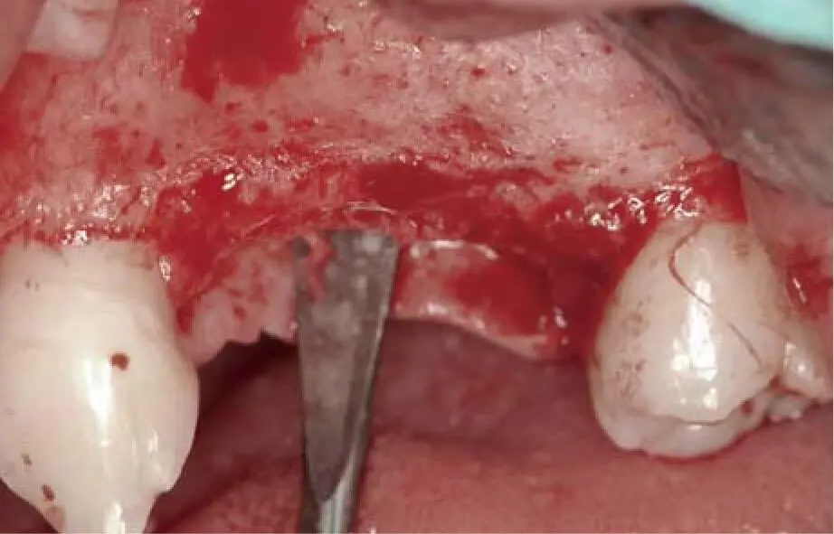
Fig 2-29kVertical bone deficit in the left maxilla.
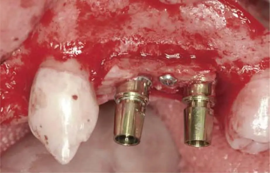
Fig 2-29lVertical bone augmentation with simultaneous insertion of two XiVE implants.
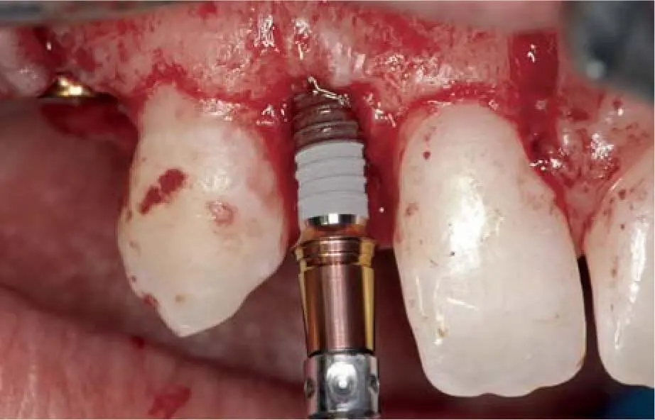
Fig 2-29mInsertion of a 3-mm XiVE implant at the area of the right lateral incisor.
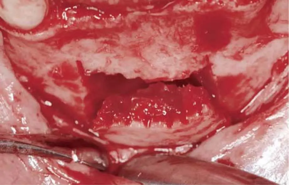
Fig 2-29nExtremely thin alveolar ridge in the anterior mandible.
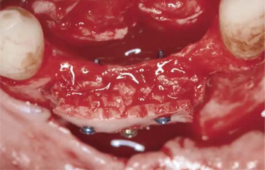
Fig 2-29oBone harvesting apical of the atrophied alveolar ridge.
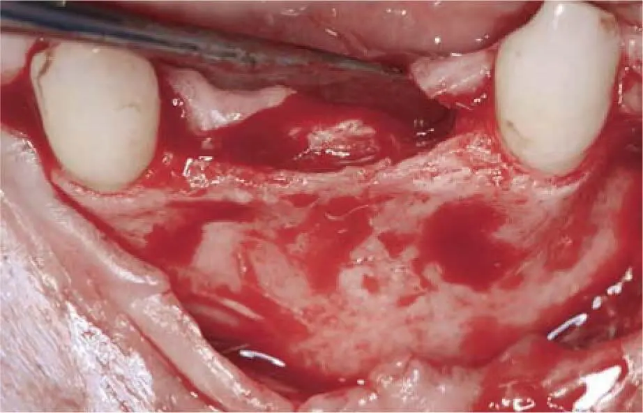
Fig 2-29pBone block grafting in the anterior mandible.
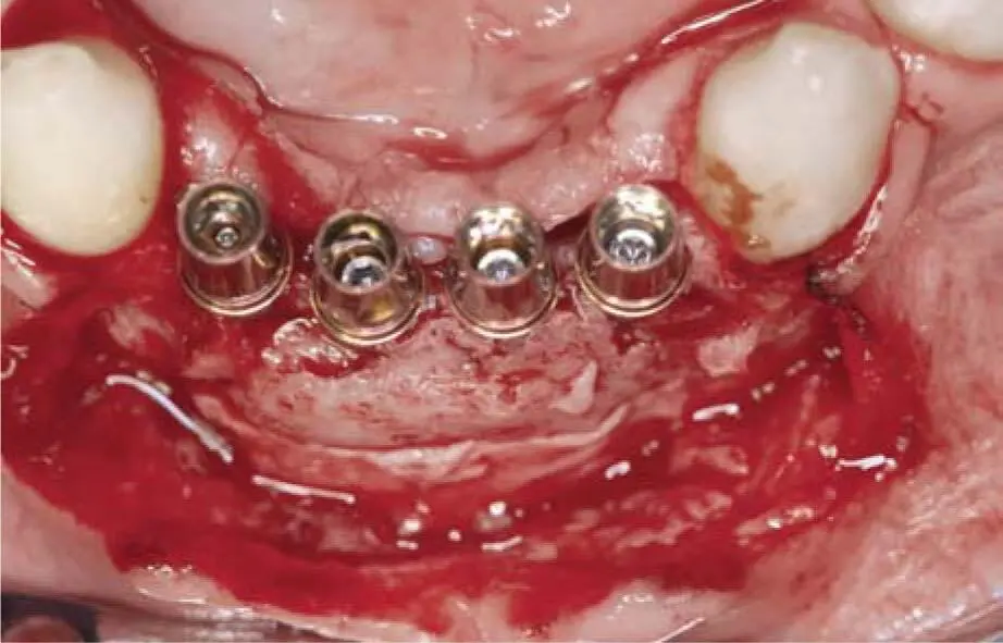
Fig 2-29qInsertion of four XiVE implants with a 3-mm diameter in the grafted area 3 months postoperatively.
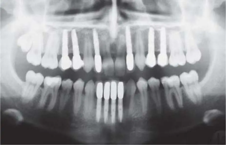
Fig 2-29rPanoramic radiograph 4 years postoperatively.
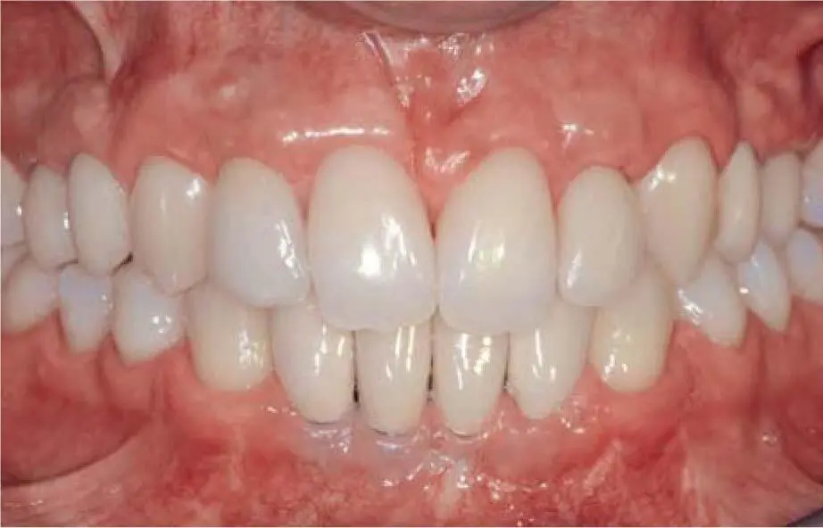
Fig 2-29sClinical situation 4 years postoperatively. Thin mucosa region (41 and 42) was later augmented using a connective tissue graft.
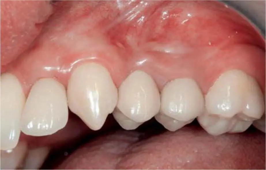
Fig 2-29tClinical situation of the restored implants in the left maxilla.
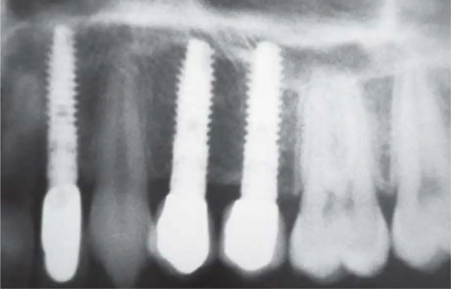
Fig 2-29uControl radiograph 4 years postoperatively of the restored implants in the vertically grafted left maxilla.
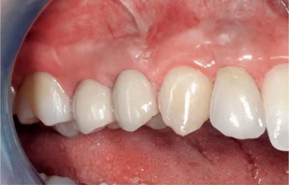
Fig 2-29vClinical situation in the right maxilla 4 years postoperatively.
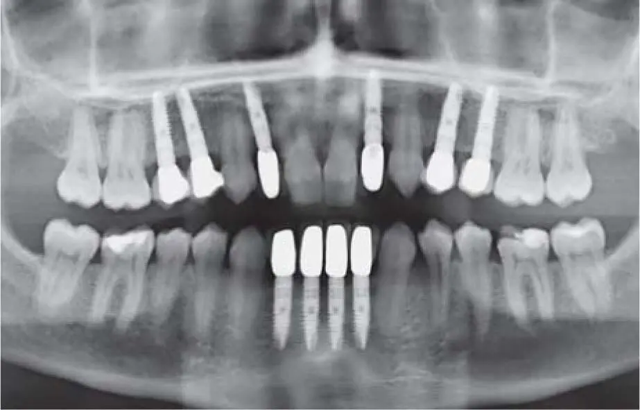
Fig 2-29wControl radiograph 16 years postoperatively demonstrating a stable peri-implant bone level.
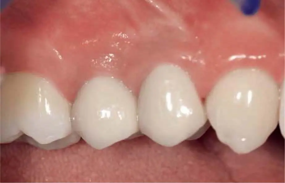
Fig 2-29xClinical appearance of the restored implants in the right maxilla 16 years postoperatively.
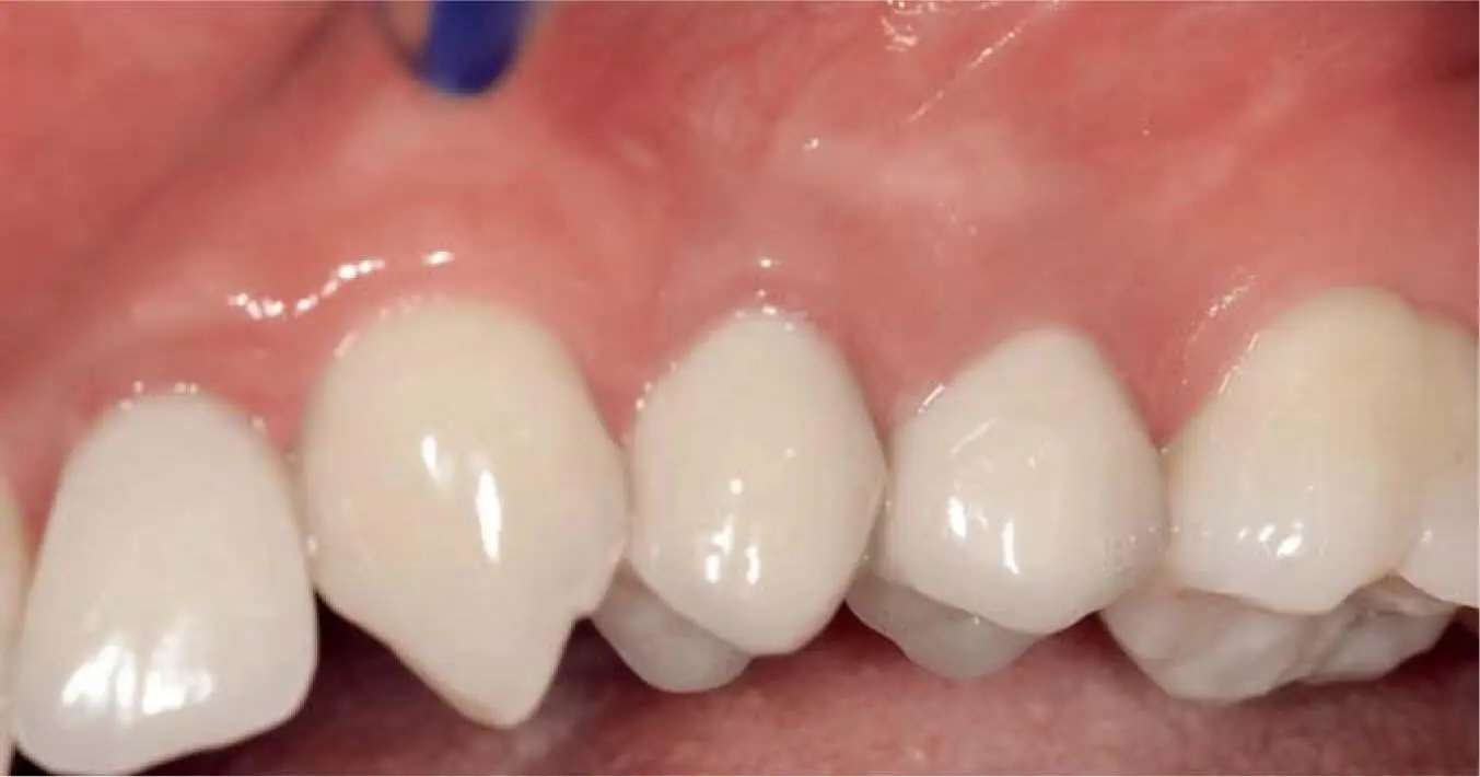
Fig 2-29ySimilar stability in the left maxilla.
2.7 References
1. Gesetz zum Schutz vor der schädlichen Wirkung ionisierender Strahlung (Strahlenschutzgesetz StrlSchG). Bundesgesetzblatt Teil 1 S 1966, 2017.
2. Verordnung zum Schutz vor der schädlichen Wirkung ionisierender Strahlung (Strahlenschutzverordnung – StrlSchV). Bundesgesetzblatt Teil 1 S 2034, 2018: 2034–2208.
3. Alfadda SA. Current evidence on dental implants outcomes in smokers and nonsmokers: a systematic review and meta-analysis. J Oral Implantol 2018;44:390–399.
4. Alsaadi G, Quirynen M, Michiles K, Teughels W, Komárek A, van Steenberghe D. Impact of local and systemic factors on the incidence of failures up to abutment connection with modified surface oral implants. J Clin Periodontol 2008;35:51–57.
5. Arai Y, Tammisalo E, Iwai K, Hashimoto K, Shinoda K. Development of a compact computed tomographic apparatus for dental use. Dentomaxillofac Radiol 1999;28: 245–248.
6. Baig MR, Rajan M. Effects of smoking on the outcome of implant treatment: a literature review. Indian J Dent Res 2007;18:190–195.
7. Barnea E, Alt I, Kolerman R, Nissan J. Accuracy of a laboratory-based computer implant guiding system. Oral Surg Oral Med Oral Pathol Oral Radiol Endod 2010; 109:e6–e10.
8. Berger C, Engels HB. Kapitel B Qualitätsleitlinie “Implantologie” des BDIZ. In BDIZ (ed) Indikation enossaler Implantate in Gutachterhandbuch Implantologie, Breisach: Med. Verl.- und Informationsdienste, 2002; 59–74.
9. Bindl A, Ritter L, Mehl A. Digital 3-D implant planning: Cerec meets Galileos. Int J Comput Dent 2010;13: 221–231.
10. Boquete-Castro A, Gómez-Moreno G, Calvo-Guirado JL, Aguilar-Salvatierra A, Delgado-Ruiz RA. Denosumab and osteonecrosis of the jaw. A systematic analysis of events reported in clinical trials. Clin Oral Implants Res 2016;27:367–375.
Читать дальшеИнтервал:
Закладка:
Похожие книги на «Bone and Soft Tissue Augmentation in Implantology»
Представляем Вашему вниманию похожие книги на «Bone and Soft Tissue Augmentation in Implantology» списком для выбора. Мы отобрали схожую по названию и смыслу литературу в надежде предоставить читателям больше вариантов отыскать новые, интересные, ещё непрочитанные произведения.


Обсуждение, отзывы о книге «Bone and Soft Tissue Augmentation in Implantology» и просто собственные мнения читателей. Оставьте ваши комментарии, напишите, что Вы думаете о произведении, его смысле или главных героях. Укажите что конкретно понравилось, а что нет, и почему Вы так считаете.