Bone and Soft Tissue Augmentation in Implantology
Здесь есть возможность читать онлайн «Bone and Soft Tissue Augmentation in Implantology» — ознакомительный отрывок электронной книги совершенно бесплатно, а после прочтения отрывка купить полную версию. В некоторых случаях можно слушать аудио, скачать через торрент в формате fb2 и присутствует краткое содержание. Жанр: unrecognised, на английском языке. Описание произведения, (предисловие) а так же отзывы посетителей доступны на портале библиотеки ЛибКат.
- Название:Bone and Soft Tissue Augmentation in Implantology
- Автор:
- Жанр:
- Год:неизвестен
- ISBN:нет данных
- Рейтинг книги:4 / 5. Голосов: 1
-
Избранное:Добавить в избранное
- Отзывы:
-
Ваша оценка:
Bone and Soft Tissue Augmentation in Implantology: краткое содержание, описание и аннотация
Предлагаем к чтению аннотацию, описание, краткое содержание или предисловие (зависит от того, что написал сам автор книги «Bone and Soft Tissue Augmentation in Implantology»). Если вы не нашли необходимую информацию о книге — напишите в комментариях, мы постараемся отыскать её.
R. Gruber, Th. Hanser, Ph. Keeve, Ch. Khoury, J. Neugebauer, J. E. Zöller
Bone and Soft Tissue Augmentation in Implantology addresses useful methods of bone grafting procedures in implant treatment based on current biologic principles and constitutes a unique reference in this field. The book describes, in over 760 pages and 2837 mostly color illustrations, the different possibilities available to augment the bone volume in width and height. The information presented includes not only the underlying scientific concepts of the different augmentation techniques with autogenous bone, but also the associated soft tissue management, from safe approaches to different possibilities for soft tissue augmentation and papilla reconstruction techniques.
The book provides surgeons with a basic understanding of the biologic response to bone grafting procedures. Experienced implantologists will benefit from the in-depth background information, details of high-level surgical techniques, and scientific results, which will enable them to optimize their surgical procedures. Each chapter offers a wealth of information on the specific topic covered, with much attention given to the scientific concepts behind each one. Extensive case reports with step-by-step documentation allow readers to gain an impression of what is possible today in the 3D reconstruction procedures of the alveolar crest. Important criteria for success are presented as well as possible complications and their treatment.
Bone and Soft Tissue Augmentation in Implantology is a must-read for every implantologist, oral and maxillofacial surgeon, and any dentist interested in surgery.
Bone and Soft Tissue Augmentation in Implantology — читать онлайн ознакомительный отрывок
Ниже представлен текст книги, разбитый по страницам. Система сохранения места последней прочитанной страницы, позволяет с удобством читать онлайн бесплатно книгу «Bone and Soft Tissue Augmentation in Implantology», без необходимости каждый раз заново искать на чём Вы остановились. Поставьте закладку, и сможете в любой момент перейти на страницу, на которой закончили чтение.
Интервал:
Закладка:
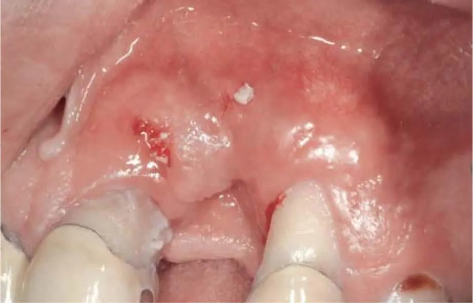
Fig 2-10bFailed augmentation with bovine bone substitute material. Part of the material spontaneously perforated the soft tissue.
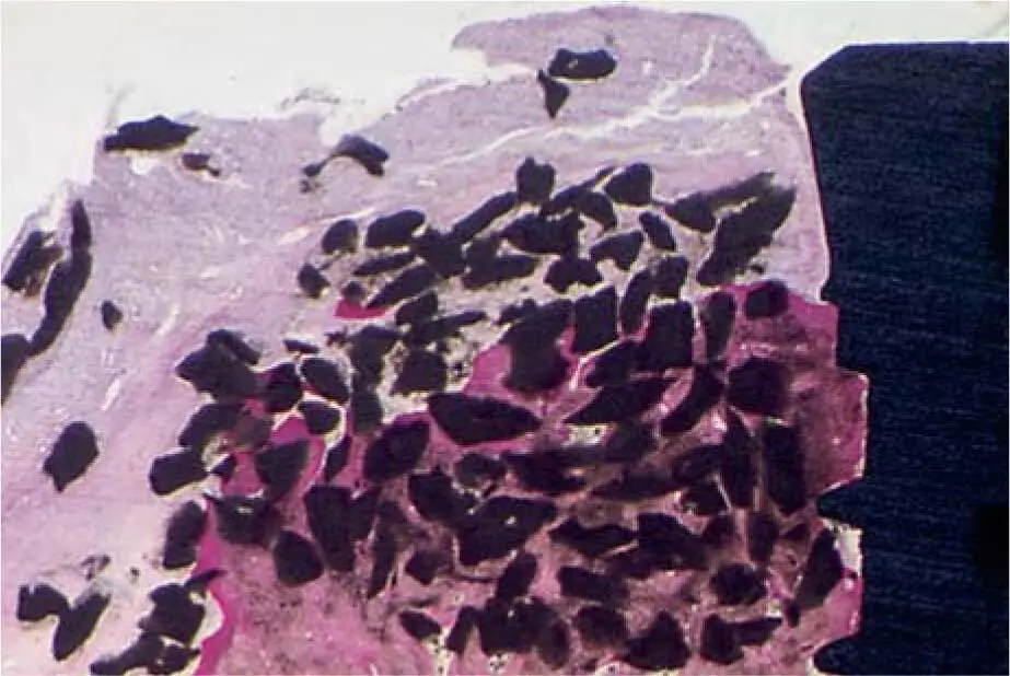
Fig 2-10cPartially regenerated bone substitute material but with important infiltration in the soft tissue area.
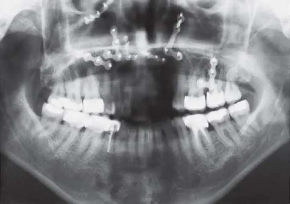
Fig 2-11aPanoramic radiograph after treatment of a complex midfacial fracture with multiple osteosynthesis in the maxilla and the remains of an important bony defect in the anterior tooth area.
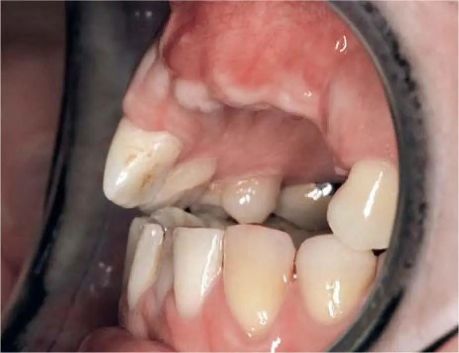
Fig 2-11bFracture-related open bite.
Fig 2-11cWax-up of the desired prosthetic result for the planning of a bone reconstruction with an autogenous graft.
2.4.3.3 Structure of the bone
The bone quality of the regenerated area is generally classified as reduced, especially when using biomaterials (xenogenic or allografts). By applying the 3D technique with an intraoral bone transplant, iliac crest reconstruction using monocortical strips, and compressed cancellous bone or distraction osteogenesis, a vital and stable bone bed can be achieved that corresponds to a bone class of D2 or D3. Depending on the bone quality, it is important that dysfunctions are detected early to prevent possible overloading of the implant site due to bruxism.
Determining the bone volume is suitable as an initial and immediately available means to so-called bone mapping. The height of the soft tissue situation is determined with a pointed needle and a rubber stopper. By transferring these measured values to a saw-cut model, the existing bone volume can then be measured ( Fig 2-12aand b). This kind of diagnostic measuring is rarely used today due to the increased use of digital diagnostic methods such as cone beam computed tomography (CBCT). The use of appropriately modified calipers has not proven to be successful in clinical practice and therefore they are only of historical significance today.
2.4.4 Radiologic findings
In the context of implant therapy, especially in the atrophic jaw, radiologic diagnosis provides the essential information for deciding on the possibilities and scope of the necessary therapy and ensuring the treatment result. Radiologic diagnosis delivers the relevant information for the protection of the anatomical structures in the surgical field. In addition to the purely volumetric assessment of the jaw, the bone structure is also evaluated.
Due to chronic infections, especially after multiple endodontic treatments with revisions of the root fillings, apical root resections or any other infection, sclerotic changes in the jawbone can be seen. As a rule, these are not relevant for further implant therapy because the body’s own immune system has healed the infection through the inflammatory process; in fact, it sometimes repairs more than necessary through new bone apposition. This increases the bone density and the possibility of achieving high primary implant stability. On the other hand, increased bone density makes implant bed preparation more difficult, and in case of insufficient cooling can increase the risk of burning the bone. In rare cases, a more or less pronounced local osteomyelitis may be present.
Osteomyelitis sclerosans Garré is an infectious bacterial chronic event that is sustained by a persistent infection or by itself. Microbiologically, bacterial growth can rarely be detected by an intraoperative swab test. If there is anamnestic evidence of a persistent bony inflammation with symptoms, it is recommended to consider a CBCT ( Fig 2-13) or a skeletal scintigraphy to exclude chronic subacute processes.
2.4.4.1 Radiologic techniques
For therapeutic planning, all radiologic procedures in dentistry are now applied on an indication-related basis depending on the different forms of implant prosthetic therapy, from single tooth replacement after traumatic tooth loss to maxillary ridge reconstruction in severely atrophied jaws.
Periapical radiographs
The use of periapical radiographs is particularly recommended to obtain clearer detail of the teeth, bone, and implants. This kind of diagnostic technique is especially recommended for the determination of bone loss around implants or teeth and for the detection of caries ( Fig 2-14aand b). In case of planning a bone augmentation, especially if using vertical bone augmentation techniques, periapical radiographs are recommended to detect bone structures covering the neighboring teeth. This information is very important, since vertical bone grafting is limited to the bone level of the neighboring teeth.
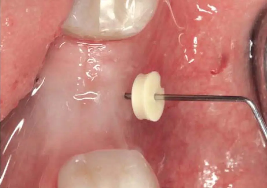
Fig 2-12aMeasurement of mucosal thickness with a thin needle with an attached endo stopper.
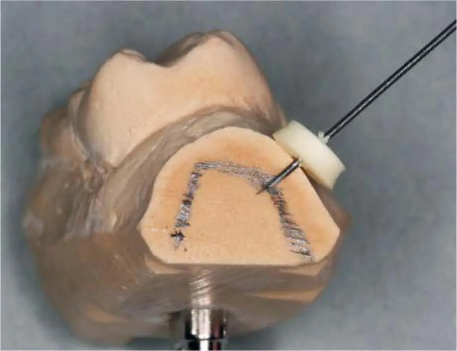
Fig 2-12bTransmission of mucosal thickness measurement on a saw-cut model.
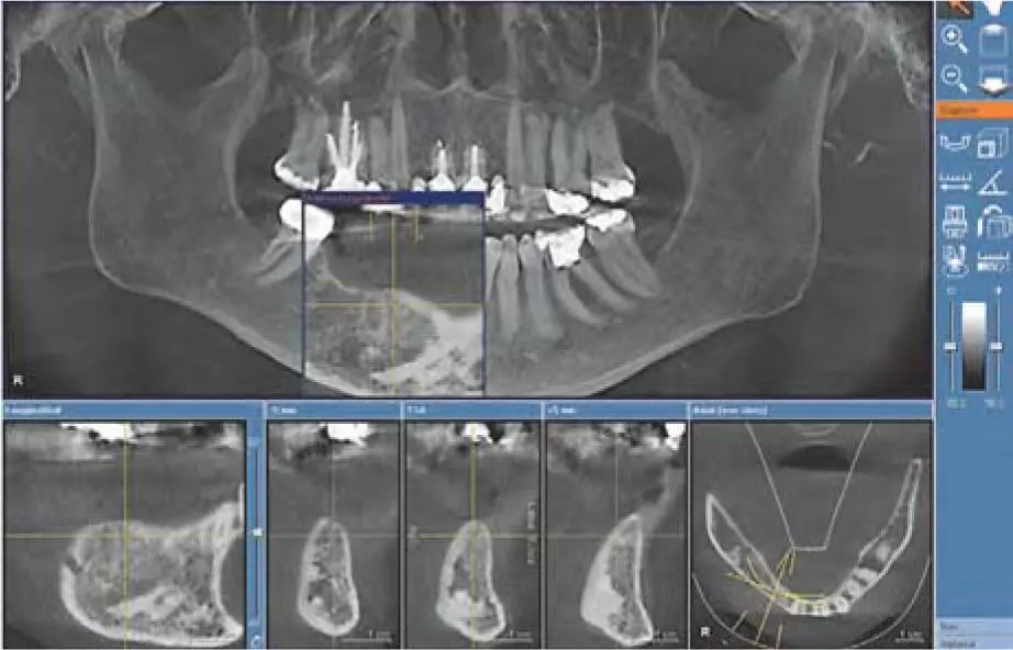
Fig 2-13Important sclerosis close to the basal areas of the edentulous part of the mandible after failure and removal of all implants inserted in grafted bone from the iliac crest. The patient’s history reveals the extraction of several teeth after multiple apicoectomies due to long-term chronic infections.
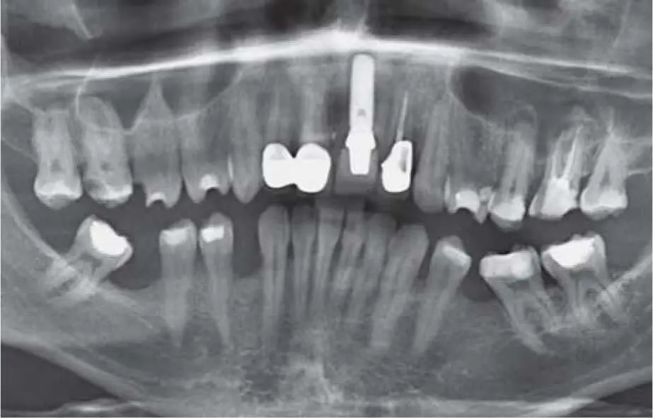
Fig 2-14aPanoramic radiograph documenting an implant at the area of the central left incisor and some apical reactions on the second premolar and first maxillary left molar. A peri-implant pathology is difficult to detect here.
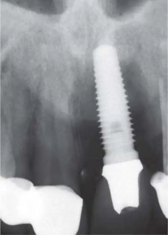
Fig 2-14bPeriapical radiograph documenting a severe peri-implant bone loss and a deep caries on the neighboring lateral incisor. A thin bone layer is still covering the neighboring roots.
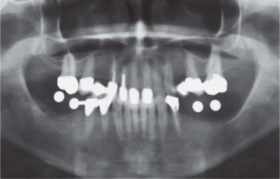
Fig 2-14cDetermination of the vertical bone dimension with reference balls.
Читать дальшеИнтервал:
Закладка:
Похожие книги на «Bone and Soft Tissue Augmentation in Implantology»
Представляем Вашему вниманию похожие книги на «Bone and Soft Tissue Augmentation in Implantology» списком для выбора. Мы отобрали схожую по названию и смыслу литературу в надежде предоставить читателям больше вариантов отыскать новые, интересные, ещё непрочитанные произведения.


Обсуждение, отзывы о книге «Bone and Soft Tissue Augmentation in Implantology» и просто собственные мнения читателей. Оставьте ваши комментарии, напишите, что Вы думаете о произведении, его смысле или главных героях. Укажите что конкретно понравилось, а что нет, и почему Вы так считаете.