Bone and Soft Tissue Augmentation in Implantology
Здесь есть возможность читать онлайн «Bone and Soft Tissue Augmentation in Implantology» — ознакомительный отрывок электронной книги совершенно бесплатно, а после прочтения отрывка купить полную версию. В некоторых случаях можно слушать аудио, скачать через торрент в формате fb2 и присутствует краткое содержание. Жанр: unrecognised, на английском языке. Описание произведения, (предисловие) а так же отзывы посетителей доступны на портале библиотеки ЛибКат.
- Название:Bone and Soft Tissue Augmentation in Implantology
- Автор:
- Жанр:
- Год:неизвестен
- ISBN:нет данных
- Рейтинг книги:4 / 5. Голосов: 1
-
Избранное:Добавить в избранное
- Отзывы:
-
Ваша оценка:
Bone and Soft Tissue Augmentation in Implantology: краткое содержание, описание и аннотация
Предлагаем к чтению аннотацию, описание, краткое содержание или предисловие (зависит от того, что написал сам автор книги «Bone and Soft Tissue Augmentation in Implantology»). Если вы не нашли необходимую информацию о книге — напишите в комментариях, мы постараемся отыскать её.
R. Gruber, Th. Hanser, Ph. Keeve, Ch. Khoury, J. Neugebauer, J. E. Zöller
Bone and Soft Tissue Augmentation in Implantology addresses useful methods of bone grafting procedures in implant treatment based on current biologic principles and constitutes a unique reference in this field. The book describes, in over 760 pages and 2837 mostly color illustrations, the different possibilities available to augment the bone volume in width and height. The information presented includes not only the underlying scientific concepts of the different augmentation techniques with autogenous bone, but also the associated soft tissue management, from safe approaches to different possibilities for soft tissue augmentation and papilla reconstruction techniques.
The book provides surgeons with a basic understanding of the biologic response to bone grafting procedures. Experienced implantologists will benefit from the in-depth background information, details of high-level surgical techniques, and scientific results, which will enable them to optimize their surgical procedures. Each chapter offers a wealth of information on the specific topic covered, with much attention given to the scientific concepts behind each one. Extensive case reports with step-by-step documentation allow readers to gain an impression of what is possible today in the 3D reconstruction procedures of the alveolar crest. Important criteria for success are presented as well as possible complications and their treatment.
Bone and Soft Tissue Augmentation in Implantology is a must-read for every implantologist, oral and maxillofacial surgeon, and any dentist interested in surgery.
Bone and Soft Tissue Augmentation in Implantology — читать онлайн ознакомительный отрывок
Ниже представлен текст книги, разбитый по страницам. Система сохранения места последней прочитанной страницы, позволяет с удобством читать онлайн бесплатно книгу «Bone and Soft Tissue Augmentation in Implantology», без необходимости каждый раз заново искать на чём Вы остановились. Поставьте закладку, и сможете в любой момент перейти на страницу, на которой закончили чтение.
Интервал:
Закладка:
In addition to the reconstructions in the classical radiologic layers, special dental CT reconstructions are also possible, which offer sections perpendicular to a panoramic curve ( Fig 2-17cto h). For dental diagnostics, the images are exported in DICOM format so that they can be read in the various viewing and planning software. 1In addition to a precise representation of the bony structures, the spiral CT is particularly distinguished in the imaging of soft tissue ( Fig 2-18ato e). Although radiation exposure has been reduced in modern devices, the spiral CT still presents a higher radiation exposure compared with digital volume tomography devices (CBCT). 56
Therefore, if CBCT is available, spiral CT should be avoided for preimplantation diagnostics or for proposed 3D surgical guides or navigation procedures. 21,23,72
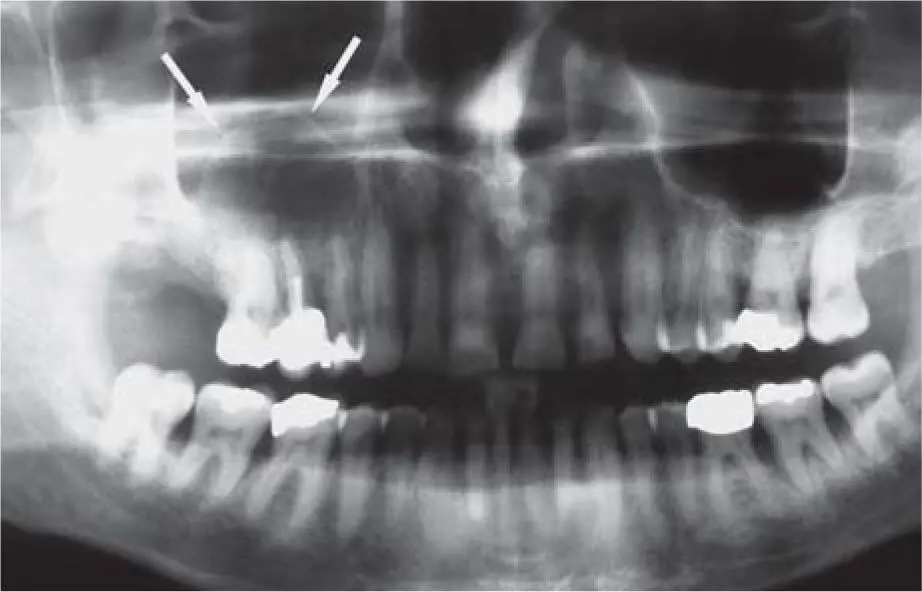
Fig 2-17aPanoramic view with suspected cystic findings in the right sinus.
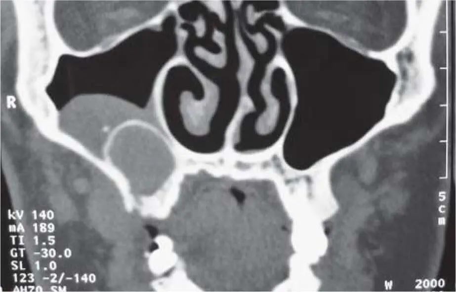
Fig 2-17bCT scan with clarification of the findings: maxillary cyst with concomitant mucocele in the maxillary right sinus.
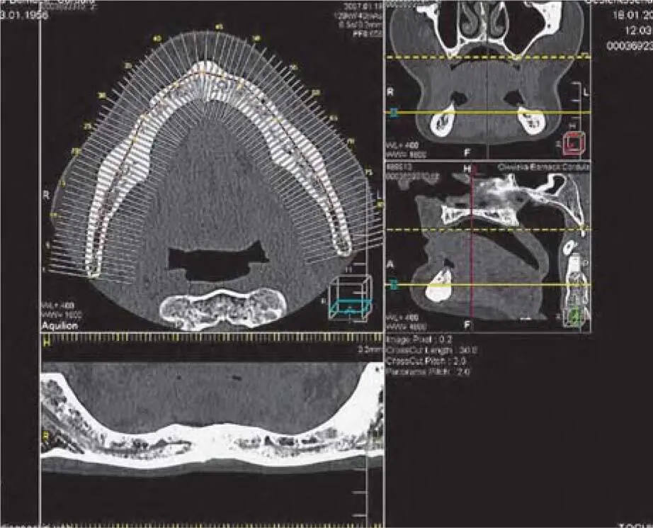
Fig 2-17cCT images of the mandible in the context of augmentation planning: the layers are 1 mm in thickness.
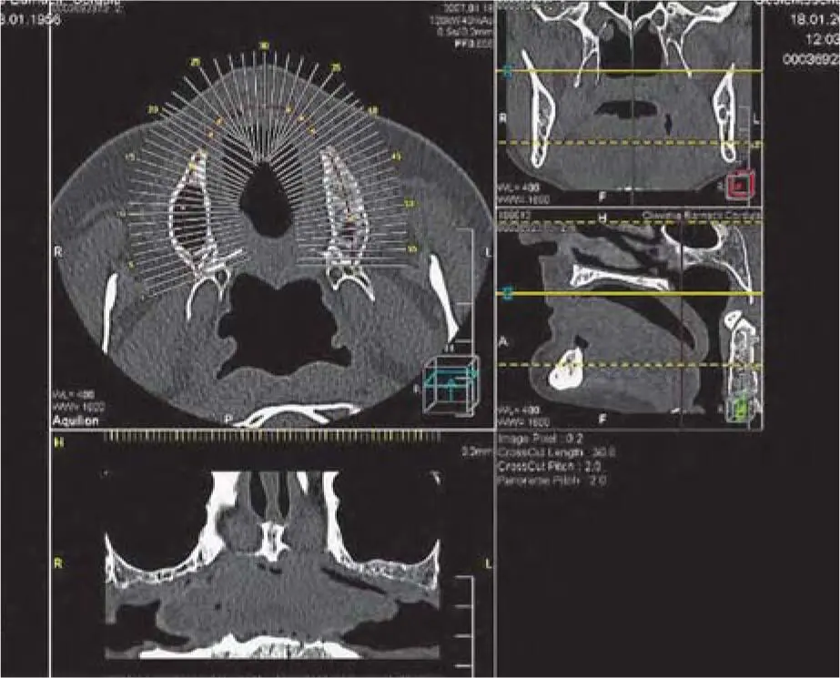
Fig 2-17dCT images of the maxilla with the same thickness.
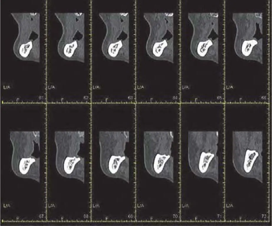
Fig 2-17eMultiple slices from the posterior region of the mandible.

Fig 2-17fMultiple slices from the posterior region of the maxilla.
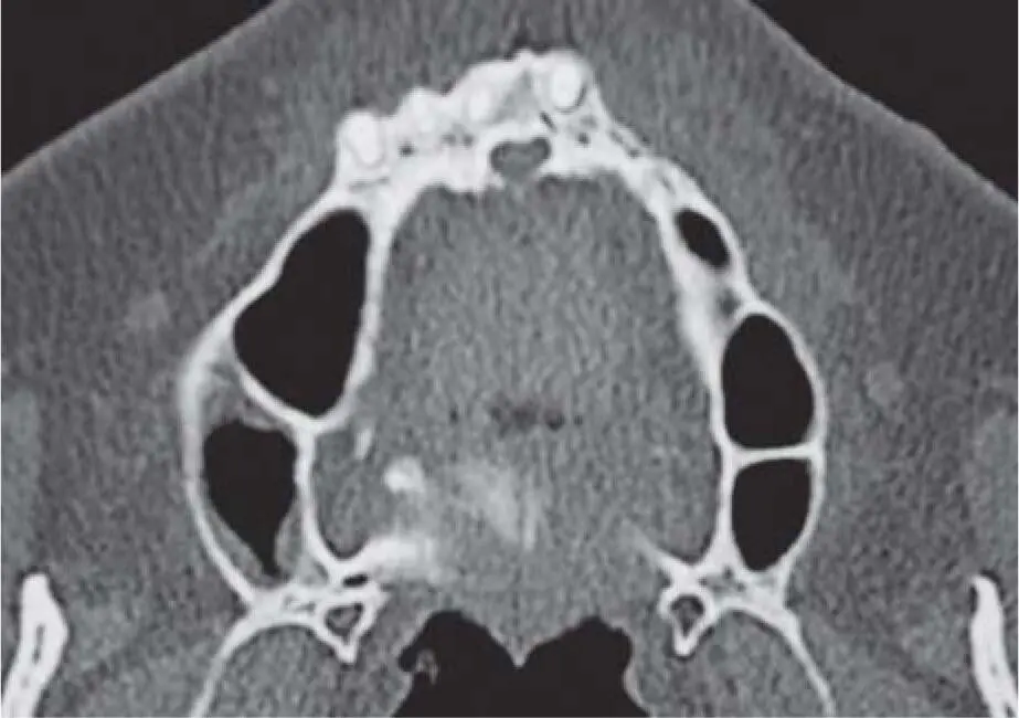
Fig 2-17gImportant septa in the basal area of both maxillary sinuses.
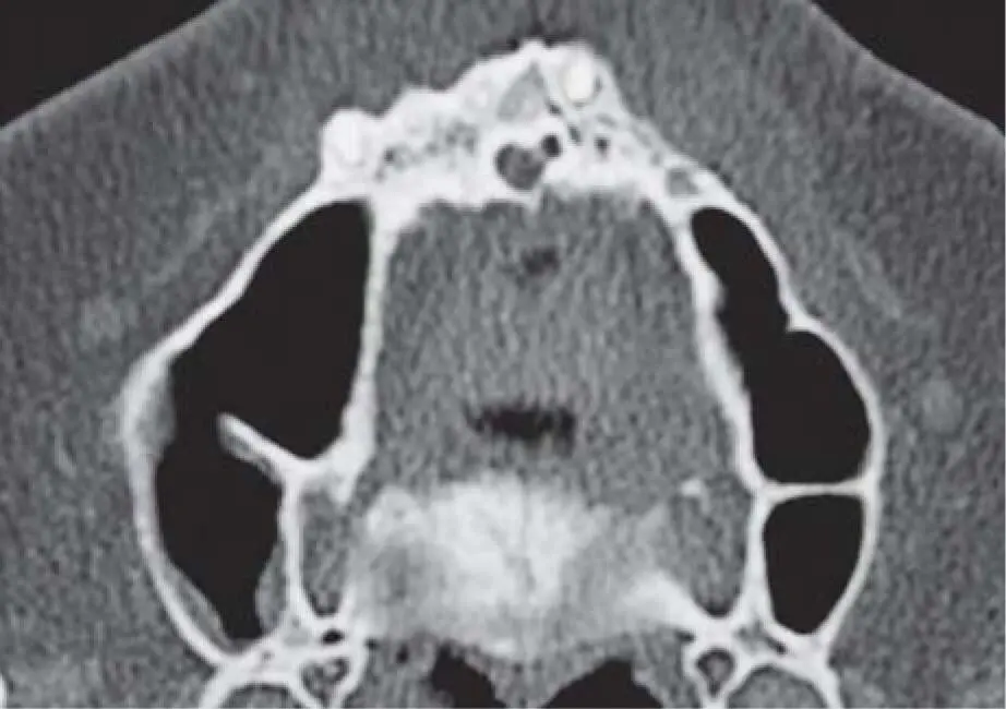
Fig 2-17hThe same septa in the middle of both maxillary sinuses.
Cone beam computed tomography
In addition to spiral CT, CBCT has been available to the dentist for more than 20 years as a continuative method of radiologic imaging. 59Today, devices from various manufacturers are offered that enable a 3D diagnosis in the oral and maxillofacial area. 66Since CBCT devices are much cheaper than CT devices, they have become increasingly common in dental practices oriented toward implantology.
In CBCT, the patient is usually positioned standing or sitting (only a few devices require a horizontal position similar to that used for spiral CT). The radiologic beam is cone shaped so that the radiation source moves on one level around the patient ( Fig 2-19aand b). The volume is then reconstructed from 200 to over 500 single images. The applied energy and the resolution of the detector determine the quality of information, with the volume size and resolution of the recording. Depending on the detector technology, the field of view, the radiation exposure with a continuous or pulsed beam, and the filters used, a large variation of the effective dose is possible (between 3 µSv and about 800 µSv).
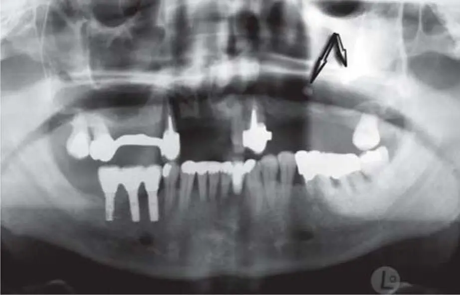
Fig 2-18aPanoramic radiograph showing two foreign bodies (suspected to be overfilled root filling mass) in the region of the maxillary left sinus: incidental findings without symptoms in the context of pre-implant diagnosis.
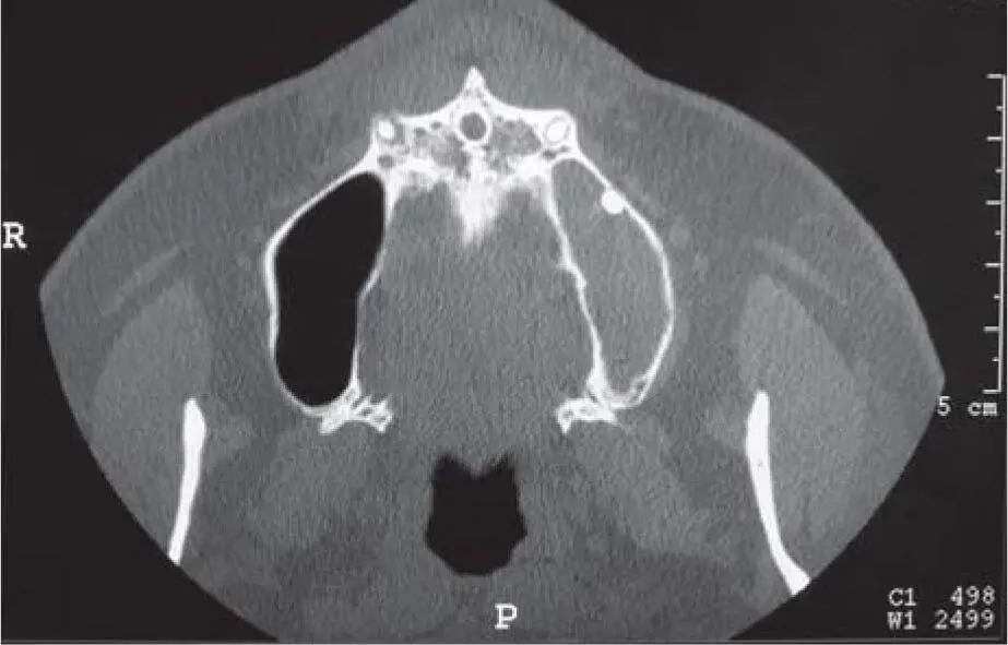
Fig 2-18bA round foreign body is clearly visible on the lower (caudal) layer of this CT image. In addition, the left maxillary sinus is completely shadowed.
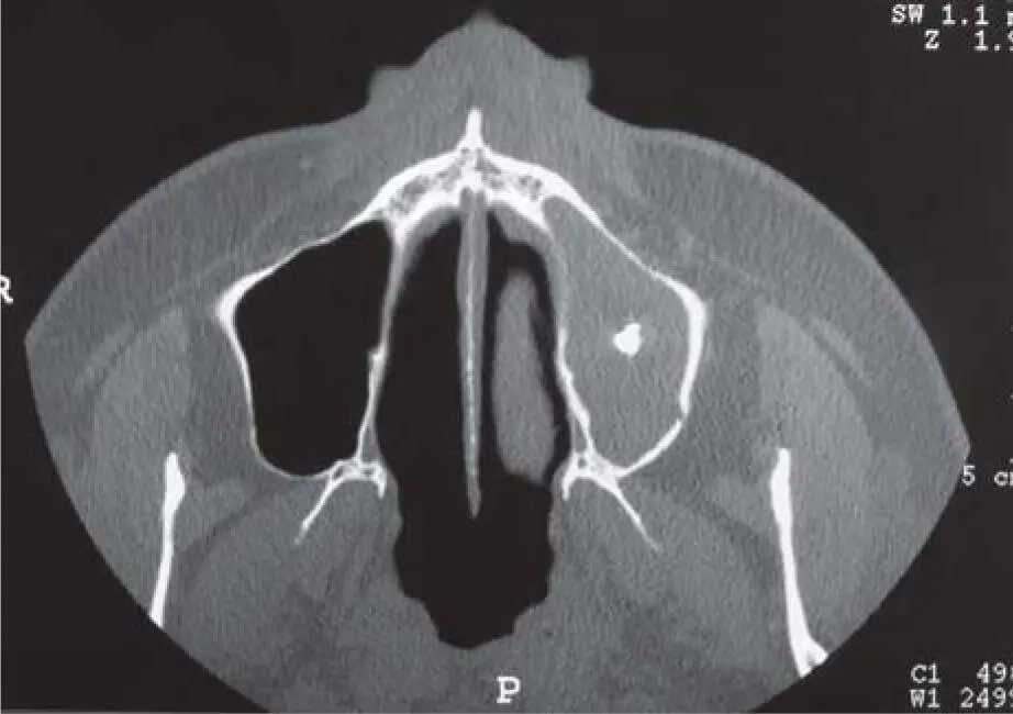
Fig 2-18cA second foreign body is also clearly visible on the higher layer. Also on this level, the complete shading of the maxillary left sinus is clearly visible.
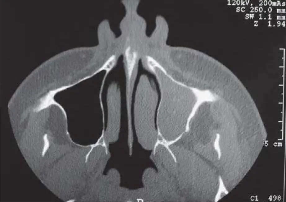
Fig 2-18dThe maxillary left sinus is also completely shadowed in this image, even in the upper area. The suspected diagnosis is aspergillosis infection due to root filling material.
Due to the specific target of CBCT – the high contrast imaging of bone – the soft tissue structures can only be evaluated to a limited extent. The disadvantage of reduced soft tissue visualization is compensated for by appropriately designed prosthetic planning templates or additionally placed cotton rolls to separate the soft tissue of the alveolar crest from the tongue or floor of the mouth ( Fig 2-19c). The diagnostic validity could be optimized by special reconstruction algorithms, so that the same diagnostic value is available when compared with CT. 59In particular, the automatic reconstruction of the known panoramic layer from the 3D volume allows for the usual dental radiologic diagnosis. 97
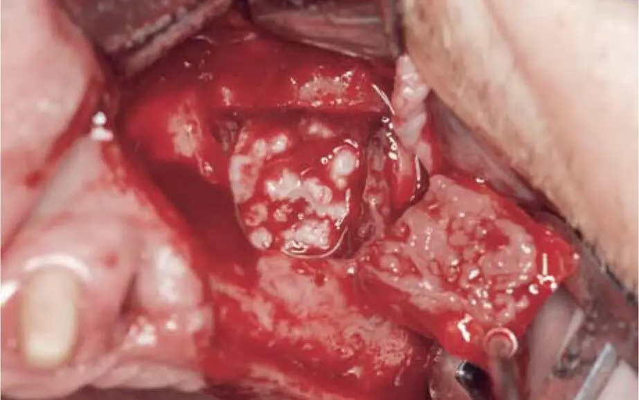
Fig 2-18eThe intraoperative findings, and later also the pathohistologic findings, confirm the suspected diagnosis.
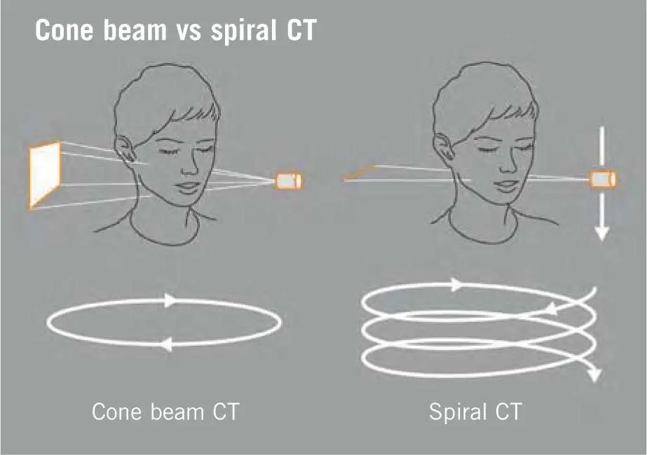
Fig 2-19aFunctional principle of cone beam technology, with a conical beam that revolves on one level, compared with a spiral CT that has a line-like and moving beam path.
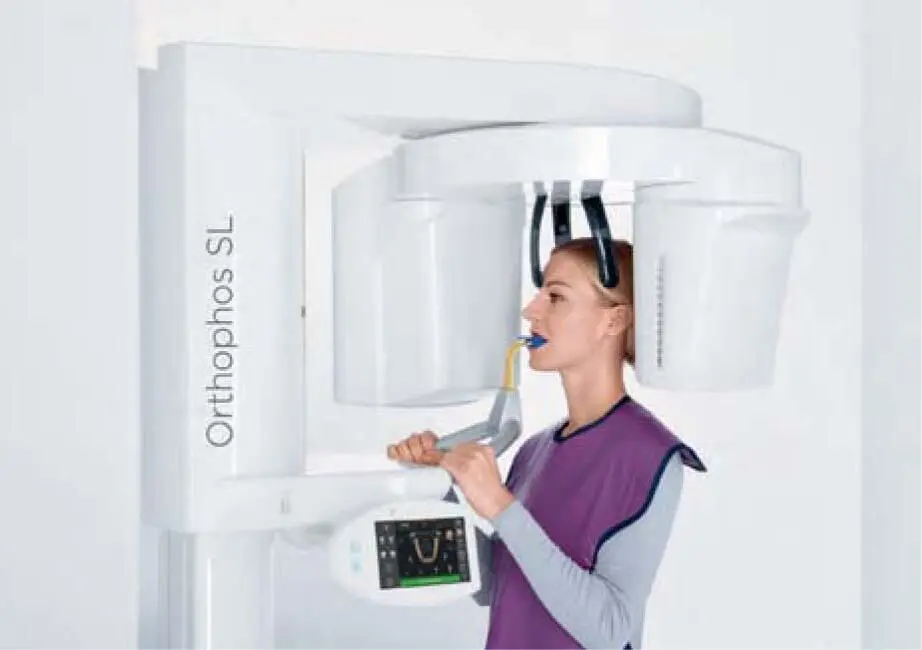
Fig 2-19bDevice for CBCT for positioning the standing or sitting patient.
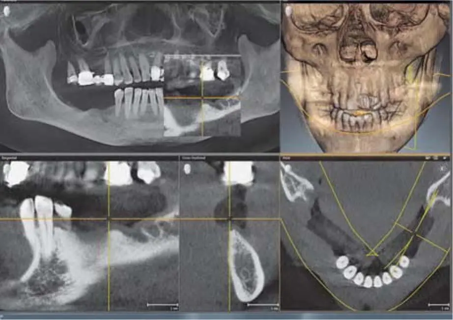
Fig 2-19cCBCT for implant planning in the posterior mandible, with cotton rolls to separate the mobile soft tissue of the mouth floor from the alveolar crest.
Читать дальшеИнтервал:
Закладка:
Похожие книги на «Bone and Soft Tissue Augmentation in Implantology»
Представляем Вашему вниманию похожие книги на «Bone and Soft Tissue Augmentation in Implantology» списком для выбора. Мы отобрали схожую по названию и смыслу литературу в надежде предоставить читателям больше вариантов отыскать новые, интересные, ещё непрочитанные произведения.


Обсуждение, отзывы о книге «Bone and Soft Tissue Augmentation in Implantology» и просто собственные мнения читателей. Оставьте ваши комментарии, напишите, что Вы думаете о произведении, его смысле или главных героях. Укажите что конкретно понравилось, а что нет, и почему Вы так считаете.