Bone and Soft Tissue Augmentation in Implantology
Здесь есть возможность читать онлайн «Bone and Soft Tissue Augmentation in Implantology» — ознакомительный отрывок электронной книги совершенно бесплатно, а после прочтения отрывка купить полную версию. В некоторых случаях можно слушать аудио, скачать через торрент в формате fb2 и присутствует краткое содержание. Жанр: unrecognised, на английском языке. Описание произведения, (предисловие) а так же отзывы посетителей доступны на портале библиотеки ЛибКат.
- Название:Bone and Soft Tissue Augmentation in Implantology
- Автор:
- Жанр:
- Год:неизвестен
- ISBN:нет данных
- Рейтинг книги:4 / 5. Голосов: 1
-
Избранное:Добавить в избранное
- Отзывы:
-
Ваша оценка:
Bone and Soft Tissue Augmentation in Implantology: краткое содержание, описание и аннотация
Предлагаем к чтению аннотацию, описание, краткое содержание или предисловие (зависит от того, что написал сам автор книги «Bone and Soft Tissue Augmentation in Implantology»). Если вы не нашли необходимую информацию о книге — напишите в комментариях, мы постараемся отыскать её.
R. Gruber, Th. Hanser, Ph. Keeve, Ch. Khoury, J. Neugebauer, J. E. Zöller
Bone and Soft Tissue Augmentation in Implantology addresses useful methods of bone grafting procedures in implant treatment based on current biologic principles and constitutes a unique reference in this field. The book describes, in over 760 pages and 2837 mostly color illustrations, the different possibilities available to augment the bone volume in width and height. The information presented includes not only the underlying scientific concepts of the different augmentation techniques with autogenous bone, but also the associated soft tissue management, from safe approaches to different possibilities for soft tissue augmentation and papilla reconstruction techniques.
The book provides surgeons with a basic understanding of the biologic response to bone grafting procedures. Experienced implantologists will benefit from the in-depth background information, details of high-level surgical techniques, and scientific results, which will enable them to optimize their surgical procedures. Each chapter offers a wealth of information on the specific topic covered, with much attention given to the scientific concepts behind each one. Extensive case reports with step-by-step documentation allow readers to gain an impression of what is possible today in the 3D reconstruction procedures of the alveolar crest. Important criteria for success are presented as well as possible complications and their treatment.
Bone and Soft Tissue Augmentation in Implantology is a must-read for every implantologist, oral and maxillofacial surgeon, and any dentist interested in surgery.
Bone and Soft Tissue Augmentation in Implantology — читать онлайн ознакомительный отрывок
Ниже представлен текст книги, разбитый по страницам. Система сохранения места последней прочитанной страницы, позволяет с удобством читать онлайн бесплатно книгу «Bone and Soft Tissue Augmentation in Implantology», без необходимости каждый раз заново искать на чём Вы остановились. Поставьте закладку, и сможете в любой момент перейти на страницу, на которой закончили чтение.
Интервал:
Закладка:
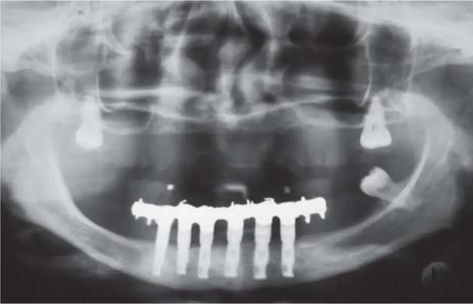
Fig 2-7cRadiologic control 12 years postoperatively.

Fig 2-7dSevere bone atrophy with hypodontia.
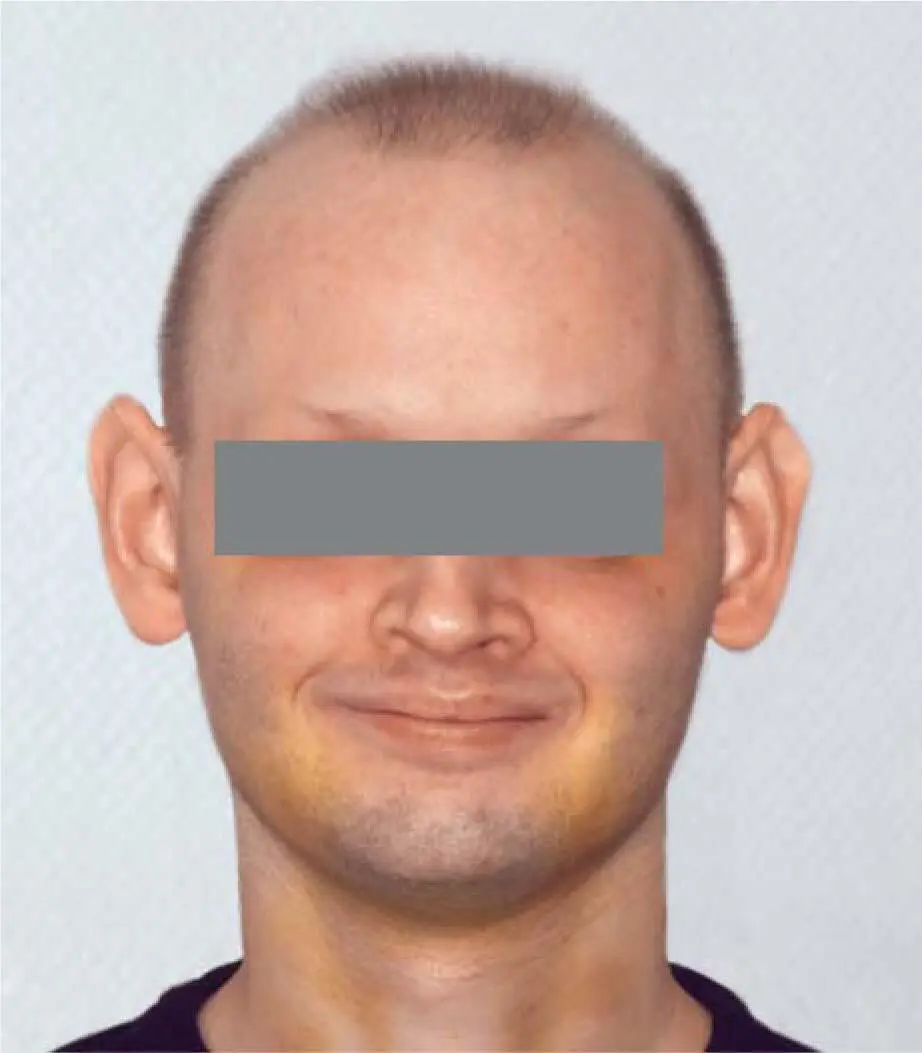
Fig 2-7eTypical appearance of a patient with moderate ectodermal dysplasia.
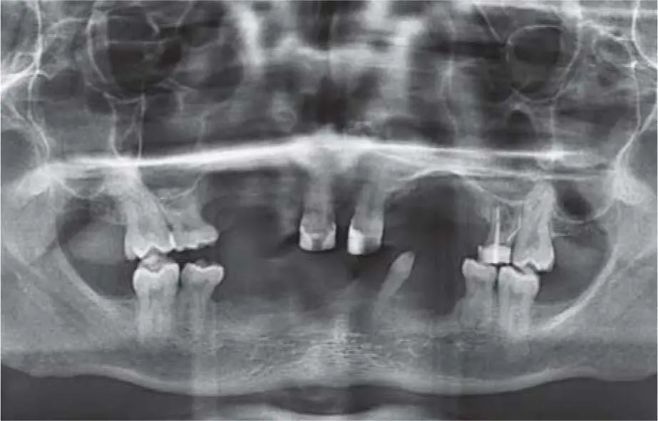
Fig 2-7fPanoramic radiograph revealing the absence of many teeth.
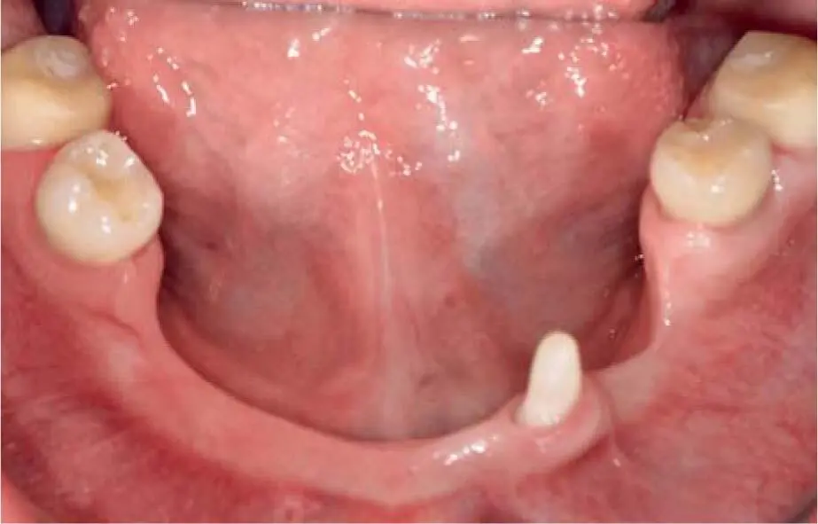
Fig 2-7gClinical situation of the mandible with hypodontia and severe bone atrophy.
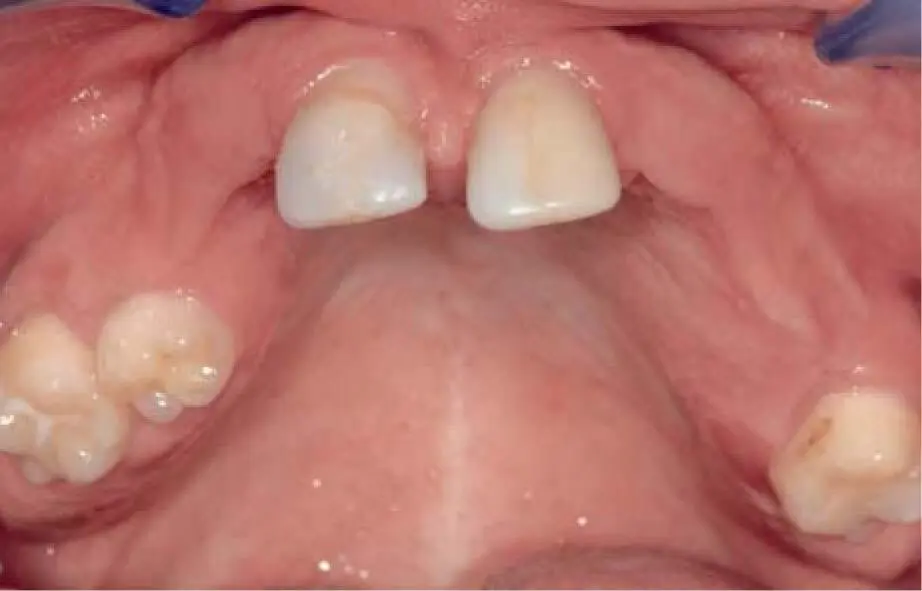
Fig 2-7hClinical aspect of the maxilla.
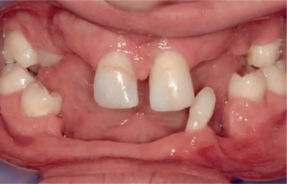
Fig 2-7iAbsence of a physiologic VDO due to missing occlusal support.
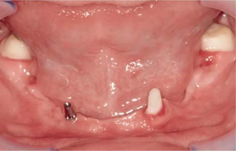
Fig 2-7jThe remaining teeth are prepared to support a fixed temporary restoration. In addition, a temporary implant is inserted in the right mandible.
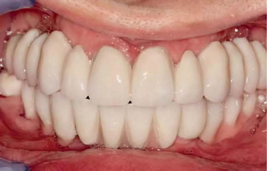
Fig 2-7kA fixed temporary restoration for the correction of the VDO.
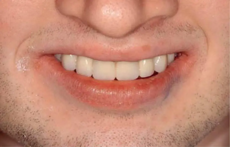
Fig 2-7lThe temporary restoration offers good lip support, improving the esthetics.
In the very rare autosomal recessive inherited Papillon-Lefèvre syndrome, the periodontal findings show severe periodontitis, leading to early loss of primary teeth usually up to the 4th year of life, and permanent teeth up to the 14th year. This exceptional periodontal disease presents a pronounced atrophy of the alveolar processes, which requires augmentative pretreatment. 86
Fig 2-7mMultiple bone block augmentation to reconstruct the missing bone.
Fig 2-7nRadiograph control after insertion of the remaining implants in the grafted bone.
2.4.2 Extraoral examination
When assessing the extraoral findings, the position of the maxilla and mandible in relation to each other should be evaluated. Due to the differing development of the position of the alveolar ridge due to the centrifugally oriented atrophy in the mandible and the centripetally oriented course in the maxilla, a pronounced prognathic position of the mandible can occur, especially in edentulous patients. The necessary grafting procedures in these cases do not only restore the vertical jaw relation but also determine the position of the alveolar ridge for the new prosthetic restoration. Due to the loss of the vertical dimension in cases of severe atrophy, the extraoral profile often shows a sunken upper lip or a reduced dimension of the lower third of the face; symptoms of this are often angular cheilitis and Candida albicans infection at the corners of the lips. A pronounced mental crease is a clinical sign.
When planning the type of dental prosthesis in the maxilla, the shape of the upper lip influences the decision about whether a fixed or removable prosthesis can be incorporated. Since even extensive augmentation procedures cannot restore the entire alveolar process, either long crowns or crowns with an attached gingiva through the use of pink ceramic or resin should be delivered. Due to the change in tension of the peri-oral soft tissue with age, the problem is less relevant for elderly patients. For younger patients with a short upper lip, this can lead to unacceptable esthetic results, in which case a removable prosthesis should be provided. In the presence of a long upper lip, the final choice between a fixed ( Fig 2-8ato c) or removable restoration should be made during the esthetic try-in with the patient.
Patients with less-pronounced atrophy but with long-term partial edentulousness with loss of vertical dimension often have functional complaints with the temporomandibular joints (TMJs). Allegedly asymptomatic findings then show a lack of acceptance after complete restoration of the support zones and optimization of the chewing behavior. After the now optimally reconstructed oral system, the risk exists for the manifestation of oromandibular dysfunctions. In these patients, early functional therapy should be initiated to assess any risk factors prior to the start of the final rehabilitation. In many cases, functional therapy could be started during the healing periods between the grafting procedures. In this way, one can avoid a situation where, in the further course of treatment, the symptoms of an oromandibular dysfunction are erroneously assigned as a concomitant or side effect of the grafting and implant treatment. If functional therapy cannot be successfully performed due to the reduced dental system and vertical dimension, the treatment procedure should be extended through the use of an implant-supported provisional for a period of time. With the aid of the provisional dental prosthesis, functional disorders could be detected before the final superstructure is fabricated, and necessary adjustments to the bite position could be performed.
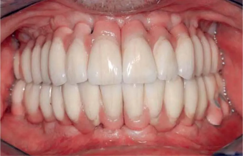
Fig 2-8aFixed implant prosthetic restoration in the mandible and maxilla.
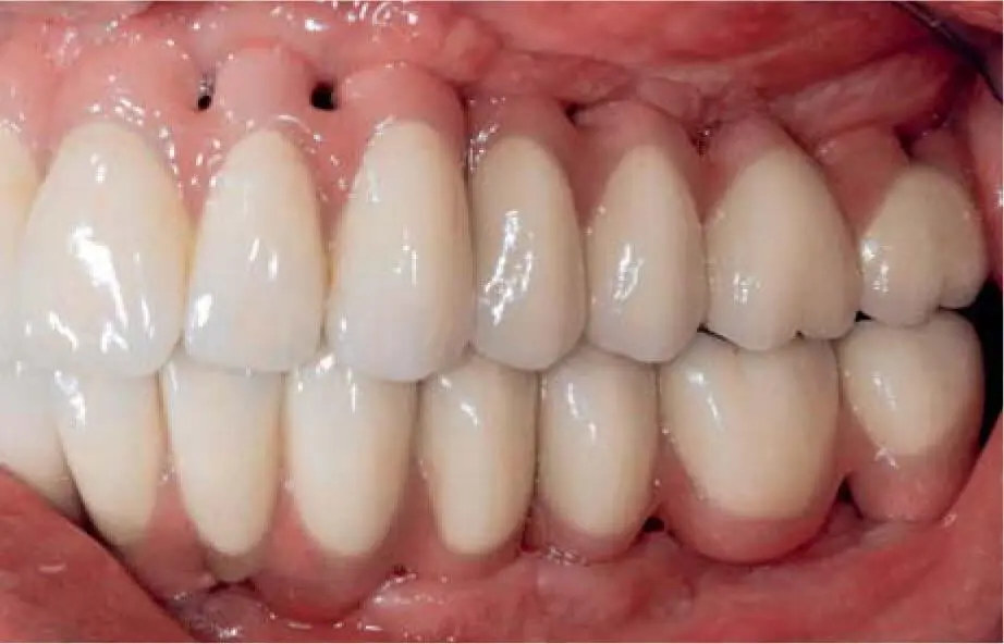
Fig 2-8bCleaning channels are important for unrestricted oral hygiene.
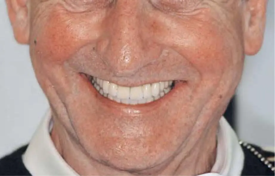
Fig 2-8cLong upper lip covers the pink ceramic with the cleaning channels.
Читать дальшеИнтервал:
Закладка:
Похожие книги на «Bone and Soft Tissue Augmentation in Implantology»
Представляем Вашему вниманию похожие книги на «Bone and Soft Tissue Augmentation in Implantology» списком для выбора. Мы отобрали схожую по названию и смыслу литературу в надежде предоставить читателям больше вариантов отыскать новые, интересные, ещё непрочитанные произведения.


Обсуждение, отзывы о книге «Bone and Soft Tissue Augmentation in Implantology» и просто собственные мнения читателей. Оставьте ваши комментарии, напишите, что Вы думаете о произведении, его смысле или главных героях. Укажите что конкретно понравилось, а что нет, и почему Вы так считаете.