Adrian Becker - Orthodontic Treatment of Impacted Teeth
Здесь есть возможность читать онлайн «Adrian Becker - Orthodontic Treatment of Impacted Teeth» — ознакомительный отрывок электронной книги совершенно бесплатно, а после прочтения отрывка купить полную версию. В некоторых случаях можно слушать аудио, скачать через торрент в формате fb2 и присутствует краткое содержание. Жанр: unrecognised, на английском языке. Описание произведения, (предисловие) а так же отзывы посетителей доступны на портале библиотеки ЛибКат.
- Название:Orthodontic Treatment of Impacted Teeth
- Автор:
- Жанр:
- Год:неизвестен
- ISBN:нет данных
- Рейтинг книги:4 / 5. Голосов: 1
-
Избранное:Добавить в избранное
- Отзывы:
-
Ваша оценка:
Orthodontic Treatment of Impacted Teeth: краткое содержание, описание и аннотация
Предлагаем к чтению аннотацию, описание, краткое содержание или предисловие (зависит от того, что написал сам автор книги «Orthodontic Treatment of Impacted Teeth»). Если вы не нашли необходимую информацию о книге — напишите в комментариях, мы постараемся отыскать её.
Provides protocols for common cases as well as complex and rare presentations Contains individual chapters on the specific aspects of the diagnosis and treatment of impaction in each of the different types of teeth Covers prevalence, etiology, diagnosis, attitudes to treatment, treatment timing, treatment methods, and prognosis Features more than 1,000 high-quality color images and illustrations
remains essential reading for all specialist orthodontists, academic researchers and instructors, oral and maxillofacial surgeons, and advanced students in orthodontics.
Orthodontic Treatment of Impacted Teeth — читать онлайн ознакомительный отрывок
Ниже представлен текст книги, разбитый по страницам. Система сохранения места последней прочитанной страницы, позволяет с удобством читать онлайн бесплатно книгу «Orthodontic Treatment of Impacted Teeth», без необходимости каждый раз заново искать на чём Вы остановились. Поставьте закладку, и сможете в любой момент перейти на страницу, на которой закончили чтение.
Интервал:
Закладка:
Partial and full‐flap closure on the palatal side
Impacted canines that are located on the palatal side are often palpable immediately beneath the palatal mucosa, which is itself firmly bound down to the underlying bone. In this situation, it is tempting to carry out the surgical removal of a circular section of the overlying mucosa and of the thin bony covering, in order to leave the tooth exposed. This has obvious advantages. In particular, the newly exposed tooth, when it finally erupts, will be favourably invested with attached gingiva. However, the palatal mucosal covering is very thick and the surgery will leave a broad cut surface, which will tend to close over unless its edges are substantially trimmed back and the dental follicle removed. Additionally, the exposure will need to be maintained using a surgical pack.
The result will be that, at the completion of the orthodontic alignment of the canine, this type of surgical approach will inevitably leave the palatal side of the tooth with a soft tissue deficiency and a long clinical crown. This is so even though, in the long term, the surrounding tissue will show the desired attached gingiva ( Figure 5.8). This method has been favoured and promoted by Schmidt and Kokich in relation to palatally impacted canines. Their rationale is based on the assumption that the canines will, in many instances, improve their position and, in the course of time, erupt autonomously in the palate [41].
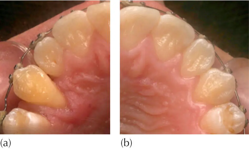
Fig. 5.8 Treatment for the right palatally impacted canine was performed with an open exposure technique. (a) The post‐treatment result shows attached gingiva of the palatal tissues covering most of the root, although the clinical crown length extends well down on the palatal side of the tooth, leaving several millimetres of root exposed on that side. The bone level is expected to be 8–10% defective compared to the untreated side. (b) The normally erupted left canine is shown for comparison.
Their descriptive study offered a retrospective evaluation of the post‐treatment periodontal status of a group of patients who had been successfully treated by this method. However, their study was not based on a control group. Additionally, there appears to be no published controlled study that investigates the reliability and predictability of this treatment protocol.
As described above in relation to labial/buccal exposure surgery, so too on the palatal side, where full‐flap closure allows the tooth to be exposed with the minimum of tissue removal and consequent reduction of surgical trauma. In addition, similar to the situation with the buccal side, it also requires the bonding of an attachment on the exposed tooth prior to suturing. When this is done and subjected to appropriate orthodontic mechanics, the final result will show that the bone support for the tooth, as well as the health and appearance of the muco‐gingival tissues, is very satisfactory, as will be demonstrated in the following chapters.
In cases where there is a deeply located impacted tooth high in the palate, there is an accumulated body of evidence supporting a full‐flap closure approach [8–12, 26–28, 30–35]. The advantages of this recommended method are both qualitative and quantitative: the excellent clinical appearance of the crown length and gingival architecture and the number of objective parameters considered in a periodontal examination. In addition, there will be a reduction in post‐surgical pain and discomfort during the healing process [42–44].
The relief of crowding to reduce canine displacement
In cases where displacement of the canine has been caused by crowding or space loss (following early extraction of deciduous teeth), it is clear that relief of the crowding will facilitate spontaneous improvement in the position of the canine. However, time may not be on the side of the clinician who opts for this approach, since if there is too much delay the tooth may erupt through the oral mucosa. Nevertheless, if it is decided to proceed with this approach, a full case analysis should be prepared, leading to a diagnosis and treatment plan for the overall malocclusion. If, while the treatment is proceeding, the crowding is to be dispersed by distal movement of the molars, it will take longer to achieve the requisite available space in the canine region to permit the spontaneous improvement of the canine position. On the other hand, a premolar extraction will provide immediate relief of the crowding and an excellent opportunity for a self‐correction of the buccal displacement, and with it the disappearance of the potential periodontal hazard.
In the treatment of a palatally impacted maxillary canine, a buccal approach to solving the crowding may sometimes be preferred, provided that its palatal displacement from the line of the arch is fairly minimal. Where the impacted tooth is vertically close to the CEJs of the adjacent teeth, the buccal approach may present a risk that interproximal bone may thereby disappear. Indeed, the greater the palatal displacement of the tooth, the greater will be the bony defect caused. Nevertheless, the buccal approach may be appropriate in cases where the teeth are marginally palatally displaced and situated higher in the maxilla. This will afford the opportunity for traction of the tooth by a more direct route to the labial archwire.
Impaction of the maxillary canine, close to the line of the dental arch, may have been caused by the mesially tipped long axis that it presents, and by the consequent direct contact of the mesial crown incline with the distal side of the root of the lateral incisor. This type of impaction is relatively simple to treat. Indeed, it should correct itself if the crown of the lateral incisor is tipped mesially, thereby closing the anterior spacing and providing room for the canine within the arch. The process will automatically cause the root apex of the incisor to move distally, coordinating the orientation of its long axis with that of the canine. If, however, the tooth maintains its stubbornness and shows no sign of erupting, then a labial surgical approach to the very mildly palatally displaced canine will often be the most suitable and, in terms of the traction, the most direct treatment.
A conservative attitude to the dental follicle
The dental sac or follicle is a fibrovascular capsule that has developed from a mesodermal condensation of cells on the outer surface of the external enamel epithelium of the enamel organ of a forming tooth. The follicle has an inner vascular plexus, through which the enamel organ is supplied with nutrients during growth and an outer vascular plexus, whose function is enlarging the bony crypt in which the tooth germ lies. This enlargement is achieved by its inherent capability to resorb the alveolar bone, notably as it begins to erupt. The follicle encompasses the entire crown of the tooth. Later, the outer surface of this sheath eventually develops into the periodontal membrane, which will connect the cementum covering of the developing root to the developing alveolar bone.
The enamel cuticle covering the crown is made up of a keratinous deposit from the ameloblasts and reduced enamel epithelium, and is contiguous with Hertwig’s epithelial root sheath. This cuticle separates the crown of the tooth from the follicle, from which the root develops and cementum forms. It is this separation that is responsible for cementum not forming on the crown of the tooth.
The resorption process of the bony crypt is accompanied by eruptive movements, which, in turn, bring the tooth follicle into close proximity to the oral mucosa. The follicular epithelium fuses with the epithelium of the oral mucosa and allows the tooth to break through an epithelium‐lined opening, with no associated bleeding. As eruption proceeds, the remainder of the follicle everts – indeed, it is turned ‘inside out’ – with the reduced enamel epithelium becoming the gingival cuff and presenting the most superficial point of attachment. When the tooth erupts in this normal manner, it is accompanied by a relatively generous band of gingival tissue, which is rounded and initially appears a little swollen. The most coronal portion of this gingival tissue is the free gingiva, which enables the attachment at its base to be direct to the cervical area of the enamel of the crown of the tooth, several millimetres coronal to the CEJ. An attachment of this nature is unusual, in the sense that the gingival band, immediately adjacent to the crown, adheres directly to the enamel through the agency of hemidesmosomes (cells that originate in the reduced enamel epithelium). Over the subsequent period of 3–4 years, this free gingival area of direct adherence to the enamel transforms into the junctional epithelium, thus providing the initial form of attachment of the gingiva to the tooth, on the cervical enamel area [45, 46].
Читать дальшеИнтервал:
Закладка:
Похожие книги на «Orthodontic Treatment of Impacted Teeth»
Представляем Вашему вниманию похожие книги на «Orthodontic Treatment of Impacted Teeth» списком для выбора. Мы отобрали схожую по названию и смыслу литературу в надежде предоставить читателям больше вариантов отыскать новые, интересные, ещё непрочитанные произведения.


Обсуждение, отзывы о книге «Orthodontic Treatment of Impacted Teeth» и просто собственные мнения читателей. Оставьте ваши комментарии, напишите, что Вы думаете о произведении, его смысле или главных героях. Укажите что конкретно понравилось, а что нет, и почему Вы так считаете.