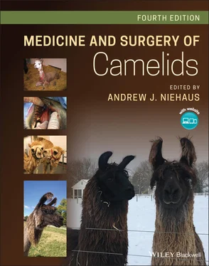
Medicine and Surgery of Camelids

Здесь есть возможность читать онлайн «Medicine and Surgery of Camelids» — ознакомительный отрывок электронной книги совершенно бесплатно, а после прочтения отрывка купить полную версию. В некоторых случаях можно слушать аудио, скачать через торрент в формате fb2 и присутствует краткое содержание. Жанр: unrecognised, на английском языке. Описание произведения, (предисловие) а так же отзывы посетителей доступны на портале библиотеки ЛибКат.

Medicine and Surgery of Camelids: краткое содержание, описание и аннотация

Предлагаем к чтению аннотацию, описание, краткое содержание или предисловие (зависит от того, что написал сам автор книги «Medicine and Surgery of Camelids»). Если вы не нашли необходимую информацию о книге — напишите в комментариях, мы постараемся отыскать её.

A thoroughly updated new edition of the classic veterinary reference In the newly revised Fourth Edition of
accomplished veterinary surgeon, Dr. Andrew J. Niehaus delivers a comprehensive reference to all aspects of camelid medicine and surgery. The book covers general husbandry, restraint, nutrition, diagnosis, anesthesia, surgery, and the treatment of specific diseases veterinarians are likely to encounter in camelid patients. Although the focus of the text remains on llamas and alpacas, camel-specific information has received more attention than in previous editions with a chapter dedicated to old-world camelids.
The editor revitalizes the emphasis on evidence-based information and pathophysiology and draws on the experience of expert contributors to provide up-to-date and authoritative material on nutrition, internal medicine, and more. A classic text of veterinary medicine, this latest edition comes complete with high-quality color photographs and access to a companion website that offers supplementary resources.
Readers will also find:
A thorough introduction to the general biology and evolution of camelids, as well as their husbandry and handling Comprehensive explorations of camelid physical exams, diagnostics, anesthesia, pain management, and surgery Topical discussions arranged by body system including the integumentary system, the musculoskeletal system and multisystem disorders Chapters dedicated to camelid radiology, parasitology, and diagnostic clinical pathology In-depth examinations of camelid toxicology, neonatology, and congenital diseases Perfect for veterinary specialists and general practitioners,
will also earn a place in the libraries of veterinary students and trainees with an interest in camelids.

Medicine and Surgery of Camelids — читать онлайн ознакомительный отрывок

Ниже представлен текст книги, разбитый по страницам. Система сохранения места последней прочитанной страницы, позволяет с удобством читать онлайн бесплатно книгу «Medicine and Surgery of Camelids», без необходимости каждый раз заново искать на чём Вы остановились. Поставьте закладку, и сможете в любой момент перейти на страницу, на которой закончили чтение.

Тёмная тема
Сбросить

Интервал:

Закладка:

Сделать

11 Chapter 11Figure 11.1 (a) Left nostril of a Dromedary camel showing narrowness of the ...Figure 11.2 Anesthetized alpaca in right lateral recumbency for passage of a...Figure 11.3 (a) Soft palate situated ventral to the epiglottis. (A) Larynx, ...Figure 11.4 (a) Skull illustrating the outlines of the maxillary (blue) and ...Figure 11.5 (a) A 12‐year‐old female alpaca presented for anorexia and abdom...Figure 11.6 Unremarkable thoracic radiograph (right lateral projection) of a...Figure 11.7 A three‐day‐old alpaca cria (male) with failure of transfer of m...Figure 11.8 (a) Standing lateral image of thorax of three‐year‐old female al...Figure 11.9 Cytospin preparation of lung lavage fluid from an alpaca with se...Figure 11.10 (a) Llama presented for swelling superficial to right mandible....

12 Chapter 12Figure 12.1 Split philtrum in an alpaca. The split philtrum present in camel...Figure 12.2 Diagram illustrating fighting teeth of male camelids. (a) Latera...Figure 12.3 Permanent incisors of a vicuña and alpaca. (a) vicuña. Incisors ...Figure 12.4 Incisor teeth of a mature llama. The incisors are spatulate and ...Figure 12.5 Occlusal surface of mandibular incisors following trimming. Note...Figure 12.6 Overgrown incisors. The teeth protrude rostrally and do not cont...Figure 12.7 Rostral teeth of a male SAC. The maxillary and mandibular canine...Figure 12.8 Diagrams of the maxillary and mandibular cheek teeth of a SAC. T...Figure 12.9 Diagram of the teeth of a premature cria. Deciduous alpaca incis...Figure 12.10 Diagram of the teeth of a newborn llama. Deciduous incisors sho...Figure 12.11 Diagram of the teeth of a five‐month‐old llama.Figure 12.12 Diagram of the teeth of a one‐year‐old llama.Figure 12.13 Diagram of the teeth of a two‐year‐old llama.Figure 12.14 Diagram of the teeth of a two‐and‐a‐half‐year‐old llama.Figure 12.15 Diagram of the teeth of a three‐year‐old llama.Figure 12.16 Diagram of the teeth of a three‐and‐a‐half‐year‐old llama.Figure 12.17 Diagram of the teeth of a four‐ to six‐year‐old llama.Figure 12.18 Diagram of the teeth of a seven‐year‐old llama (left) and an 11...Figure 12.19 Cadaver alpaca specimen with a catheter inserted through the st...Figure 12.20 Diagram of the intestinal tract of a camelid (left) and that of...Figure 12.21 Relative volume of the gastric compartments of adult cattle, sh...Figure 12.22 Comparative origins of different regions of the monogastric sto...Figure 12.23 Stomach of a neonate llama. Similar to the rumen true ruminants...Figure 12.24 Diagram of the cross section of an alpaca abdomen taken at the ...Figure 12.25 Stomach of a llama as viewed from the right lateral aspect. The...Figure 12.26 Stomach of a llama. The left side of the image is cranial and t...Figure 12.27 Glandular saccules of C1.Figure 12.28 Mucosal surface of C2. Saccules along the majority of the surfa...Figure 12.29 C3: (A) mucinous glandular tissue lines the proximal (orad) 4/5...Figure 12.30 Serosal view of C3. The acid secreting portion is the terminal ...Figure 12.31 Camel stomach, C1.Figure 12.32 Camel stomach, C2.Figure 12.33 Camel stomach, C3.Figure 12.34 Homogeneous ingesta, characteristic of C1. The contents of the ...Figure 12.35 Camelids' esophageal groove has one lip and extends from C1 int...Figure 12.36 Intestines of a llama. (A) Ileum, (B) cecum, (C) proximal loop ...Figure 12.37 Diagram of the spiral colon of a llama. The centripetal loops a...Figure 12.38 Dung pile used by llamas for urination and defecation. SACs wil...Figure 12.39 Liver of a llama.Figure 12.40 Fimbriated caudal border of a SAC liver.Figure 12.41 Decision‐making algorithm for performing abdominal surgery in c...Figure 12.42 Abdominal distension in an alpaca. (a) Left Lateral view. (b) T...Figure 12.43 Pressing and abnormal position of a llama with colic.Figure 12.44 Depression and recumbency of a llama with colic.Figure 12.45 Fractured mandibular cheek tooth.Figure 12.46 Alpaca cria with superior brachygnathism. This congenital defec...Figure 12.47 Diagram for dental examination using Tridan nomenclature.Figure 12.48 A ventrolateral skin incision is made around a draining tract. ...Figure 12.49 The soft tissues are elevated from the mandibular bone to expos...Figure 12.50 The lateral alveolar bone plate has been removed with a pneumat...Figure 12.51 Curettage of the socket is performed following removal of the t...Figure 12.52 Overcrowding of mandibular incisors of an alpaca. The third inc...Figure 12.53 The front view of an alpaca. The rostral pressure on the first ...Figure 12.54 Strategic gaps are created between the incisors so that the cau...Figure 12.55 Second incisor is lateral to the first incisor without contact....Figure 12.56 The third incisor is growing horizontally and pushing rostrally...Figure 12.57 The same alpaca as in Figure 12.5. The length of the third inci...Figure 12.58 A bright light is used to illuminate the translucent pulp canal...Figure 12.59 Gaps are created in between the incisors allowing them to more ...Figure 12.60 (a) Incisors of an alpaca following corrective trimming. Note t...Figure 12.61 Lateral radiograph of dilated thoracic esophagus (arrows outlin...Figure 12.62 A necropsy photo of a llama with megaesophagus. (a). A dilated ...Figure 12.63 Mucosal ulceration of C1 in a llama.Figure 12.64 Mucosal ulceration of C2 in a llama.Figure 12.65 Linear mucosal ulceration of C3 in a llama.Figure 12.66 Serosal surface of C1 with gastric ulcers.Figure 12.67 Ulceration of C1.Figure 12.68 Abdominal contamination from perforating gastric ulcer in C3.Figure 12.69 Acute, fibrinous peritonitis caused by perforating gastric ulce...Figure 12.70 Diagram of spiral colon impaction in a llama.Figure 12.71 Gastroliths in the glandular saccules of C1 in a llama.Figure 12.72 Postmortem examination revealing jejunal torsion.Figure 12.73 Multiple trichophytobezoars removed from C1 of an alpaca cria. ...Figure 12.74 Lateral abdominal radiograph of a llama revealing gastroliths i ...Figure 12.75 Transabdominal ultrasound image in an adult alpaca. Excessive a...Figure 12.76 Acute peritonitis, with fibrin tags.Figure 12.77 Chronic peritonitis, with fibrous bands that may incarcerate a ...Figure 12.78 Gross postmortem examination of a camel revealing hepatitis.Figure 12.79 Gross postmortem examination of a llama revealing hepatic necro...Figure 12.80 Gross postmortem examination of a llama revealing hepatic lipid...Figure 12.81 Position of the internal abdominal oblique muscle. (A) Tuber co...Figure 12.82 Lateral abdomen of a llama demonstrating the position of the tr...Figure 12.83 Preputial muscles of the llama.Figure 12.84 Cradle used to restrain llama in dorsal recumbency.Figure 12.85 Diagram of camelid cradle. (A) 30 cm, (B) 38 cm, (C) 125 cm.Figure 12.86 Diagram of the placement of an inverted cruciate suture for a v...Figure 12.87 Alpaca following repair of atresia ani.

13 Chapter 13Figure 13.1 Camelid liver with severe hepatic lipidosis. The yellow discolor...

14 Chapter 14Figure 14.1 Blood from a healthy alpaca. There are numerous elliptical RBCs,...Figure 14.2 Blood from a llama. There is a large granular lymphocyte in the ...Figure 14.3 Blood from an alpaca. Numerous Mycoplasma haemolamae organisms a...Figure 14.4 Blood from an alpaca that was markedly anemic and was treated wi...Figure 14.5 Blood from a llama with marked neutrophilic inflammation. These ...Figure 14.6 Blood from an alpaca. There are two neutrophils, one monocyte (a...Figure 14.7 Blood from an alpaca. There are two neutrophils and a monocyte (...Figure 14.8 Blood from an alpaca. There is a neutrophil, a small lymphocyte,...Figure 14.9 Blood from an alpaca. Though this is a thicker area than would b...Figure 14.10 Blood from a normal alpaca. Individual platelets are small with...Figure 14.11 Abdominal fluid from an alpaca with lymphoma. There are three n...Figure 14.12 Abdominal fluid from a four‐year‐old male alpaca with septic pe...Figure 14.13 CSF from a seven‐year‐old male castrated alpaca with neurologic...

Читать дальше
Тёмная тема
Сбросить

Интервал:

Закладка:

Сделать

Похожие книги на «Medicine and Surgery of Camelids»

Представляем Вашему вниманию похожие книги на «Medicine and Surgery of Camelids» списком для выбора. Мы отобрали схожую по названию и смыслу литературу в надежде предоставить читателям больше вариантов отыскать новые, интересные, ещё непрочитанные произведения.


Отзывы о книге «Medicine and Surgery of Camelids»

Обсуждение, отзывы о книге «Medicine and Surgery of Camelids» и просто собственные мнения читателей. Оставьте ваши комментарии, напишите, что Вы думаете о произведении, его смысле или главных героях. Укажите что конкретно понравилось, а что нет, и почему Вы так считаете.

x