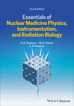
Rachel A. Powsner - Essentials of Nuclear Medicine Physics, Instrumentation, and Radiation Biology
Здесь есть возможность читать онлайн «Rachel A. Powsner - Essentials of Nuclear Medicine Physics, Instrumentation, and Radiation Biology» — ознакомительный отрывок электронной книги совершенно бесплатно, а после прочтения отрывка купить полную версию. В некоторых случаях можно слушать аудио, скачать через торрент в формате fb2 и присутствует краткое содержание. Жанр: unrecognised, на английском языке. Описание произведения, (предисловие) а так же отзывы посетителей доступны на портале библиотеки ЛибКат.
- Название:Essentials of Nuclear Medicine Physics, Instrumentation, and Radiation Biology
- Автор:
- Жанр:
- Год:неизвестен
- ISBN:нет данных
- Рейтинг книги:4 / 5. Голосов: 1
-
Избранное:Добавить в избранное
- Отзывы:
-
Ваша оценка:
- 80
- 1
- 2
- 3
- 4
- 5
Essentials of Nuclear Medicine Physics, Instrumentation, and Radiation Biology: краткое содержание, описание и аннотация
Предлагаем к чтению аннотацию, описание, краткое содержание или предисловие (зависит от того, что написал сам автор книги «Essentials of Nuclear Medicine Physics, Instrumentation, and Radiation Biology»). Если вы не нашли необходимую информацию о книге — напишите в комментариях, мы постараемся отыскать её.
Essentials of Nuclear Medicine Physics, Instrumentation, and Radiation Biology — читать онлайн ознакомительный отрывок
Ниже представлен текст книги, разбитый по страницам. Система сохранения места последней прочитанной страницы, позволяет с удобством читать онлайн бесплатно книгу «Essentials of Nuclear Medicine Physics, Instrumentation, and Radiation Biology», без необходимости каждый раз заново искать на чём Вы остановились. Поставьте закладку, и сможете в любой момент перейти на страницу, на которой закончили чтение.
Интервал:
Закладка:
Table of Contents
1 Cover
2 Dedication Page
3 Title Page
4 Copyright Page
5 Preface
6 Acknowledgments
7 CHAPTER 1: Basic Nuclear Medicine PhysicsProperties and structure of matter Radioactivity Questions Answers
8 CHAPTER 2: Interaction of Radiation with Matter Interaction of photons with matter Interaction of charged particles with matter Reference Questions Answers
9 CHAPTER 3: Formation of Radionuclides Generators Cyclotrons Reactors Questions Answers
10 CHAPTER 4: Nonscintillation Detectors Gas‐filled detectors Semiconductor detectors Photographic and luminescent detectors Questions Answers
11 CHAPTER 5: Scintillation Detectors Structure Sodium iodide detector energy spectrum Characteristics of scintillation detectors Types of scintillation‐based detectors Questions Answers
12 CHAPTER 6: Imaging InstrumentationTheory and structure Planar imaging Questions Answers
13 CHAPTER 7: Single‐photon Emission Computed Tomography (SPECT) Equipment Acquisition Dedicated cardiac SPECT cameras Quantitation of lesion activity in SPECT studies Questions Answers
14 CHAPTER 8: Positron Emission Tomography (PET) Advantages of PET imaging PET camera components Factors affecting resolution in PET imaging Attenuation in PET imaging Standard uptake values References Questions Answers
15 CHAPTER 9: X‐ray Computed Tomography (CT) X‐ray production X‐ray imaging Computed tomography Questions Answers
16 CHAPTER 10: Magnetic Resonance Imaging (MRI) Background Introduction to MRI References Questions Answers
17 CHAPTER 11: Hybrid Imaging Systems PET‐CT and SPECT‐CT imaging Current limitations in SPECT‐CT and PET‐CT hybrid imaging PET‐MR imaging Questions Answers
18 CHAPTER 12: Image Reconstruction, Processing, and Display Reconstruction Postreconstruction image processing Questions Answers
19 CHAPTER 13: Information Technology Network DICOM PACS Information systems Additional DICOM capabilities Questions Answers
20 CHAPTER 14: Quality Control Nonimaging devices Imaging Reference Questions Answers
21 CHAPTER 15: Radiation Biology Radiation Units The effects of radiation on living organisms References Questions Answers
22 CHAPTER 16: Radiation Dosimetry Nuclear medicine dosimetry CT dosimetry Reference Questions Answers
23 CHAPTER 17: Radiation SafetyRationale Dose limits Methods for limiting exposure Regulations References Questions
24 CHAPTER 18: Radiopharmaceutical Therapy Introduction Tissue‐specific radiopharmaceutical treatments Radiation protection References Questions Answers
25 CHAPTER 19: Management of Nuclear Event Casualties Interaction of radiation with tissue Radionuclides Hospital response to a radiation accident Exposure and contamination Hospital facilities External decontamination Personnel Evaluation of the radiation accident victim Acute radiation sickness Local radiation injury to the skin Medical and industrial accidental overexposure References Questions Answers
26 Appendix A: Common Nuclides
27 Appendix B: Major Dosimetry for Common Pharmaceuticals
28 Appendix C: Guide to Nuclear Regulatory Commission (NRC) PublicationsTitle 10, “Energy”, Code of Federal Regulations (10CFR) [1] NUREG – 1556, Vol. 9, Revision 3, Consolidated Guidance About Materials Licenses [2] References
29 Appendix D: Recommended Reading by TopicReview of Basic Physics Nuclear Medicine Basic Nuclear Medicine Physics and Instrumentation PET Technology CT Technology MRI Technology DICOM and Information Technology Nuclear Medicine Quality Control Radiobiology Radiation Dosimetry NRC Regulations Therapeutics in Nuclear Medicine
30 Index
31 End User License Agreement
List of Tables
1 Chapter 1 Table 1.1 Properties of the subatomic particles Table 1.2 Conversion values for units of radioactivity
2 Chapter 2 Table 2.1 Predominant photon interactions in common materials at diagnostic... Table 2.2 HVL and TVL of lead for photons of common medical nuclides
3 Chapter 3 Table 3.1 Characteristics of three commonly used generators
4 Chapter 8Table 8.1 Common positron‐emitting nuclides [1]Table 8.2 Some of the available PET radiopharmaceuticalsTable 8.3 Properties of crystals used for PET imaging
5 Chapter 15Table 15.1 Radiation weighting factors ( W R) for ionizing radiationTable 15.2 Quantities and units used in nuclear medicineTable 15.3 Radiosensitivity of cell typesTable 15.4 Organ toxicity from acute exposureTable 15.5 Radiosensitivity of the human embryo and fetus up to week 25
6 Chapter 16Table 16.1 Part of the estimated dosimetry for 18F‐FDGTable 16.2 Hypothetical physical, biologic, and effective half‐livesTable 16.3 Sample physical, biologic, and effective half‐livesTable 16.4 Tissue weighting factors ( W T)
7 Chapter 17Table 17.1 Terms used to describe radiation exposure aTable 17.2 NRC radiation dose equivalent limits for occupational exposure (...Table 17.3 General guidance for differentiating minor versus major spills a...Table 17.4 Sample recommended times to discontinue breast‐feeding following...
8 Chapter 19Table 19.1 Selected radionuclides used for occupational applications and/or...Table 19.2 Signs, symptoms, and recommended disposition of exposed patientsTable 19.3 Estimated local radiation dose and recommended patient managemen...
9 3Table D.1 Selected sections of 10CFR Parts 19, 20, 35, and NUREG‐1556, Vol. 9
List of Illustrations
1 Chapter 1 Figure 1.1 Electrostatic charge. Figure 1.2 The NaCl molecule is the smallest unit of salt that retains the c... Figure 1.3 Periodic table. Figure 1.4 Flat atom. The standard two‐dimensional drawing of atomic structu... Figure 1.5 An electron shell is a representation of the energy level associa... Figure 1.6 K, L, and M electron shells. Figure 1.7 Electron orbitals and sub‐orbitals. (a) s orbital, (b) p suborbit... Figure 1.8 The nucleus of an atom is composed of protons and neutrons. Figure 1.9 Nuclear binding force is strong enough to overcome the electrical... Figure 1.10 Standard atomic notation. Figure 1.11 Nuclides of the same atomic number but different atomic mass are... Figure 1.12 Combinations of neutrons and protons that can coexist in a stabl... Figure 1.13 Alpha decay. Figure 1.14 Fission of a 235U nucleus. Figure 1.15 β –(negatron) decay. Figure 1.16 β +(positron) decay. Figure 1.17 Beta emissions (both β –and β +) are ejected from the nucle... Figure 1.18 Electron capture. Figure 1.19 Isomeric transition. Excess nuclear energy is carried off as a g... Figure 1.20 Internal conversion. As an alternative to gamma emission, it can... Figure 1.21 Decay schematics. Figure 1.22 Decay schemes showing principal transitions for technetium‐99m, ... Figure 1.23 Decay curve. Note the progressive replacement of radioactive ato...
2 Chapter 2 Figure 2.1 Predominant type of interaction for various combinations of incid... Figure 2.2 Compton scattering. Figure 2.3 Angle of photon scattering. Figure 2.4 Photoelectric effect. Figure 2.5 Attenuation. Figure 2.6 The amount of attenuation of a photon beam is dependent on the ph... Figure 2.7 Penetrating radiation and nonpenetrating radiation. Figure 2.8 Excitation and de‐excitation. Figure 2.9 Ionization. The ejected electron and the positively charged atom ... Figure 2.10 Particle range in an absorber. Figure 2.11 Annihilation reaction. Figure 2.12 Einstein’s theory of the equivalence of energy and mass. Figure 2.13 Bremsstrahlung. Beta particles (β –) and positrons (β +) tha... Figure 2.14 Bremsstrahlung X‐ray energies increase with increasing proximity... Figure 2.15 Bremsstrahlung X‐ray energies vary from near zero to the maximum...
Читать дальшеИнтервал:
Закладка:
Похожие книги на «Essentials of Nuclear Medicine Physics, Instrumentation, and Radiation Biology»
Представляем Вашему вниманию похожие книги на «Essentials of Nuclear Medicine Physics, Instrumentation, and Radiation Biology» списком для выбора. Мы отобрали схожую по названию и смыслу литературу в надежде предоставить читателям больше вариантов отыскать новые, интересные, ещё непрочитанные произведения.
Обсуждение, отзывы о книге «Essentials of Nuclear Medicine Physics, Instrumentation, and Radiation Biology» и просто собственные мнения читателей. Оставьте ваши комментарии, напишите, что Вы думаете о произведении, его смысле или главных героях. Укажите что конкретно понравилось, а что нет, и почему Вы так считаете.


