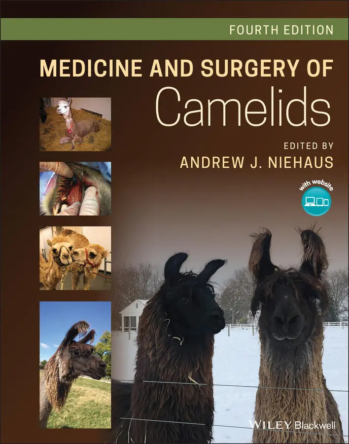
Medicine and Surgery of Camelids
Здесь есть возможность читать онлайн «Medicine and Surgery of Camelids» — ознакомительный отрывок электронной книги совершенно бесплатно, а после прочтения отрывка купить полную версию. В некоторых случаях можно слушать аудио, скачать через торрент в формате fb2 и присутствует краткое содержание. Жанр: unrecognised, на английском языке. Описание произведения, (предисловие) а так же отзывы посетителей доступны на портале библиотеки ЛибКат.
- Название:Medicine and Surgery of Camelids
- Автор:
- Жанр:
- Год:неизвестен
- ISBN:нет данных
- Рейтинг книги:3 / 5. Голосов: 1
-
Избранное:Добавить в избранное
- Отзывы:
-
Ваша оценка:
Medicine and Surgery of Camelids: краткое содержание, описание и аннотация
Предлагаем к чтению аннотацию, описание, краткое содержание или предисловие (зависит от того, что написал сам автор книги «Medicine and Surgery of Camelids»). Если вы не нашли необходимую информацию о книге — напишите в комментариях, мы постараемся отыскать её.
accomplished veterinary surgeon, Dr. Andrew J. Niehaus delivers a comprehensive reference to all aspects of camelid medicine and surgery. The book covers general husbandry, restraint, nutrition, diagnosis, anesthesia, surgery, and the treatment of specific diseases veterinarians are likely to encounter in camelid patients. Although the focus of the text remains on llamas and alpacas, camel-specific information has received more attention than in previous editions with a chapter dedicated to old-world camelids.
The editor revitalizes the emphasis on evidence-based information and pathophysiology and draws on the experience of expert contributors to provide up-to-date and authoritative material on nutrition, internal medicine, and more. A classic text of veterinary medicine, this latest edition comes complete with high-quality color photographs and access to a companion website that offers supplementary resources.
Readers will also find:
A thorough introduction to the general biology and evolution of camelids, as well as their husbandry and handling Comprehensive explorations of camelid physical exams, diagnostics, anesthesia, pain management, and surgery Topical discussions arranged by body system including the integumentary system, the musculoskeletal system and multisystem disorders Chapters dedicated to camelid radiology, parasitology, and diagnostic clinical pathology In-depth examinations of camelid toxicology, neonatology, and congenital diseases Perfect for veterinary specialists and general practitioners,
will also earn a place in the libraries of veterinary students and trainees with an interest in camelids.
Medicine and Surgery of Camelids — читать онлайн ознакомительный отрывок
Ниже представлен текст книги, разбитый по страницам. Система сохранения места последней прочитанной страницы, позволяет с удобством читать онлайн бесплатно книгу «Medicine and Surgery of Camelids», без необходимости каждый раз заново искать на чём Вы остановились. Поставьте закладку, и сможете в любой момент перейти на страницу, на которой закончили чтение.
Интервал:
Закладка:
8 Chapter 8Figure 8.1 Lymphosarcoma of abdominal organs.Figure 8.2 Hyperthermic alpaca cooling herself in a pond.Figure 8.3 Bilateral scrotal edema caused by hyperthermia (a) compared to un...
9 Chapter 9Figure 9.1 Diagram of the skin. (A) Laceration extending into the dermis, (B...Figure 9.2 Handmade rope of llama fiber from Peru.Figure 9.3 Q'aras' type llamas at Machu Picchu in Peru.Figure 9.4 Chaku type llama. Photo courtesy of Donna Moore, Timberlane Llama...Figure 9.5 (a) Huacaya fiber showing crimp. (b) Huacaya in full fleece. Phot...Figure 9.6 (a) Suri fiber showing locks.(b) Suri in Full fleece. Photo c...Figure 9.7 Llama with lion cut. Photo courtesy of Barb Baker, Baker & Compan...Figure 9.8 Alpaca “slick‐shorn” Photo courtesy of Lindsay Warne, The Alpacas...Figure 9.9 (a) Biopsy of alpaca skin with low‐density follicles present with...Figure 9.10 (a) Metatarsal gland in a llama. (b) Metatarsal gland in an alpa...Figure 9.11 Metatarsal gland demonstrating excretions flaking off.Figure 9.12 (a) Interdigital gland in a llama foot. (b) Interdigital gland i...Figure 9.13 (a). Unknown lesion in mouth, probably due to irritation from fe...Figure 9.14 Chronic nonspecific dermatitis. The animal pictured in (b), (c),...Figure 9.15 (a) Dark nose and ear “dermatitis” in a black alpaca. (b) Dark n...Figure 9.16 Camelpox, acute, disseminated.Figure 9.17 Camelpox, acute, scrotum.Figure 9.18 Camelpox, acute, lung lesions.Figure 9.19 Camelpox, subacute.Figure 9.20 Camelpox, healing lesions.Figure 9.21 Camelpox experimentally produced in a guanaco.Figure 9.22 Individual pox lesion.Figure 9.23 Contagious ecthyma (Orf) is a zoonotic disease.Figure 9.24 (a and b) Contagious ecthyma (orf) lesion in an alpaca.Figure 9.25 Diagrams for identification of Trichophyton verrucosum . (A) Fibe...Figure 9.26 Diagrams for identification of Trichophyton mentagrophytes . (A) ...Figure 9.27 (a) Ringworm in a llama. (b) Ringworm in an alpaca.Figure 9.28 Life cycle of Coccidioides immitis . (A) Arthroconidia, (B) emer...Figure 9.29 (a) Dermal coccidioidomycosis. (b) Dermal coccidioidomycosis. (c...Figure 9.30 Culture of Coccidioides immitis grown from swab of submandibular...Figure 9.31 Large abscess on shoulder of an alpaca. Red triangle illustrates...Figure 9.32 (a) Corynebacterium pseudotuberculosis abscess on a camel's leg,...Figure 9.33 (a and b) Botryomycosis (Staphylococcus) in an alpaca.Figure 9.34 (a) Staphylococcus aureus dermatitis (severe) involving the enti...Figure 9.35 Acute facial zinc‐responsive dermatitis.Figure 9.36 (a) Follicular/sebaceous gland cyst. (b) Site of surgical remova...Figure 9.37 Typical follicular cyst.Figure 9.38 Follicular cyst after shearing ruptured the cyst.Figure 9.39 (a and b) Mild cases of munge in crias.Figure 9.40 (a) Moderately severe munge in a cria. Note the fissures and inf...Figure 9.41 (a) Severe case of munge in a yearling alpaca. (b) Same yearling...Figure 9.42 Acute nonspecific dermatitis.Figure 9.43 Nonspecific dermatitis, seen during reproductive exams in otherw...Figure 9.44 (a) Alpaca burn wound on footpad resulting from a forest fire. T...Figure 9.45 Photosensitization of the muzzle associated with fascioliasis.Figure 9.46 Photosensitization of the ears of a llama cria.Figure 9.47 (a and b) Ichthyosis in an adult alpaca.Figure 9.48 Bottom view of llama foot.Figure 9.49 Bottom view of adult alpaca foot.Figure 9.50 Diagram of SAC foot and pastern. (N) Toenail, (DC) digital cushi...Figure 9.51 Lateral radiograph of the foot, pastern, and fetlock of a llama....Figure 9.52 Normal llama foot, lateral view.Figure 9.53 Normal alpaca foot, lateral view.Figure 9.54 Normal llama foot, dorsal view.Figure 9.55 Normal alpaca foot, dorsal view.Figure 9.56 Digital cushion, lateral view.Figure 9.57 (a) Toenail of a camel. (b) Lamina of a toenail of a camel.Figure 9.58 Lamina of toenail of a llama.Figure 9.59 Bottom of a camel foot.Figure 9.60 Camel foot dorsal view.Figure 9.61 Elongated toenails on a llama.Figure 9.62 Elongated toenails on a llama.Figure 9.63 Elongated toenails on an alpaca in Peru. Extreme inward curling ...Figure 9.64 Elongated toenails, both curved outward.Figure 9.65 Elongated toenails, both curved inward.Figure 9.66 Saboten hoof‐trimming shears.Figure 9.67 ARS sheep hoof‐trimming shears.Figure 9.68 Equine hoof nippers.Figure 9.69 (a) Obstetrical wire is being used to trim an overgrown toenail ...Figure 9.70 Avulsed toenail of a llama. (a) Dorsal view, (b) Palmar view, (c...Figure 9.71 (a and b) Infectious pododermatitis.Figure 9.72 Interdigital dermatitis with abundant foot fiber.Figure 9.73 (a) Trauma to foot after degloving injury to leg and foot. (b) S...Figure 9.74 (a and b) Waxy accumulations and crusts of the interdigital tiss...Figure 9.75 Diagram of a camelid teat and collecting system. (A) Streak cana...Figure 9.76 Latex cast of mammary gland‐collecting system.Figure 9.77 Diagrams of the side view of teats: (A) Normal, (B) alternate or...Figure 9.78 Diagram of the ventral view of teats of a camelid: (A) normal co...Figure 9.79 Double teat in a llama.Figure 9.80 Diagrams of early embryologic development of the bovine mammary ...Figure 9.81 Acute mastitis and dermatitis caused by E. coli infection.Figure 9.82 Acute mastitis with abscessation.Figure 9.83 Necrotic mastitis caused by E. coli infection.Figure 9.84 (a) Pinnae of a llama, banana ears. (b) Pinnae of a llama, round...Figure 9.85 (a) Pinnae of alpaca ears – cria.(b) Pinnae of alpaca ears –...Figure 9.86 Diagram of the relationship of the pinna, external ear canal, ty...Figure 9.87 Dorsoventral radiograph of the skull with sclerosis of the tympa...Figure 9.88 Facial paralysis.Figure 9.89 Facial paralysis due to inner ear infection.Figure 9.90 Head tilt due to middle ear infection.Figure 9.91 Chronic facial paralysis.Figure 9.92 Diagrams of llama ear shapes. (A) Normal banana ear, (B) sharp‐p...Figure 9.93 Diagrams of alpaca ear shapes. (A) Normal spear‐shaped ears, (B)...Figure 9.94 (a) Gopher ear of a llama in Peru. (b) Gopher ear of a llama in ...Figure 9.95 Pinna defect, one short and one normal ear.Figure 9.96 (a) Pinna in a newborn alpaca, temporarily curled tips but norma...Figure 9.97 Frostbite ears.Figure 9.98 Deep, contaminated wound, distal to the hock involving multiple ...Figure 9.99 This male alpaca was attacked by a dog and sustained multiple wo...Figure 9.100 (a) Second‐intention wound healing of a left flank wound result...Figure 9.101 Alpaca with extensive head wound. (a) There is not enough surro...Figure 9.102 Llama with chemically induced keratitis secondary to chlorhexid...Figure 9.103 Llama with chronic wound over the carpus. Wounds on the distal ...
10 Chapter 10Figure 10.1 Diagrams of camelid skeletons. (a) Llama, (b) Alpaca, (c). Bactr...Figure 10.2 Diagrams of pastern angulation. (a) Appropriate angulation for a...Figure 10.3 Hyperextension of fetlock.Figure 10.4 Hind limb, lateral view. (a) Straight leg, (b) excessive angulat...Figure 10.5 Treatment of a radius fracture with a Thomas splint/cast combina...Figure 10.6 A transfixation pin cast is used to stabilize a metacarpal fract...Figure 10.7 Radiograph of a fracture of the dens of C2.Figure 10.8 Diagram of the use of wire to stabilize a rostral mandibular fra...Figure 10.9 Radiograph of a llama fetlock with fetlock angle measuring appro...Figure 10.10 Radiograph of llama with severe hyperextension of the fetlock. ...Figure 10.11 Medial to lateral projection of a luxated scapulohumeral joint ...Figure 10.12 Postoperative radiograph of an alpaca following open reduction ...Figure 10.13 Lateral radiograph of an alpaca six‐months following scapulohum...Figure 10.14 A lateral to medial projection of an alpaca with a ruptured cra...Figure 10.15 Bilateral medial patellar luxation in a llama neonate. Cria has...Figure 10.16 Imbricating sutures being placed alongside the patella and pate...Figure 10.17 Lateral radiograph of tibiotarsal luxation. Note that the tibio...Figure 10.18 A dorso‐plantar radiographic projection showing sequestrum in t...Figure 10.19 A juvenile llama cria with ligamentous laxity that could be str...Figure 10.20 A juvenile alpaca cria with flexural contracture in the hind li...
Читать дальшеИнтервал:
Закладка:
Похожие книги на «Medicine and Surgery of Camelids»
Представляем Вашему вниманию похожие книги на «Medicine and Surgery of Camelids» списком для выбора. Мы отобрали схожую по названию и смыслу литературу в надежде предоставить читателям больше вариантов отыскать новые, интересные, ещё непрочитанные произведения.


Обсуждение, отзывы о книге «Medicine and Surgery of Camelids» и просто собственные мнения читателей. Оставьте ваши комментарии, напишите, что Вы думаете о произведении, его смысле или главных героях. Укажите что конкретно понравилось, а что нет, и почему Вы так считаете.