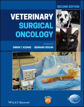
Veterinary Surgical Oncology
Здесь есть возможность читать онлайн «Veterinary Surgical Oncology» — ознакомительный отрывок электронной книги совершенно бесплатно, а после прочтения отрывка купить полную версию. В некоторых случаях можно слушать аудио, скачать через торрент в формате fb2 и присутствует краткое содержание. Жанр: unrecognised, на английском языке. Описание произведения, (предисловие) а так же отзывы посетителей доступны на портале библиотеки ЛибКат.
- Название:Veterinary Surgical Oncology
- Автор:
- Жанр:
- Год:неизвестен
- ISBN:нет данных
- Рейтинг книги:5 / 5. Голосов: 1
-
Избранное:Добавить в избранное
- Отзывы:
-
Ваша оценка:
- 100
- 1
- 2
- 3
- 4
- 5
Veterinary Surgical Oncology: краткое содержание, описание и аннотация
Предлагаем к чтению аннотацию, описание, краткое содержание или предисловие (зависит от того, что написал сам автор книги «Veterinary Surgical Oncology»). Если вы не нашли необходимую информацию о книге — напишите в комментариях, мы постараемся отыскать её.
The new edition of the most comprehensive resource on surgical oncology, covering both basic and advanced surgical oncology procedures in small animals Veterinary Surgical Oncology
Veterinary Surgical Oncology
Veterinary Surgical Oncology, Second Edition
Veterinary Surgical Oncology — читать онлайн ознакомительный отрывок
Ниже представлен текст книги, разбитый по страницам. Система сохранения места последней прочитанной страницы, позволяет с удобством читать онлайн бесплатно книгу «Veterinary Surgical Oncology», без необходимости каждый раз заново искать на чём Вы остановились. Поставьте закладку, и сможете в любой момент перейти на страницу, на которой закончили чтение.
Интервал:
Закладка:
5 Chapter 5 Figure 5.1 Squamous cell carcinoma of the nasal planum in a golden retriever... Figure 5.2 Preoperative appearance of a nasal planum SCC in a cat. The deepl... Figure 5.3 The typical appearance of an SCC of the pinna in a cat. Note the ... Figure 5.4 Immediate postoperative appearance following nasal planum resecti... Figure 5.5 Cosmetic appearance six weeks after nasal planum resection in a c... Figure 5.6 Cosmetic appearance following nasal planum resection in a golden ... Figure 5.7 A MCT along the medial border of the helix of the pinna. Incision... Figure 5.8 (a) Pinnectomy in a cat with multifocal head and neck SCC. Note t... Figure 5.9 Immediate postoperative appearance of the reconstructed pinna fol... Figure 5.10 Partial pinnectomy of the affected pinna in Figure 5.3. Minimum ... Figure 5.11 (a) A 1.7 × 1.9 × 1 cm fluid‐filled mass on the convex surface o...Figure 5.12 (a) Ceruminous gland adenocarcinoma in the horizontal ear canal ...Figure 5.13 (a) Ceruminous gland cysts affecting the pinna and external ear ...Figure 5.14 (a) Vertical ear canal resection for ceruminous adenoma of the v...Figure 5.15 Traction‐avulsion of a nasopharyngeal polyp extending into the e...Figure 5.16 Traction‐avulsion of a large nasopharyngeal polyp in a cat. Note...Figure 5.17 (a, b) Salivary adenoma of a minor salivary gland in the lip of ...Figure 5.18 (a) MCT affecting the cheek of a seven‐year‐old female spayed Be...Figure 5.19 (a) Local recurrence of an SCC on the dorsolateral lip of an 11‐...Figure 5.20 (a) A MCT involving the rostral lip of an adult mixed breed dog....Figure 5.21 (a) A buccal rotation flap was used to repair the lip of a seven...Figure 5.22 Superficial temporal axial pattern flap. (a) A cutaneous fistula...Figure 5.23 (a, b) A 3.2 × 1.8 × 1.2 cm squamous cell carcinoma confined to ...Figure 5.24 (a) Buccal SCC in an 11‐year‐old male neutered west highland whi...
6 Chapter 6Figure 6.1 Computed tomography scan of a dog with a zygomatic squamous cell ...Figure 6.2 Computed tomography scan of a dog with a multilobular osteochondr...Figure 6.3 Sentinel lymph node mapping and biopsy as described by Brissot an...Figure 6.4 Bone tunnels have been drilled into the hard palate to provide a ...Figure 6.5 Anatomy of the mandible. The inferior alveolar artery, vein, and ...Figure 6.6 Anatomy of the mandible showing the muscles of mastication. (a) P...Figure 6.7 Unilateral rostral mandibulectomy. (a) The labial and gingival mu...Figure 6.8 Bilateral rostral mandibulectomy. (a) A malignant melanoma involv...Figure 6.9 Segmental mandibulectomy. (a) The labial and gingival mucosa are ...Figure 6.10 Rim mandibulectomy. (a) The labial and gingival mucosa are incis...Figure 6.11 Subtotal and total hemimandibulectomy. (a) A skin incision may b...Figure 6.12 Ventral approach for segmental mandibulectomy in a dog with a ma...Figure 6.13 Vertical ramus or caudal mandibulectomy. (a) The dotted line rep...Figure 6.14 It is important that a feeding tube be inserted following any ma...Figure 6.15 (a and b) The typical postoperative appearance of a dog followin...Figure 6.16 The typical postoperative appearance of a dog following bilatera...Figure 6.17 The typical postoperative appearance of a dog following subtotal...Figure 6.18 Reconstruction of a segmental mandibulectomy in a cat with a cus...Figure 6.19 A ranula‐like lesion (arrow) in a dog one day following subtotal...Figure 6.20 Dehiscence of the commissure of the lips and rostral end of the ...Figure 6.21 Mandibular drift following subtotal hemimandibulectomy for a man...Figure 6.22 Anatomy of the maxilla and skull. (a and b) A number of maxillec...Figure 6.23 (a) A benign acanthomatous ameloblastoma arising from the period...Figure 6.24 Unilateral rostral maxillectomy. (a) The labial and gingival muc...Figure 6.25 Bilateral rostral maxillectomy. (a) Bilateral rostral maxillecto...Figure 6.26 (a and b) Typical drooped nose cosmetic appearance following bil...Figure 6.27 (a) For the cantilever suture technique, a 3–5 cm skin incision ...Figure 6.28 (a) Bilateral rostral maxillectomy combined with nasal planum re...Figure 6.29 An 18‐day postoperative image of a dog with a nasal planum resec...Figure 6.30 Radical maxillectomy. (a and b) The labial and gingival mucosal ...Figure 6.31 (a) Caudal maxillectomy through an intraoral approach is recomme...Figure 6.32 (a) A caudal maxillectomy through combined approach is recommend...Figure 6.33 Segmental maxillectomy in a dog with a biologically high‐grade, ...Figure 6.34 Caudal maxillectomy through a combined approach in a cat with a ...Figure 6.35 (a) The typical appearance of a dog following caudal maxillectom...Figure 6.36 (a) The postoperative appearance of a dog following unilateral r...Figure 6.37 The typical appearance of a dog following bilateral rostral maxi...Figure 6.38 (a–c) The typical postoperative appearance following radical max...Figure 6.39 Dehiscence of the intraoral incision following caudal maxillecto...Figure 6.40 The oronasal fistula has been debrided and repaired with a trans...Figure 6.41 A free auricular cartilage autograft for treatment of a central ...Figure 6.42 (a) A caudal midline oronasal fistula following segmental maxill...Figure 6.43 Ulceration of the upper lip in a cat following unilateral hemime...Figure 6.44 (a) A melanotic malignant melanoma of the caudal mandible in a d...Figure 6.45 Squamous cell carcinoma can have a variable gross appearance. Ul...Figure 6.46 A fibrosarcoma in the caudal maxilla of a dog. Note the typical ...Figure 6.47 An acanthomatous ameloblastoma arising from the periodontal liga...Figure 6.48 A peripheral odontogenic fibroma arising from the periodontal li...Figure 6.49 (a) A squamous cell carcinoma arising from the alveolar ridge of...Figure 6.50 An inductive fibroameloblastoma arising from the rostral maxilla...Figure 6.51 An eosinophilic granuloma in a cat. These lesions are non‐neopla...Figure 6.52 (a) Full‐thickness excision of the hard palate is indicated for ...Figure 6.53 (a) A fibrosarcoma on the free portion of the tongue of a dog ha...Figure 6.54 (a) A primary glossal soft tissue sarcoma in a dog; (b) The soft...Figure 6.55 (a and b). The tumor is removed with a wedge of the tongue. The ...Figure 6.56 (a) A benign extramedullary plasmacytoma of the tongue in a dog;...Figure 6.57 (a and b) A full‐thickness incision is made transversely across ...Figure 6.58 (a) A full‐thickness longitudinal incision is made along the mid...Figure 6.59 (a) A benign extramedullary plasmacytoma of the tongue in a dog;...
7 Chapter 7Figure 7.1 (a) Lateral and (b) ventrodorsal radiograph of a dog with an esop...Figure 7.2 CT of an esophageal tumor located in the region of the cardia....Figure 7.3 Esophagoscopy of a carcinoma diagnosed by endoscopic‐assisted bio...Figure 7.4 Endoscopic view of an esophageal leiomyosarcoma.Figure 7.5 An abdominal approach has been used for a leiomyoma involving the...Figure 7.6 (a) Resection and anastomosis of an esophageal leiomyoma depicted...Figure 7.7 Precontrast helical CT image of a dog with a midbody gastric neop...Figure 7.8 (a) Intraoperative image of the initial dissection for a gastric ...Figure 7.9 Postcontrast helical CT image of a dog with a multilobular, cavit...Figure 7.10 (a) Illustration demonstrating the guillotine technique for proc...Figure 7.11 (a, b) Intraoperative image of TA‐90 linear stapling device used...Figure 7.12 Postoperative image of a central division liver lobectomy after ...Figure 7.13 Picture of inside of the vena cava seen from the right side. The...Figure 7.14 Schematic representation of canine hepatic venous system. The gr...Figure 7.15 (a, b) Postoperative image of canine (a) and feline (b) HCC, bot...Figure 7.16 (a, b) Postmortem images of a cat with a massive cystadenoma ori...Figure 7.17 Intraoperative image of a cat with bile duct carcinoma and secon...Figure 7.18 Anatomy of the canine pancreatic duct system and vascular system...Figure 7.19 Enucleation of a pancreatic mass (insulinoma) using blunt dissec...Figure 7.20 (a) Insulinoma within the body of the pancreas (black arrow) and...Figure 7.21 (a) Intraoperative image of a dog undergoing a functional end‐to...Figure 7.22 Phases of FNA of enlarged sublumbar lymph nodes. (a) After the l...Figure 7.23 The rectal mass is exposed by traction on four stay sutures appl...Figure 7.24 Endoscopic view of (a) a rectal leiomyosarcoma (the same as in F...Figure 7.25 (a) Megacolon caused a colorectal adenocarcinoma in a dog; (b) b...Figure 7.26 (a) Intraoperative view of an infiltrative colorectal adenocarci...Figure 7.27 (a) Intraoperative view of a small cecal gastrointestinal stroma...Figure 7.28 (a) Colocolonic end‐to‐end anastomosis obtained in a dog using a...Figure 7.29 Pull‐out procedure. (a) Application of a stay suture 1–2 cm cran...Figure 7.30 Endoscopic polypectomy. The polypectomy snare is opened and the ...Figure 7.31 Following pull‐out (on the left) and exteriorization of a relati...Figure 7.32 Pull‐out (prolapse) and stapling procedure. (a) The rectum has b...Figure 7.33 Transanal pull‐through procedure. (a) Eversion of the rectum by ...Figure 7.34 a) and (b) CT scan appearance of a leiomyoma dorsal to the rectu...Figure 7.35 a) Blunt undermining of a leiomyoma in a dog through a dorsal ap...Figure 7.36 Rectal pull‐through procedure. (a) Circumferential skin incision...Figure 7.37 Rectal pull‐through procedure for a rectal adenocarcinoma 2 cm f...Figure 7.38 Green line shows site of osteotomy for symphyseal separation. Re...Figure 7.39 Both the pubis and ischium are spread apart with a Finocchietto ...Figure 7.40 The phases of the osteotomy of both the pubis and ischium to rem...Figure 7.41 Reconstruction following pelvic osteotomy and colorectal resecti...Figure 7.42 (Same case as Figure 7.23; see also Figures 7.26d and 7.49.) (a)...Figure 7.43 Illustration of Swenson's pull‐through (a) an intra‐abdominal ap...Figure 7.44 (See also Figure 7.26) (a) Enlarged metastatic sublumbar lymph n...Figure 7.45 (a) The clinical appearance of the perianal/perineal region in a...Figure 7.46 Balloon dilation in a small dog that developed a stricture follo...Figure 7.47 In this case, the pull‐through procedure was performed for an in...Figure 7.48 (a) The clinical appearance of a prolapsed rectal mast cell tumo...Figure 7.49 Phases of colorectal resection for a colorectal adenocarcinoma i...Figure 7.50 Three different clinical presentations of canine hepatoid adenom...Figure 7.51 (a and b) Two different presentations of canine perianal adenoca...Figure 7.52 Hepatoid perianal adenocarcinoma in an 11‐year‐old male German s...Figure 7.53 (a) Right anal sac adenocarcinoma in a dog, and (b) in a cat....Figure 7.54 Lateral abdominal radiograph which shows an enlargement of the s...Figure 7.55 Multiple lung metastases from an anal sac adenocarcinoma in a do...Figure 7.56 The CT appearance of an extensive iliac internal lymphoadenopath...Figure 7.57 (a) CT scan of a large sublumbar lymph node that caused a signif...Figure 7.58 (a) CT scan of a large medial iliac lymph node secondary to a pe...Figure 7.59 (a–d) Marginal excision of a large perianal adenoma.Figure 7.60 (a) Multiple perianal adenomas. (b) Postoperative view after mar...Figure 7.61 Phases of anoplasty. (a) The anal region, together with both ana...Figure 7.62 Phases of the excision of a perianal soft tissue sarcoma in a do...Figure 7.63 (a) The clinical appearance of an anal carcinoma causing fecal o...Figure 7.64 (a) The clinical presentation of a recurrent perianal adenocarci...Figure 7.65 Advancement flap in a rectocutaneous plasty. These flaps have th...Figure 7.66 (a) Marginal excision of an anal sac adenocarcinoma in a dog. (b...Figure 7.67 Clinical (a) and CT view (b) of a large anal sac adenocarcinoma ...Figure 7.68 Wide excision of an anal sac adenocarcinoma in a cat. (a) The ar...Figure 7.69 Macroscopic view of several sublumbar lymph nodes after excision...Figure 7.70 (a) Intraoperative appearance of an enlarged medial iliac node i...Figure 7.71 The region of both the medial iliac (on the lateral aspect of th...Figure 7.72 (a) This male German shepherd dog experienced a wound dehiscence...Figure 7.73 (a) Anoplasty; (b) following a partial dehiscence, healing has o...Figure 7.74 Incontinent dog two years after surgery.Figure 7.75 Anoplasty in a German shepherd dog 10 days after surgery. In thi...Figure 7.76 This dog has been operated several times for bilateral perineal ...Figure 7.77 (a) Ulcerated hepatoid adenoma of the “tail gland” in a dog. The...Figure 7.78 The clinical appearance of a preputial hepatoid adenoma at the l...Figure 7.79 Perianal adenoma in a spayed bitch. This followed a similar exci...Figure 7.80 Ventral vertebral changes caused by an adjacent metastatic sublu...Figure 7.81 Humeral metastasis secondary to an anal sac adenocarcinoma in a ...Figure 7.82 Squamous cell carcinoma of the anal region in two different Germ...Figure 7.83 The clinical (a) and post‐excisional (b) appearance of a melanom...Figure 7.84 Lymphoma of the anal region in a seven‐year‐old female mongrel d...Figure 7.85 Large hemangioma located between the anus and the tail base in a...Figure 7.86 (a) The clinical appearance of an ventrally located plasma cell ...Figure 7.87 (a) The clinical appearance of a large soft tissue sarcoma invol...
Читать дальшеИнтервал:
Закладка:
Похожие книги на «Veterinary Surgical Oncology»
Представляем Вашему вниманию похожие книги на «Veterinary Surgical Oncology» списком для выбора. Мы отобрали схожую по названию и смыслу литературу в надежде предоставить читателям больше вариантов отыскать новые, интересные, ещё непрочитанные произведения.
Обсуждение, отзывы о книге «Veterinary Surgical Oncology» и просто собственные мнения читателей. Оставьте ваши комментарии, напишите, что Вы думаете о произведении, его смысле или главных героях. Укажите что конкретно понравилось, а что нет, и почему Вы так считаете.
