Magnetic Resonance Microscopy
Здесь есть возможность читать онлайн «Magnetic Resonance Microscopy» — ознакомительный отрывок электронной книги совершенно бесплатно, а после прочтения отрывка купить полную версию. В некоторых случаях можно слушать аудио, скачать через торрент в формате fb2 и присутствует краткое содержание. Жанр: unrecognised, на английском языке. Описание произведения, (предисловие) а так же отзывы посетителей доступны на портале библиотеки ЛибКат.
- Название:Magnetic Resonance Microscopy
- Автор:
- Жанр:
- Год:неизвестен
- ISBN:нет данных
- Рейтинг книги:4 / 5. Голосов: 1
-
Избранное:Добавить в избранное
- Отзывы:
-
Ваша оценка:
Magnetic Resonance Microscopy: краткое содержание, описание и аннотация
Предлагаем к чтению аннотацию, описание, краткое содержание или предисловие (зависит от того, что написал сам автор книги «Magnetic Resonance Microscopy»). Если вы не нашли необходимую информацию о книге — напишите в комментариях, мы постараемся отыскать её.
Explore the interdisciplinary applications of magnetic resonance microscopy in this one-of-a-kind resource Magnetic Resonance Microscopy: Instrumentation and Applications in Engineering, Life Science and Energy Research,
Magnetic Resonance Microscopy: Instrumentation and Applications in Engineering, Life Science and Energy Research
Magnetic Resonance Microscopy: Instrumentation and Applications in Engineering, Life Science and Energy Research
Magnetic Resonance Microscopy — читать онлайн ознакомительный отрывок
Ниже представлен текст книги, разбитый по страницам. Система сохранения места последней прочитанной страницы, позволяет с удобством читать онлайн бесплатно книгу «Magnetic Resonance Microscopy», без необходимости каждый раз заново искать на чём Вы остановились. Поставьте закладку, и сможете в любой момент перейти на страницу, на которой закончили чтение.
Интервал:
Закладка:
3.4.3 Assessing Pediatric Hydrocephalus in the Developing World
A low-cost and readily accessible POC MRI could be invaluable to guide and assess surgical treatment of pediatric hydrocephalus. In hydrocephalus, the brain’s ventricular system is blocked, which in turn causes swelling of the head to an abnormal size and increased cranial pressure. In Africa this is usually caused by a bacterial infection during the early months of life [64]. Untreated hydrocephalus can hamper brain development and incur a significant disease burden, including brain damage, blindness, and death [65]. Treatments include introducing a shunt to reestablish the cerebrospinal fluid (CSF) flow; however, this must be monitored and possibly replaced if clogged. More minimally invasive shuntless approaches [66] are becoming increasingly common in Africa, where follow-up assessment is difficult.
Imaging of the brain’s ventricular system is needed at all stages, from diagnosis to surgical follow-ups. Fortunately, the ventricular system is a large structure that dominates routine T 1- or T 2-weighted MR images and is perhaps one of the easiest intracranial structures to image with MRI and does not require high spatial resolution or sensitivity. It is also well depicted on CT, but the focus on a pediatric population and the need for repeated follow-up imaging creates a concern about the effects of CT’s ionizing radiation. This places MRI as the first choice for assessing and following this disorder. The combination of disease severity and prevalence, the available treatment, the limited spatial resolution needed, and remote distribution of the patients makes low-cost, portable MRI an ideal tool for pediatric hydrocephalus assessment [67–69]. Figure 3.4 shows a low-cost 50-mT permanent magnet-based system under development for this purpose [68,70].
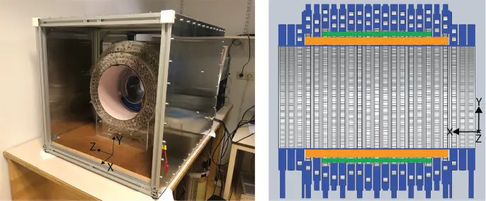
Figure 3.4 A low-cost, lightweight scanner under development for pediatric hydrocephalus monitoring. The system uses a rare-earth permanent magnet “Halbach” arrangement producing a 50-mT transverse field. Magnet bore is 27 cm diameter × 50 cm length.
3.4.3 Mass-effect Monitor in ED or ICU Setting
Similar to the hydrocephalus application, knowledge of a mass effect (compression or displacement of the brain by a pathological source) is critical in acute care because it alerts the care team to the development of significant ICP (which is life-threatening). Mass effects can arise in a wide spectrum of conditions associated with hemorrhage (e.g. traumatic brain injury, rupture of an aneurysm or vascular malformation, hemorrhagic conversion of acute stroke, and hemorrhage resulting from a postsurgical event). Other sources include alterations of CSF dynamics (e.g. obstructive and nonobstructive hydrocephalus, intracranial hypertension, CSF leaks, and other disorders of CSF flow), and cerebral edema (e.g. from demyelinating disease, cerebral infections, or cytotoxic edema due to acute stroke). Finally ICP can be induced by tumors (primary brain tumors, metastases, and extra-axial masses). The presence of ICP is frequently detected by looking for a shift in mid-line structures of the brain, ventricular effacement (one ventricle bigger than the other), or sulcal effacement (CSF spaces in sulci smaller in one hemisphere).
While CT (including portable CT) is effective at identifying a mass effect, it is less useful for answering the second most important clinical question: “Is it getting worse?” Because the mass effect and thus increasing pressure could be developing for hours post injury (e.g. in the case of a hemorrhage after trauma), a single imaging time point is insufficient. Multiple time points are desirable and would ideally be acquired with a wearable monitor. CT is still relatively cumbersome to use repeatedly in a POC setting and the ionizing radiation makes it ill-suited for anything resembling continuous monitoring. Instead, compact, single-sided “MR monitors” could fill this gap. This is a potential new role for MR technology in medicine: a real-time monitor of ventricular/CSF asymmetry to provide an early warning sign of impending herniation, particularly in patients where clinical exam is difficult (e.g. sedated patients).
In a true monitoring scenario, the patient will have the MR monitor on for hours, perhaps days, likely precluding traditional architectures where the patient goes “inside” the bore (of even a small magnet). Instead, the magnet is “worn” or simply adjacent to the head. Such a device will not provide a homogeneous field, or even allow imaging over the whole head. Instead, the clinical information must be extracted from a 3D image of only a region of brain, or perhaps a 2D or 1D image. Nonetheless, this line of instrumentation can follow the extensive development effort previously applied to single-sided MR devices for spectroscopy and materials evaluation [49,71,72], and relaxometry and diffusion measurements in oil well logging [73].
3.4.4 Neonatal Intensive Care Unit (NICU)
A scanner situated in an NICU or capable of being brought to the NICU (beside incubator/isolette) could have many uses. Here we focus on one: HIE to provide a general picture of the uses and issues of MR in a NICU.
HIE and resulting hypoxic-ischemic injury (HII) is the leading cause of neonatal mortality and morbidity with an occurrence of 1 to 8 out of 1000 live births in the United States and up to 26 out of 1000 births in developing countries [74,75]. In survivors, HIE is a predictor of permanent neurodevelopmental disability. The significance of early diagnosis and intervention in HIE is profound, particularly when considering the long-term consequences in remaining years of life.
Once HIE is identified, immediate supportive care is applied to encourage brain perfusion, including intubation and mechanical ventilation for more severely affected neonates, titration of intravenous fluids, and blood pressure support [76]. Continuous electroencephalogram (EEG) monitoring is used to monitor background brain electrical activity and seizures. Expedient intervention with hypothermia neural rescue therapy within the first few hours of life is vital, improving survival rates and reducing future disabilities [77]. Thus, moderate to severe cases undergo neuroprotective interventions with therapeutic hypothermia (also known as hypothermia neural rescue therapy or “cooling”) where a cooling blanket and temperature monitor maintain a body temperature decrease of ~3°C for 72 h. Successful treatment of neonatal brain injury could eliminate many decades of disease burden and healthcare needs.
As the gold standard for brain imaging, MRI is the best imaging technique for diagnosing and staging HIE, particularly with diffusion-weighted contrast [78]. CT is less suitable due to its limited soft tissue contrast and the risks of exposing neonates to ionizing radiation. Although the preferred imaging method, MRI currently requires transport of the fragile patients to a radiology suite. This introduces safety concerns, since it requires a full hand-off of patient care responsibilities and exposes the neonate to stress from transport and acoustic noise. In particular, HIE patients are often not stable enough to transfer to the scanner for several days after birth. Although safety is the number one concern, we must also consider the burden on the hospital workflow. Transfer of critical care patients requires tremendous teamwork and coordination from multiple sources. For example, a neonatologist, nurse, and respiratory therapist must accompany the infant to the scanner and remain with the patient for the duration of the study. The time required for the scan and transfer to/from the NICU is around three hours and NICU staffing coverage must be implemented during their absence. Currently, bedside cranial imaging is available with ultrasound, but provides low sensitivity for early abnormalities associated with HIE [78].
Читать дальшеИнтервал:
Закладка:
Похожие книги на «Magnetic Resonance Microscopy»
Представляем Вашему вниманию похожие книги на «Magnetic Resonance Microscopy» списком для выбора. Мы отобрали схожую по названию и смыслу литературу в надежде предоставить читателям больше вариантов отыскать новые, интересные, ещё непрочитанные произведения.


Обсуждение, отзывы о книге «Magnetic Resonance Microscopy» и просто собственные мнения читателей. Оставьте ваши комментарии, напишите, что Вы думаете о произведении, его смысле или главных героях. Укажите что конкретно понравилось, а что нет, и почему Вы так считаете.