Paul M. Speight - Shear's Cysts of the Oral and Maxillofacial Regions
Здесь есть возможность читать онлайн «Paul M. Speight - Shear's Cysts of the Oral and Maxillofacial Regions» — ознакомительный отрывок электронной книги совершенно бесплатно, а после прочтения отрывка купить полную версию. В некоторых случаях можно слушать аудио, скачать через торрент в формате fb2 и присутствует краткое содержание. Жанр: unrecognised, на английском языке. Описание произведения, (предисловие) а так же отзывы посетителей доступны на портале библиотеки ЛибКат.
- Название:Shear's Cysts of the Oral and Maxillofacial Regions
- Автор:
- Жанр:
- Год:неизвестен
- ISBN:нет данных
- Рейтинг книги:5 / 5. Голосов: 1
-
Избранное:Добавить в избранное
- Отзывы:
-
Ваша оценка:
Shear's Cysts of the Oral and Maxillofacial Regions: краткое содержание, описание и аннотация
Предлагаем к чтению аннотацию, описание, краткое содержание или предисловие (зависит от того, что написал сам автор книги «Shear's Cysts of the Oral and Maxillofacial Regions»). Если вы не нашли необходимую информацию о книге — напишите в комментариях, мы постараемся отыскать её.
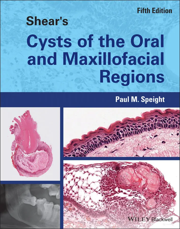
Shear’s Cysts of the Oral and Maxillofacial Regions
Shear’s Cysts of the Oral and Maxillofacial Regions Fifth Edition
Shear's Cysts of the Oral and Maxillofacial Regions — читать онлайн ознакомительный отрывок
Ниже представлен текст книги, разбитый по страницам. Система сохранения места последней прочитанной страницы, позволяет с удобством читать онлайн бесплатно книгу «Shear's Cysts of the Oral and Maxillofacial Regions», без необходимости каждый раз заново искать на чём Вы остановились. Поставьте закладку, и сможете в любой момент перейти на страницу, на которой закончили чтение.
Интервал:
Закладка:
Slabbert et al. (1995 ) studied 154 mandibular radicular and residual cysts and found unequivocal mucous metaplasia in 15 (10%). In many cases they found that the mucous cells were associated with vacuolated cells, many of which were empty, but some contained fine granules or networks of periodic acid–Schiff (PAS)–positive material. The authors observed that the vacuolated cells resembled those described by Fell (1957 ) in the process of metaplasia from stratified squamous to ciliated epithelium in explants of chick embryo skin grown under the influence of excess vitamin A. By analogy to Fell's findings, Slabbert et al. (1995 ) suggested that the vacuolated cells represented an intermediate stage in the process of mucous metaplasia. Takeda et al. (2005 ) found similar clear cells and agreed with Slabbert et al. (1995 ) that mucous cell differentiation is a process of metaplasia. This view is also supported by the fact that mucous cells are found in mandibular lesions. Some have suggested that an origin from antral mucosa (see below) may explain mucous cells in radicular cysts. This may happen on occasion, but the mucous cells in mandibular lesions are almost certainly explained by true metaplasia, and provide good evidence for the metaplastic potential of odontogenic epithelium. Browne (1972 ) also found mucous cells in dentigerous cysts and lateral periodontal cysts, with a similar frequency between mandibular and maxillary lesions.
Cilia are found in radicular cysts with reported frequencies of 0.7% (Browne 1972 ), 11.4% (Takeda et al. 2005 ), 4.8% (Tsesis et al. 2016 ), and 8.2% (Ricucci et al. 2014 ). In his careful ultrastructural studies, Nair examined 39 cysts and found 3 (7.6%) that were lined by ciliated columnar epithelium (Nair et al. 2002 ). All were found in the maxilla and he suggested that the cyst linings were derived in part from cell rests of Malassez, but also from antral mucosa. However, although cilia do appear to be more common in the maxilla, ciliated epithelium has also been found in cysts in the anterior and posterior regions of the mandible. In the study of Takeda et al. (2005 ), ciliated cells were found overall in 11% of radicular cysts, but in 12% and 9% of maxillary and mandibular lesions, respectively. Tsesis et al. (2016 ) found cilia in 4.8% of 711 cysts, but only in 2 (0.2%) mandibular lesions compared to 32 (8.9%) maxillary lesions. Furthermore, 16 were found in the maxillary molar regions, 12 in the anterior region, and 4 associated with premolars. Browne (1972 ) also found that cilia were more frequently encountered in the maxilla, with 2 of 3 being of maxillary origin.
Gao et al. (1988b ) and Lu et al. (2002 ) investigated cytokeratin (CK) expression in radicular cysts. Gao et al. showed strong CK19 expression in rest cells of Malassez and in the epithelium of periapical granulomas and radicular cysts, supporting an odontogenic origin for the cyst lining. As an early change, proliferating epithelium in periapical granulomas also uniformly and strongly expressed CK14 and subsequently CK13 and CK4. Further epithelial changes to form a cyst lining were associated with a more clearly differentiated phenotype of non‐keratinised stratified squamous epithelium expressing CK8 and CK18. Lu et al. (2002 ) confirmed some of these findings and also showed that most cysts expressed CK8 and CK18, as well as CK13. Of relevance to the above discussion, they compared keratin protein and mRNA expression in radicular cysts to normal nasal and oral epithelium. They showed that a CK18+/CK8+/CK13– phenotype was only found in nasal epithelium. Only three cysts showed this phenotype and all three were large maxillary lesions protruding into the maxillary sinus. They concluded that occasional maxillary radicular cysts may not be odontogenic in origin, but that their epithelium may derive from nasal or antral respiratory mucosa.
From these studies, it seems certain that some maxillary cysts may derive at least part of their epithelial lining from the antral mucosa, and this may explain the occurrence of mucous and ciliated cells or respiratory‐type epithelium found in maxillary cysts. Such an occurrence could also be deduced from the radiological appearance of large maxillary cysts, which often clearly protrude into the maxillary sinus. However, the presence of secretory and ciliated epithelium in mandibular radicular cysts also confirms that mucous and ciliated cells may arise as a result of metaplasia.
Hyaline Bodies
In approximately 10% of radicular cysts, hyaline bodies are found in the epithelial linings ( Figure 3.15). Although initially noted by Dewey in 1918, they were first described in detail by Rushton (1955 ) and are often referred to as Rushton bodies . They measure up to about 0.1 mm and are described as having two morphological patterns. The first pattern appears like linear, straight, or curved rods, sometimes with a ‘hairpin’ shape ( Figure 3.15b). These may have a pale centre and a darker eosinophilic or basophilic periphery. Serial sections, however, will show that these are not actually rods, but are folded sheets. The second pattern is of circular or polycyclic bodies ( Figure 3.15), which often have a clear or pale centre, surrounded by concentric laminations that are variably eosinophilic or basophilic. Both patterns may exist together, as can be seen in Figure 3.15. Hyaline bodies are brittle and frequently fracture during histological processing, so that only small fragments may be seen. Occasionally they may form masses that protrude into the cyst lumen ( Figure 3.15a), but only very rarely are they seen in the fibrous capsule. The presence of hyaline bodies may be suspected if, on examination of the gross specimen, the pathologist sees small, smooth, white, dome‐shaped swellings of the epithelial surface protruding into the cyst cavity.
Although they are typically seen in radicular cysts, they may also be encountered in dentigerous cysts or odontogenic keratocysts, but are always associated with areas of inflammation. Lam and Chan (2000 ) reported hyaline bodies in 7% of a series of 69 odontogenic keratocysts and Lin et al. (2013 ) found them in 11% of dentigerous cysts. Ide et al. (1996 ) reported an unusual glandular odontogenic cyst with hyaline bodies, and Takeda et al. (1985 ) described their presence in a plexiform ameloblastoma. They are, however, specific to odontogenic epithelium and have not been reported in non‐odontogenic cysts. Occasionally similar structures may be found associated with epithelial rests in dental follicles.
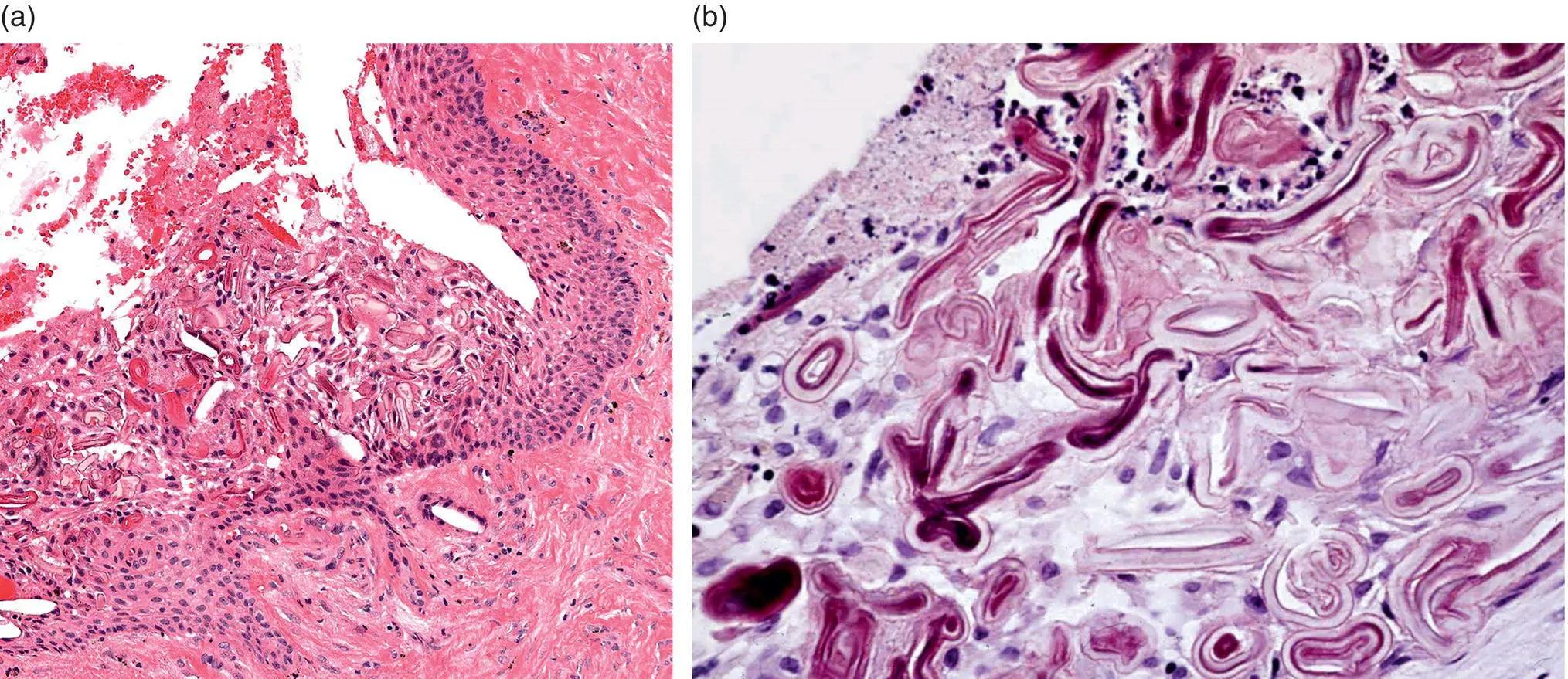
Figure 3.15 Hyaline bodies in the epithelial lining of a radicular cyst. (a) The bodies accumulate in the epithelial lining and form nodules that protrude into the lumen. (b) The bodies are eosinophilic and form circular, folded, and curved ‘rod’ shapes.
Hyaline bodies are intriguing and enigmatic, but they have little diagnostic or prognostic significance. More than 100 years after they were first noted, the origin and pathogenesis of these bodies is still debated and no consensus has been reached. There have been two competing theories of their origin: first, that they derive from epithelium; and second, that they are of haematogenous origin. Rushton (1955 ) believed that the hyaline bodies resembled, in appearance and the liability to fracture, the keratinised secondary enamel cuticle of Gottlieb. Wertheimer (Wertheimer et al. 1962 ; Wertheimer 1966 ) directly compared enamel cuticle and hyaline bodies and showed similar staining for a number of histochemical stains, also concluding that the bodies represented a keratin‐like epithelial product equivalent to dental cuticle.
Читать дальшеИнтервал:
Закладка:
Похожие книги на «Shear's Cysts of the Oral and Maxillofacial Regions»
Представляем Вашему вниманию похожие книги на «Shear's Cysts of the Oral and Maxillofacial Regions» списком для выбора. Мы отобрали схожую по названию и смыслу литературу в надежде предоставить читателям больше вариантов отыскать новые, интересные, ещё непрочитанные произведения.


Обсуждение, отзывы о книге «Shear's Cysts of the Oral and Maxillofacial Regions» и просто собственные мнения читателей. Оставьте ваши комментарии, напишите, что Вы думаете о произведении, его смысле или главных героях. Укажите что конкретно понравилось, а что нет, и почему Вы так считаете.