Jane Flint - Principles of Virology
Здесь есть возможность читать онлайн «Jane Flint - Principles of Virology» — ознакомительный отрывок электронной книги совершенно бесплатно, а после прочтения отрывка купить полную версию. В некоторых случаях можно слушать аудио, скачать через торрент в формате fb2 и присутствует краткое содержание. Жанр: unrecognised, на английском языке. Описание произведения, (предисловие) а так же отзывы посетителей доступны на портале библиотеки ЛибКат.
- Название:Principles of Virology
- Автор:
- Жанр:
- Год:неизвестен
- ISBN:нет данных
- Рейтинг книги:3 / 5. Голосов: 1
-
Избранное:Добавить в избранное
- Отзывы:
-
Ваша оценка:
Principles of Virology: краткое содержание, описание и аннотация
Предлагаем к чтению аннотацию, описание, краткое содержание или предисловие (зависит от того, что написал сам автор книги «Principles of Virology»). Если вы не нашли необходимую информацию о книге — напишите в комментариях, мы постараемся отыскать её.
Volume I: Molecular Biology
Volume II: Pathogenesis and Control
Principles of Virology, Fifth Edition
Principles of Virology — читать онлайн ознакомительный отрывок
Ниже представлен текст книги, разбитый по страницам. Система сохранения места последней прочитанной страницы, позволяет с удобством читать онлайн бесплатно книгу «Principles of Virology», без необходимости каждый раз заново искать на чём Вы остановились. Поставьте закладку, и сможете в любой момент перейти на страницу, на которой закончили чтение.
Интервал:
Закладка:
As early efforts to study virus infections in single cells were hampered by technical difficulties, the field failed to progress. This situation has changed with the development of flow cytometry and microfluidics and the adaptation of highthroughput methods, such as genome sequencing and mass spectrometry, to single cells.
Initially, micropipettes were used to aspirate a single cell at a time from a population, using a microscope. This labor-intensive method was supplanted by fluorescence-activated cell sorting to allow isolation of up to millions of cells in a few hours, according to size, morphology, or synthesis of specific proteins. More recently, automated microfluidic devices have been developed to allow automated capture of single cells using integrated fluidic circuits. Infection, cell lysis, reverse transcription, and amplification are all performed in these systems before high-throughput sequencing.
The study of virus infections in single cells is expected to provide information that explains why some cells are not infected, why the kinetics of viral reproduction may be so different, and how genomes change in a single cell. An example is the study of poliovirus infection of single cells, using a microfluidics platform installed on a fluorescent microscope ( Fig. 2.22). This approach revealed observations otherwise masked in population-based studies, including the unique and independent contribution of viral and cell parameters to reproduction kinetics, the wide variation in reproduction start times, and the finding that reproduction begins later and with greater speed in single cells than in populations. A study of influenza virus infection of single cells revealed a wide variation in the yield, from 1 to 970 PFU per cell. Furthermore, the amounts of viral RNAs within individual cells varied by three orders of magnitude.
BOX 2.12
WARNING
Determining a role for cellular proteins in viral reproduction can be quite difficult
Understanding the roles of both viral and cellular proteins at various stages of viral reproduction is essential for elucidating molecular mechanisms and for developing strategies for blocking pathogenic infections. As viral genomes have a limited set of genes, the viral proteins or genetic elements that are essential at each step can be deduced by introducing mutations and observing phenotypes. Identifying critical cellular genes begins with the identification of cellular proteins that are included in virus particles and/or bind to viral proteins ( in vitro or in cells).
Once candidates are identified, the contribution of the cellular protein to viral reproduction may be evaluated by observing the effects of
specific small-molecule inhibitors of the protein’s function (inhibitory drugs)
synthesis of an altered protein, known to have a dominant negative effect on its normal function
treatment with small RNAs that induce mRNA degradation (see Chapter 10) and reduce the concentration of the cellular protein
reproduction in cells in which the candidate gene has been mutated or deleted
Even after applying the multiple approaches and methods described above, identifying relevant cellular proteins and evaluating their roles in viral reproduction is seldom easy. The problems encountered include the following.
More than one protein may provide the required function (redundancy).
The function of the protein might be essential to the cell, and mutation of the gene that encodes it (or inhibition of protein production) could be lethal.
Only small quantities of the protein might be required, and reducing its activity with an inhibitor, or its concentration may be insufficient to induce a defect in viral reproduction.
The cellular protein might provide a slight enhancement to viral reproduction that could be difficult to detect but may be physiologically significant.
Synthesis of an altered cellular gene or overexpression of a normal cellular gene may produce changes that affect virus reproduction for reasons that are irrelevant to the natural infection (artifacts).

Given these difficulties, it is not surprising that the literature in this area is sometimes contradictory and the results can be controversial.
Infection of single cells with vesicular stomatitis virus identified 496 mutations that arose in 24 hours during genome replication within 90 cells. The rates of mutation varied among individual cells, and this high value represents an average for all of the cells. In addition, preexisting viral genetic diversity was used to track infection in single cells. These investigations revealed that even though viruses were added at a low multiplicity of infection, most cells had acquired more than one virus particle. The results suggested that virus particles have a tendency to stick to one another, raising further challenges to determining multiplicities of infection.
Single-cell studies have demonstrated that measurements of virus reproduction in populations of cells do not represent the diversity that exists among individual cells. Consequently, they will likely become a complementary tool to the one-step growth experiment for studying virus infection.
Perspectives
One-step growth analysis, while simple, remains a powerful tool for studying virus reproduction. When cells are infected at a high multiplicity of infection, sufficient viral nucleic acid or protein can be isolated to allow a study of their production during the infectious cycle. The ability to synchronize infection is the key to this approach. Many of the experimental results discussed in subsequent chapters of this book were obtained using such one-step growth analysis. The power of this approach is such that it reports on all stages of the reproduction cycle in a simple and quantitative fashion. With modest expenditure of time and reagents, virologists can deduce a great deal about viral translation, genome replication, and assembly. It has long been assumed from such one-step growth analyses that the same steps of the viral reproduction cycle occur at the same time in every infected cell. However, results from analyses of single infected cells demonstrate that the same steps can take place at vastly different times in individual cells in the population. We now understand that results from population-based studies of viral reproduction comprise an average of events occurring in individual cells. One-step growth analyses with single cells have the potential of unraveling the viral and cellular basis for such individual heterogeneity.
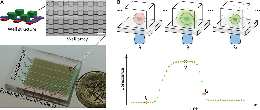
Figure 2.22 Single-cell virology. (A)A microfluidic device with 6,400 wells is fitted with four separate sample inlets (green) and pneumatic control lines (red) that permit each well to be sealed and isolated. A small part of the device is magnified at the top, showing an array of 24 wells, and four wells are further magnified to the left. (B)The device can be used to measure real-time fluorescence in cells infected with a virus encoding a fluorescent reporter. The production of fluorescence is shown in the graph and illustrated in the views of single cells in individual cells above. There is a lag in the detection of fluorescence in an infected cell (t i), followed by virus reproduction (t j) and a decline in fluorescence caused by cell lysis (t k). Reprinted from Guo F et al. 2017. Cell Rep 21:1692-1704, with permission.
Читать дальшеИнтервал:
Закладка:
Похожие книги на «Principles of Virology»
Представляем Вашему вниманию похожие книги на «Principles of Virology» списком для выбора. Мы отобрали схожую по названию и смыслу литературу в надежде предоставить читателям больше вариантов отыскать новые, интересные, ещё непрочитанные произведения.


Обсуждение, отзывы о книге «Principles of Virology» и просто собственные мнения читателей. Оставьте ваши комментарии, напишите, что Вы думаете о произведении, его смысле или главных героях. Укажите что конкретно понравилось, а что нет, и почему Вы так считаете.