Point-of-Care Ultrasound Techniques for the Small Animal Practitioner
Здесь есть возможность читать онлайн «Point-of-Care Ultrasound Techniques for the Small Animal Practitioner» — ознакомительный отрывок электронной книги совершенно бесплатно, а после прочтения отрывка купить полную версию. В некоторых случаях можно слушать аудио, скачать через торрент в формате fb2 и присутствует краткое содержание. Жанр: unrecognised, на английском языке. Описание произведения, (предисловие) а так же отзывы посетителей доступны на портале библиотеки ЛибКат.

- Название:Point-of-Care Ultrasound Techniques for the Small Animal Practitioner
- Автор:
- Жанр:
- Год:неизвестен
- ISBN:нет данных
- Рейтинг книги:5 / 5. Голосов: 1
-
Избранное:Добавить в избранное
- Отзывы:
-
Ваша оценка:
Point-of-Care Ultrasound Techniques for the Small Animal Practitioner: краткое содержание, описание и аннотация
Предлагаем к чтению аннотацию, описание, краткое содержание или предисловие (зависит от того, что написал сам автор книги «Point-of-Care Ultrasound Techniques for the Small Animal Practitioner»). Если вы не нашли необходимую информацию о книге — напишите в комментариях, мы постараемся отыскать её.
New chapters in
cover ultrasound-guided nerve blocks, musculoskeletal, brain imaging, and applications of focused ultrasound techniques in cats, exotics and marine mammals—making it an essential purchase for veterinarians wanting to incorporate point-of-care ultrasound techniques into their veterinary practices.
Presents a standardized approach to point-of-care ultrasound as an extension of the physical exam, including trauma, non-trauma, and monitoring applications Includes coverage of new techniques for focused ultrasound exams, including lung, anesthesia and ultrasound guided nerve blocks, transcranial brain imaging, musculoskeletal, volume status evaluation, and rapid assessment for treatable forms of shock Adds cats, exotic and wildlife mammals, and marine mammals to the existing canine coverage Emphasizes the integration of point-of-care ultrasound techniques for optimizing patient care and accurate patient assessment Offers access to a companion website with supplementary video clips showing many clinically relevant didactic examples The second edition of
is an excellent resource for veterinary practitioners, ranging from the general practitioner to nearly all clinical specialists, including internal medicine, oncology, cardiology, emergency and critical care, anesthesiology, ophthalmology, exotics, and zoo medicine specialists, and veterinary students.
Point-of-Care Ultrasound Techniques for the Small Animal Practitioner — читать онлайн ознакомительный отрывок
Ниже представлен текст книги, разбитый по страницам. Система сохранения места последней прочитанной страницы, позволяет с удобством читать онлайн бесплатно книгу «Point-of-Care Ultrasound Techniques for the Small Animal Practitioner», без необходимости каждый раз заново искать на чём Вы остановились. Поставьте закладку, и сможете в любой момент перейти на страницу, на которой закончили чтение.
Интервал:
Закладка:
Hepatic hematomas may be variable in appearance and initially are generally anechoic or hypoechoic to normal hepatic parenchyma. Hematomas may be difficult to distinguish from other mass lesions, including neoplasia (hemangiosarcoma). Frequently, large hematomas usually form with ruptured hepatic masses and can complicate diagnosis ( Figure 8.8A,B; see also Figure 8.7B). Fine needle aspiration of hematomas generally yields nonspecific cytological results and the absence of neoplastic cells on cytology does not rule out neoplasia. Figure 8.6. Hepatic cyst and biliary cysts. (A) Hepatic cyst (arrow) with distal acoustic enhancement (seen as a hyperechoic, bright enhancement extending from the far margin of the hepatic cyst and marked with subtle black cursors [> <] on both sides of the hyperechoic track). Hepatic cysts are typically thin‐walled and filled with anechoic fluid. A fluid‐filled structure can be differentiated from a solid nodule by looking in the far‐field for acoustic enhancement artifact (the bright track extending from the far field of the cyst to the text L LIVER). A solid or mineralized structure will conversely have acoustic shadowing artifact in the far‐field (a dark track extending from the far‐field of the solid or mineralized structure). (B) Multiple benign biliary cystadenomas in a cat (labeled liver masses by the sonographer). This can be mistaken for malignant cystic neoplasia. (C) Large hepatic cyst in a dog. Note the large anechoic structure with distal acoustic enhancement artifact extending from the far border of the cyst (labeled > <). Cysts can be complex in architecture and within this cyst is a large, echogenic, ovoid to amorphous structure. Irregularly marginated cysts or those containing echogenic material can be malignant. An additional differential for the structure within the cyst includes fibrinous material or hemorrhage and may represent abscessation, inflammation or neoplastic change. Fine needle aspiration may allude to the diagnosis. Alternatively, excisional biopsy may be needed.
Neoplastic lesions have a wide range of morphological changes. Again, it should be emphasized that it is not possible to distinguish between benign and malignant lesions based on ultrasonographic appearance alone. There are a variety of primary liver tumors including hepatocellular adenoma and adenocarcinoma as well as mesenchymal tumors (see Figure 8.7B–D). Lymphosarcoma can appear as either focal or multifocal mass lesions or as diffuse parenchymal disease. Hemangiosarcoma generally appears as complex mass lesions with mixed echogenicity and cavitations (see Figures 8.7B and 8.8A,B). Hematomas can have a similar appearance. Metastatic neoplasia is common in the liver ( Figure 8.9A,B). Hepatic mass lesions must be characterized by cytology or histopathology for a definitive diagnosis. Ultrasound‐guided fine needle or Tru‐Cut biopsy of mass lesions can usually be accomplished with minimal difficulty and risk, depending on the location of the mass. Figure 8.7. Various liver masses. Benign and malignant liver masses cannot be differentiated based on ultrasound characteristic alone. (A) Semisolid hepatic mass in a cat confirmed to be a hepatic abscess. The gallbladder (GB) is juxtaposed to the mass and may be confused with free fluid on this still image, highlighting the value of a several‐second video when images are submitted to the specialist for interpretation. Although not always present, the hallmark of an abscess regardless of location is the presence of gas within the mass effect as evidenced by comet tails and other air‐related artifacts caused by gas‐producing bacteria (such artifacts are not evident here). Diagnosis was made by liver biopsy. (B) A large, mixed echogenicity mass (MASS). It can be difficult to determine the origin of large masses. The liver, spleen, kidney or other adjacent structures should be considered if a region of organ confluence cannot be identified. The histopathological diagnosis in this case was hepatic hemangiosarcoma. (C) Another example of mixed echogenicity with a hepatic mass adjacent to the diaphragm. Note the abrupt change of echogenicity between the mass and normal hepatic parenchyma. Percutaneous biopsy result was hepatocellular carcinoma. (D) Large hyperechoic, lobulated mass with mild peritoneal effusion within the gallbladder fossa. The lesion was confirmed to be hepatocellular carcinoma. Note the differences between sonographic appearances of the same tumor type in (C) and (D) demonstrating the poor specificity of ultrasonographic findings with regard to histopathological diagnosis. Additional differentials might have included a hepatoma (benign liver mass) or other types of solid parenchymal neoplasia (GB, gallbladder; RLiver, right liver; RK, right kidney). Figure 8.8. Liver and splenic masses. The origin of large masses can be challenging to determine ultrasonographically and is sometimes not possible. (A) and (B) show the same large mass in a dog with mixed echogenicity (unlabeled and labeled). Careful evaluation showed confluence of the tumor with the splenic parenchyma. (C,D) Images of a cat (same patient) with hepatic mast cell tumor. Note that a well‐defined mass is not seen. Rather, the parenchymal changes are subtle and the liver is coarse in echotexture. This case highlights the importance of fine needle aspirate and/or liver biopsy in obtaining the patient’s definitive diagnosis.
Target lesions consist of alternating layers of different echogenicities, giving the appearance of a target or bull's eye. Single target lesions have a high predictive value for malignancy. Multiple target lesions increase the positive predictive value of malignancy up to 74–81% (Cuccovillo and Lamb 2002) (see Figure 8.9A,B). Regardless, target lesions are nonspecific and a biopsy is required for further characterization. If target lesions are detected, then the authors recommend screening for additional evidence of neoplasia with a complete detailed abdominal ultrasound, thoracic radiographs, and lung ultrasound. However, performing a Global FAST immediately following your POCUS examination is helpful for a quick survey of the remaining abdominal structures, the thorax (TFAST), and lung surface, especially for nodules (Vet BLUE).
Liver Parenchymal Disease
Ultrasonography is not always sensitive for evaluating diffuse parenchymal disease, and diffuse disease may be more challenging to assess than focal disease because sonographic changes are more easily overlooked or may not be present. When there is a high index of suspicion for diffuse hepatic disease, fine needle aspiration or biopsy is indicated to confirm a diagnosis, and ultrasound guidance can assist with these procedures. Despite sonographic limitations, some generalizations can be made regarding changes in echogenicity.
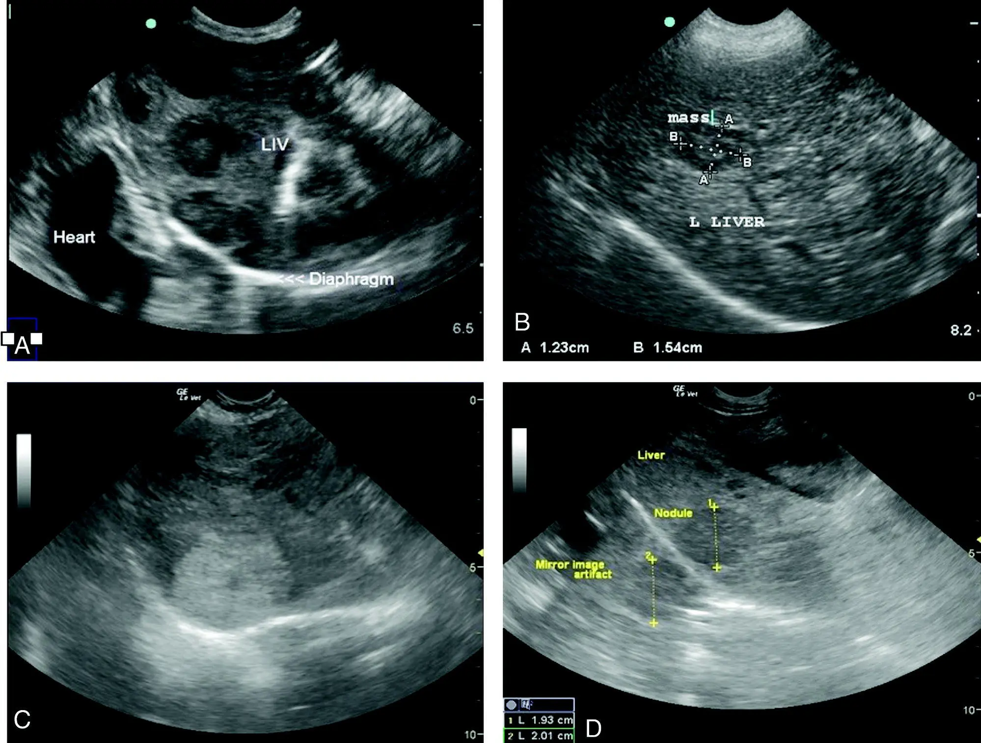
Figure 8.9. Liver masses. (A) Image of a dog with target lesions of the hepatic parenchyma. These lesions are generally considered malignant and may be consistent with metastatic disease. Less commonly, this appearance may be seen with primary hepatic neoplasia, granulomatous disease, chronic active hepatitis or even benign hyperplasia. This canine patient also had a large cavitary splenic mass supportive of metastatic disease, pericardial effusion and lung nodules emphasizing the importance of the Global FAST approach for staging. (B) An example of a small hepatic target lesion in a dog with splenic hemangiosarcoma. One must keep in mind that sonographic visualization of a target lesion is nonspecific and a biopsy is required; however, the Global FAST approach can help stage and guide diagnostic testing. (C) A large hyperechoic, well‐marginated mass in a dog that was percutaneously biopsied as hepatocellular carcinoma. (D) Another example of hepatocellular carcinoma. Note in both (C) and (D) the commonly encountered mirror image artifact because of the strong, reflective soft tissue–air interface of the liver, diaphragm, and lung.
Читать дальшеИнтервал:
Закладка:
Похожие книги на «Point-of-Care Ultrasound Techniques for the Small Animal Practitioner»
Представляем Вашему вниманию похожие книги на «Point-of-Care Ultrasound Techniques for the Small Animal Practitioner» списком для выбора. Мы отобрали схожую по названию и смыслу литературу в надежде предоставить читателям больше вариантов отыскать новые, интересные, ещё непрочитанные произведения.


Обсуждение, отзывы о книге «Point-of-Care Ultrasound Techniques for the Small Animal Practitioner» и просто собственные мнения читателей. Оставьте ваши комментарии, напишите, что Вы думаете о произведении, его смысле или главных героях. Укажите что конкретно понравилось, а что нет, и почему Вы так считаете.