Point-of-Care Ultrasound Techniques for the Small Animal Practitioner
Здесь есть возможность читать онлайн «Point-of-Care Ultrasound Techniques for the Small Animal Practitioner» — ознакомительный отрывок электронной книги совершенно бесплатно, а после прочтения отрывка купить полную версию. В некоторых случаях можно слушать аудио, скачать через торрент в формате fb2 и присутствует краткое содержание. Жанр: unrecognised, на английском языке. Описание произведения, (предисловие) а так же отзывы посетителей доступны на портале библиотеки ЛибКат.

- Название:Point-of-Care Ultrasound Techniques for the Small Animal Practitioner
- Автор:
- Жанр:
- Год:неизвестен
- ISBN:нет данных
- Рейтинг книги:5 / 5. Голосов: 1
-
Избранное:Добавить в избранное
- Отзывы:
-
Ваша оценка:
Point-of-Care Ultrasound Techniques for the Small Animal Practitioner: краткое содержание, описание и аннотация
Предлагаем к чтению аннотацию, описание, краткое содержание или предисловие (зависит от того, что написал сам автор книги «Point-of-Care Ultrasound Techniques for the Small Animal Practitioner»). Если вы не нашли необходимую информацию о книге — напишите в комментариях, мы постараемся отыскать её.
New chapters in
cover ultrasound-guided nerve blocks, musculoskeletal, brain imaging, and applications of focused ultrasound techniques in cats, exotics and marine mammals—making it an essential purchase for veterinarians wanting to incorporate point-of-care ultrasound techniques into their veterinary practices.
Presents a standardized approach to point-of-care ultrasound as an extension of the physical exam, including trauma, non-trauma, and monitoring applications Includes coverage of new techniques for focused ultrasound exams, including lung, anesthesia and ultrasound guided nerve blocks, transcranial brain imaging, musculoskeletal, volume status evaluation, and rapid assessment for treatable forms of shock Adds cats, exotic and wildlife mammals, and marine mammals to the existing canine coverage Emphasizes the integration of point-of-care ultrasound techniques for optimizing patient care and accurate patient assessment Offers access to a companion website with supplementary video clips showing many clinically relevant didactic examples The second edition of
is an excellent resource for veterinary practitioners, ranging from the general practitioner to nearly all clinical specialists, including internal medicine, oncology, cardiology, emergency and critical care, anesthesiology, ophthalmology, exotics, and zoo medicine specialists, and veterinary students.
Point-of-Care Ultrasound Techniques for the Small Animal Practitioner — читать онлайн ознакомительный отрывок
Ниже представлен текст книги, разбитый по страницам. Система сохранения места последней прочитанной страницы, позволяет с удобством читать онлайн бесплатно книгу «Point-of-Care Ultrasound Techniques for the Small Animal Practitioner», без необходимости каждый раз заново искать на чём Вы остановились. Поставьте закладку, и сможете в любой момент перейти на страницу, на которой закончили чтение.
Интервал:
Закладка:
Source: (B) and (D) courtesy of Dr Gregory Lisciandro, Hill Country Veterinary Specialists and FASTVet.com, Spicewood, TX, and (C) courtesy of Dr Terri DeFrancesco, Raleigh, NC.
The gallbladder is surrounded by hepatic parenchyma on the right side of the liver. The size is variable depending on when gallbladder contraction last occurred. In the typical normal state, the gallbladder is an anechoic structure with a uniformly thin echogenic wall. Normal gallbladder wall thickness is less than 1 mm in cats and less than 3 mm in dogs (Hittmair et al. 2001; Spaulding 1993); however, for all intents and purposes, the gallbladder wall should be a thin white line in both species ( Figure 8.4A–C; see also Figure 8.11). Gallbladder sludge (echogenic gravity‐dependent material within the lumen) is common in dogs and generally a benign finding in the absence of other significant ultrasonographic and clinical findings although the presence of “normal” gallbladder sludge has been more recently questioned (Tsukagoshi et al. 2012) (see Figure 8.12A–D). The finding may be an indication of cholestasis. The intrahepatic bile ducts are not seen unless there is extrahepatic biliary obstruction of at least five days’ duration when the fluid dilated ducts will have the appearance of hepatic veins (without the hyperechoic walls of portal veins) (see Figure 8.15A,B). If available, color flow Doppler is useful for differentiating the lack of flow in a dilated biliary duct from the normal blood flow in hepatic and portal vessels; however, tracing structures in real time is often adequate. The common bile duct is small and difficult to see in the normal state in dogs and cats, but is generally easier to identify in the cat.
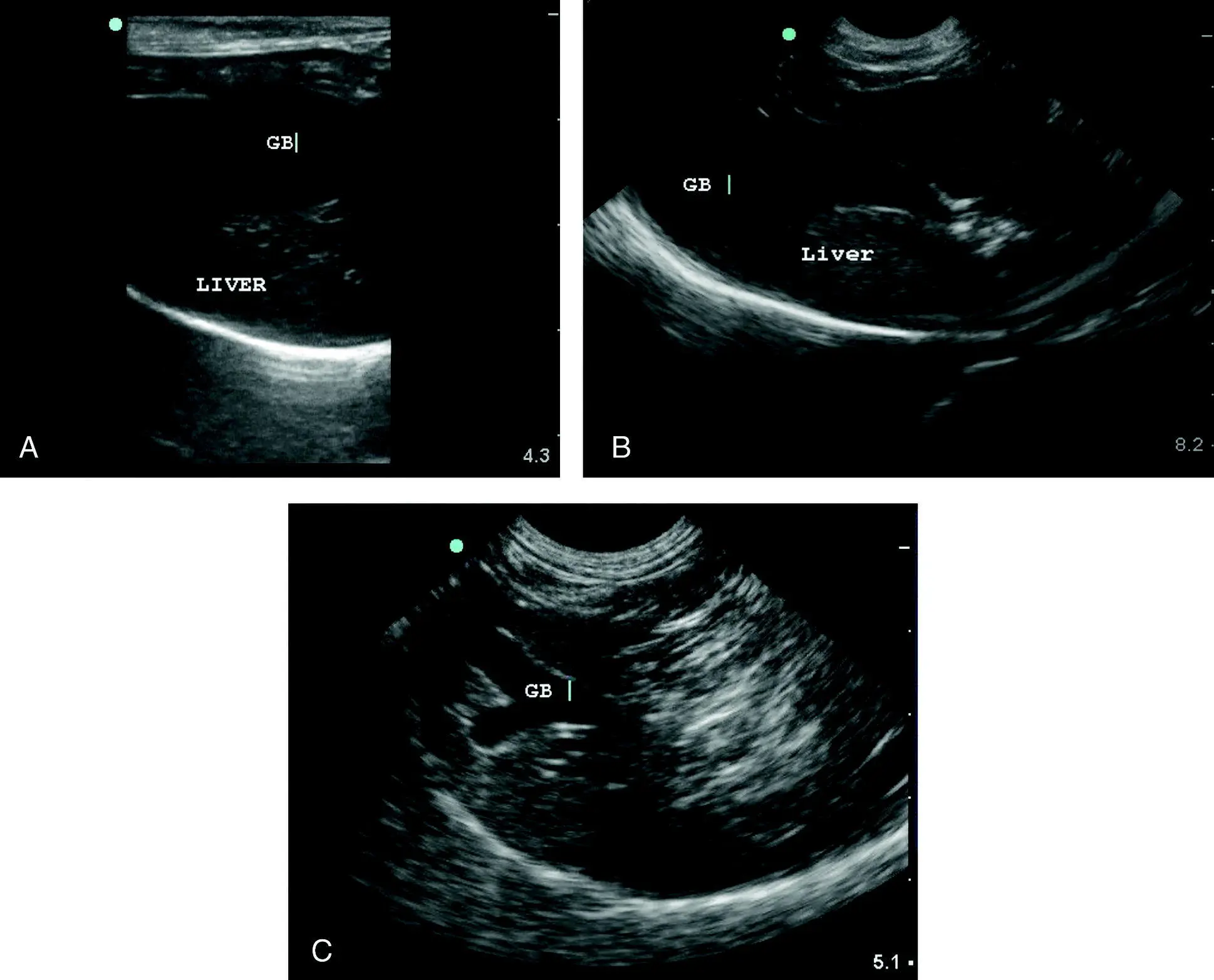
Figure 8.4. Normal gallbladder and variations. (A) The normal gallbladder wall (not labeled) is inconspicuous with normal thickness <1 mm in cats and <3 mm in dogs (GB, gallbladder). However, normal wall thickness does not rule out mild, low‐grade or chronic inflammation. (B) Similarly labeled image showing a normal canine gallbladder wall. Note the anechoic structure is marginated ultrasonographically by a thin hyperechoic (bright white) line. Evaluation of the margin of the gallbladder in real time provides a global view of the gallbladder wall and can aid in determining gallbladder wall integrity. The curvilinear line in the far‐field is created by the interface between the diaphragm and lung (air). (C) Image of a bilobed gallbladder may be seen in both dogs and cats but is more common in cats, as was seen in this normal feline patient (GB, gallbladder).
Pearl:It may be difficult to tell the difference between a dilated biliary tract (no flow) and hepatic veins (flow), in which case color flow Doppler may be helpful.
Artifacts are commonly encountered when performing the POCUS liver and gallbladder examination and include mirror image, acoustic enhancement, side‐lobe, slice thickness, and edge shadowing. These are discussed in more detail in Chapter 3and 5.
Mirror image. The most common of these artifacts results in a mirror image of the liver and gallbladder being seen on the far side of the lung–diaphragm interface. This should not be mistaken for a diaphragmatic hernia (see Figure 8.9D).
Acoustic enhancement. Acoustic enhancement is associated with a fluid‐filled gallbladder that causes an increase in echogenicity of the hepatic parenchyma on the far side of the gallbladder. Acoustic enhancement should not be mistaken for increased liver echogenicity.
Side‐lobe and slice thickness. These artifacts involve fluid‐filled organs like the gallbladder and give the false impression of the presence of luminal sediment.
Edge shadowing. This artifact also involves fluid‐filled organs like the gallbladder and gives the appearance of a defect in the wall that can be mistaken for gallbladder rupture or gallbladder wall mineralization.
Pearl:Common artifacts in the liver include mirror image, acoustic enhancement, side‐lobe, slice thickness, and edge shadow.
Clinical Significance and Implications of Abnormal Focused Liver and Gallbladder Findings
Goals of POCUS liver and gallbladder examination include recognition of liver masses (single, multifocal), obvious changes in liver echogenicity, hepatic venous congestion, identification of gallbladder wall abnormalities and gallbladder luminal conditions, and recognition of signs of biliary obstruction. Such findings should prompt referral for a complete detailed abdominal ultrasound as interventions may be indicated.
Liver Masses
Identification of focal liver masses is the easiest task to learn with the POCUS examination. It is important to remember that is not possible to differentiate between benign and malignant processes based on ultrasound alone. Focal lesions may be caused by regenerative nodules or nodular hyperplasia, benign or malignant neoplasia, focal inflammatory disease (e.g., abscesses and granulomas), cysts, and hematomas.
The origin of a large midabdominal mass can sometimes be difficult to determine when the mass contacts multiple origins. In this case, try repositioning the patient from dorsal to lateral recumbency. This will often separate the liver and spleen from one another and help determine the origin of the mass. Generally, the spleen is differentiated from the liver by its hyperechoic capsule (bright white) and its vasculature splitting its capsule unlike the liver.
Benign hepatic nodular disease is common and nodules associated with nodular hyperplasia may be variable in echogenicity when compared to normal hepatic parenchyma (hyperechoic, hypoechoic, isoechoic or mixed echogenicity). Generally, well‐defined hyperechoic homogeneous nodules are more likely to be benign but cytology or histopathology is necessary for definitive evaluation. Nodular hyperplasia is common in older dogs (Nyland et al. 2002) ( Figure 8.5).
Hepatic cysts consist of round to oval anechoic (fluid‐filled) structures with well‐defined walls. These are generally benign unless a significant amount of hepatic architecture is disrupted and there is evidence of hepatic dysfunction (polycystic disease). Cysts may be parenchymal or biliary in nature, and may be congenital or acquired. Cysts that have irregular margins or contain echogenic material are less likely to be benign and may represent abscesses, inflammation or neoplastic changes ( Figure 8.6A–C).
Other parenchymal masses may have variable appearances and include abscess, hematoma, nodular hyperplasia and neoplasia.
Hepatic abscesses are uncommon, usually indistinguishable from nodules and mass lesions of other etiology, and may have variable echogenicity, appearance, and margination ( Figure 8.7A). Most commonly, abscesses are relatively hypoechoic, thick‐walled lesions which may have cystic or cavitary components or mixed echogenicity. A more definitive finding is the presence of gas (due to gas‐forming bacteria) within the lesion, which appears as focal areas of hyperechogenicity with distal edge shadowing, comet tailing, and reverberation. Fine needle aspiration with cytology and microbial culture and sensitivity is helpful in confirming the diagnosis. However, neoplastic lesions can have necrotic centers that can be mistaken for abscesses and therefore, diagnosis should always be in conjunction with additional clinical findings that support the diagnosis. The Global FAST approach is helpful as a quick screening test for disseminated versus localized disease and the presence or absence of lung nodules (quick pulmonary metastasis check). Figure 8.5. Nodular hyperplasia. (A) Small hyperplastic nodule as demarcated by the caliper marks made by the sonographer, consistent histopathologically with benign nodular hyperplasia. This is a common finding in older dogs. Generally, nodules are seen as discrete, well‐demarcated hyper‐ or hypoechoic homogeneous nodules and can be singular or multiple and poorly demarcated. Nodules can be any size <3 cm (mass defined as >3 cm). Hence, findings are nonspecific and fine needle aspiration and/or biopsy is needed to differentiate these nodules from malignant processes. (B) Nodular hyperplasia with multiple varying sized, homogeneous, hypoechoic nodules with the largest marked with calipers (+). (C) Example of an older dog with multiple hypoechoic nodules confirmed to be benign nodular hyperplasia. (D) In contrast to (B), an example of a severely nodular liver in a 10‐year‐old male castrated Chihuahua with poorly controlled diabetes and pancreatitis. The process is diffuse and the liver margins are severely rounded. The calipers (+) demonstrate a hypoechoic cyst. Notice its distal acoustic enhancement and the multiple, hypoechoic (dark) nodules throughout the parenchyma. Fine needle aspiration results were consistent with a vacuolar degeneration and lipidosis. Benign hyperplasia and neoplasia would be considered additional differentials. A biopsy would be more definitive in this case.
Читать дальшеИнтервал:
Закладка:
Похожие книги на «Point-of-Care Ultrasound Techniques for the Small Animal Practitioner»
Представляем Вашему вниманию похожие книги на «Point-of-Care Ultrasound Techniques for the Small Animal Practitioner» списком для выбора. Мы отобрали схожую по названию и смыслу литературу в надежде предоставить читателям больше вариантов отыскать новые, интересные, ещё непрочитанные произведения.


Обсуждение, отзывы о книге «Point-of-Care Ultrasound Techniques for the Small Animal Practitioner» и просто собственные мнения читателей. Оставьте ваши комментарии, напишите, что Вы думаете о произведении, его смысле или главных героях. Укажите что конкретно понравилось, а что нет, и почему Вы так считаете.